University of Nebraska - Lincoln University of Nebraska - Lincoln DigitalCommons@University of Nebraska - Lincoln DigitalCommons@University of Nebraska - Lincoln Faculty Publications from the Harold W. Manter Laboratory of Parasitology Parasitology, Harold W. Manter Laboratory of 2003 The Blue Crab: Diseases, Parasites and Other Symbionts The Blue Crab: Diseases, Parasites and Other Symbionts Jeffrey D. Shields Virginia Institute of Marine Science, [email protected]Robin M. Overstreet Gulf Coast Research Laboratory, [email protected]Follow this and additional works at: https://digitalcommons.unl.edu/parasitologyfacpubs Part of the Parasitology Commons Shields, Jeffrey D. and Overstreet, Robin M., "The Blue Crab: Diseases, Parasites and Other Symbionts" (2003). Faculty Publications from the Harold W. Manter Laboratory of Parasitology. 426. https://digitalcommons.unl.edu/parasitologyfacpubs/426 This Article is brought to you for free and open access by the Parasitology, Harold W. Manter Laboratory of at DigitalCommons@University of Nebraska - Lincoln. It has been accepted for inclusion in Faculty Publications from the Harold W. Manter Laboratory of Parasitology by an authorized administrator of DigitalCommons@University of Nebraska - Lincoln.
Transcript
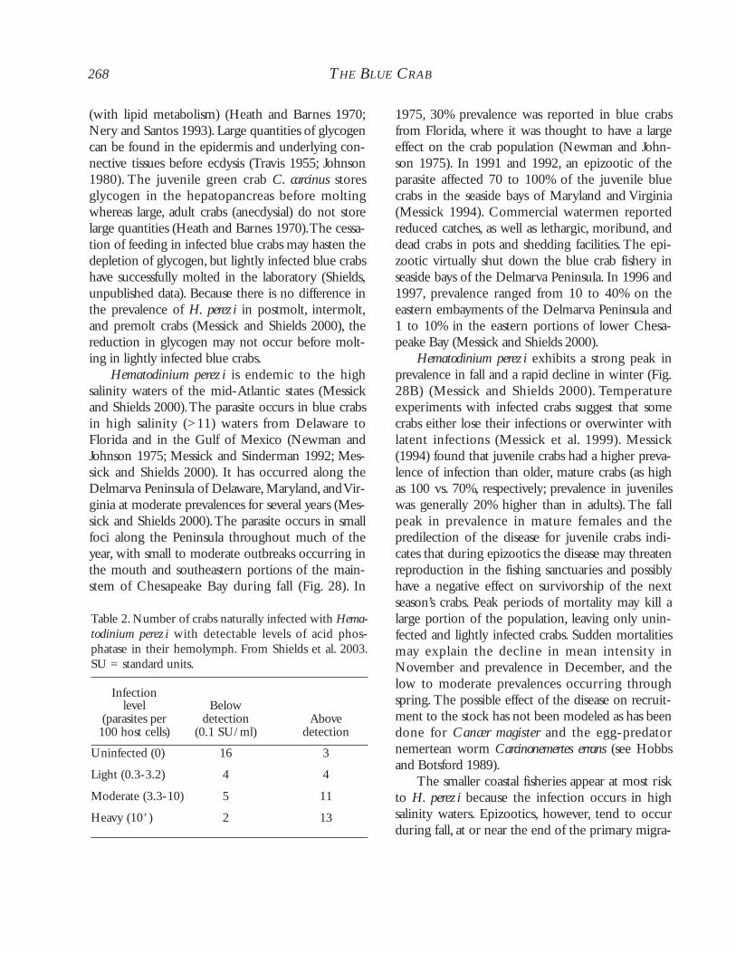
University of Nebraska - Lincoln University of Nebraska - Lincoln
DigitalCommons@University of Nebraska - Lincoln DigitalCommons@University of Nebraska - Lincoln
Faculty Publications from the Harold W. Manter Laboratory of Parasitology Parasitology, Harold W. Manter Laboratory of
2003
The Blue Crab: Diseases, Parasites and Other Symbionts The Blue Crab: Diseases, Parasites and Other Symbionts
Jeffrey D. Shields Virginia Institute of Marine Science, [email protected]
Robin M. Overstreet Gulf Coast Research Laboratory, [email protected]
Follow this and additional works at: https://digitalcommons.unl.edu/parasitologyfacpubs
Part of the Parasitology Commons
Shields, Jeffrey D. and Overstreet, Robin M., "The Blue Crab: Diseases, Parasites and Other Symbionts" (2003). Faculty Publications from the Harold W. Manter Laboratory of Parasitology. 426. https://digitalcommons.unl.edu/parasitologyfacpubs/426
This Article is brought to you for free and open access by the Parasitology, Harold W. Manter Laboratory of at DigitalCommons@University of Nebraska - Lincoln. It has been accepted for inclusion in Faculty Publications from the Harold W. Manter Laboratory of Parasitology by an authorized administrator of DigitalCommons@University of Nebraska - Lincoln.
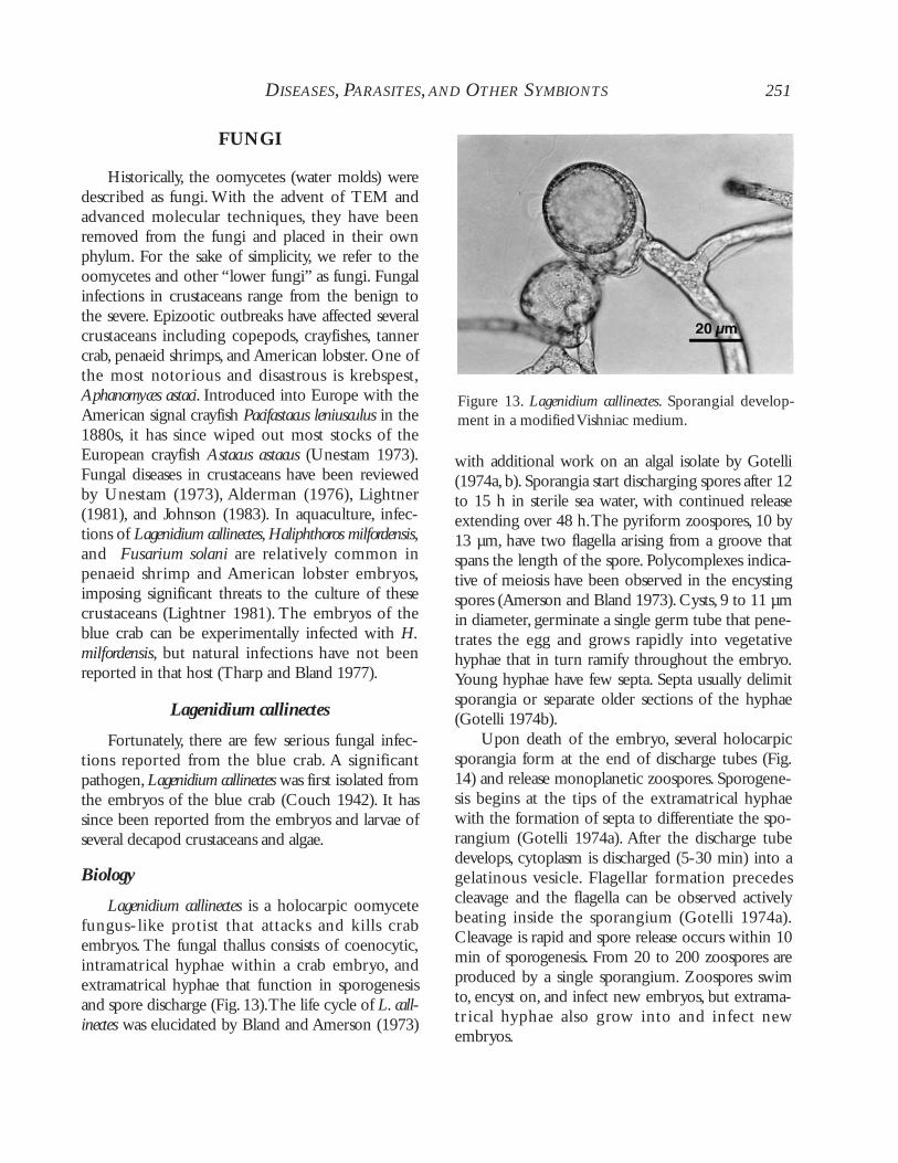
We present a critical review of the microbial dis-eases, parasites, and other symbionts of the blue crab.Previous reviews have provided brief synopses of thediseases of the blue crab (Messick and Sinderman1992; Noga et al. 1998), overviews and synthesis ofcrustacean diseases in general (Couch 1983; Johnson1983; Overstreet 1983; Brock and Lightner 1990;Meyers 1990), or aspects of specific parasitic taxa(Couch and Martin 1982; Overstreet 1982; Brad-bury 1994). Infectious diseases of blue crabs havereceived far less attention than those of the inten-sively cultured eastern oyster or the penaeid shrimps,primarily because of differences in resource manage-ment as well as the dramatic detrimental influencesof protozoal and viral diseases in the latter hosts,respectively. Nonetheless, given the appropriateenvironmental conditions, several pathogenic agents(e.g., viruses, Vibrio spp., Hematodinium perezi, Para-moeba perniciosa, Loxothylacus texanus) have the capac-ity to severely affect blue crab fisheries. Several bac-teria and a few parasites (e.g.,Vibrio spp.,microphallidtrematodes) represent minor human health con-cerns, and a few bacteria (Listeria monocytogenes,Clostridium botulinum) represent safety hazards to theseafood industry. In addition, several symbionts (e.g.,
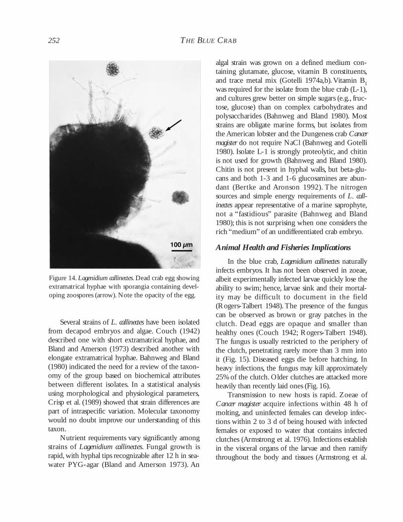
Carcinonemertes carcinophila, Octolasmis muelleri) mayserve as markers of host biology by indicatingmigration or molting patterns, and one syndrome,shell disease, may even serve as a useful indicator ofpoor water quality associated with pollution.
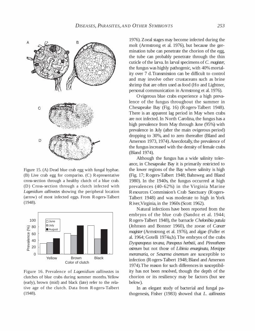
Our synthesis is meant to show the gaps in ourunderstanding of the primary diseases of the bluecrab and guide future work on their ecologicalinfluences and their pathological processes in thehost.Aspects of the immune system of the blue crabare discussed in relation to selected diseases with thecaveat that immune functions are poorly understoodin the Crustacea in general.
Throughout the chapter the term “symbiont” isused broadly as an organism in some form of closeor intimate association with its host (Overstreet1978). “Symbiosis” means “living together,” and weview it as spanning the gamut of disease, parasitic,commensalistic, mutualistic, and phoretic, but notpredatory, relationships. A “disease” imparts abnor-mal function within the host.“Pathogens” cause dis-ease by damaging physiological functions within thehost. “Parasites” may or may not be pathogens thatcause disease, but they have the potential to producea negative effect on the host, especially in heavyinfections. “Hyperparasitism,” a second order para-sitism, signifies the condition when one parasite
Chapter 8
Diseases, Parasites,and Other SymbiontsJ.D. SHIELDS AND R.M. OVERSTREET
And this great structural diversity [of marine life] is paralleled by habits and ways of life which are oftenbizarre to the point of fantasy. Against such a background, it is not surprising that the whole spectrum ofanimal associations, from the obviously casual to the intimately complex, can be seen.
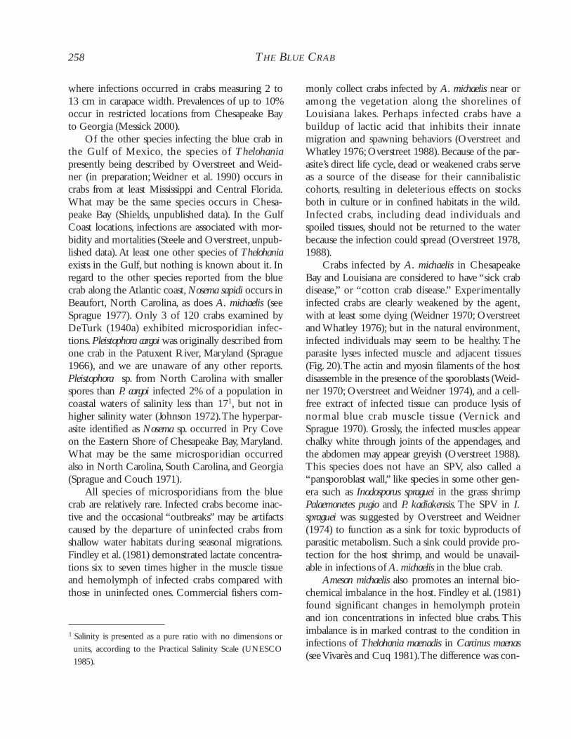
R.V. Gotto (1969)
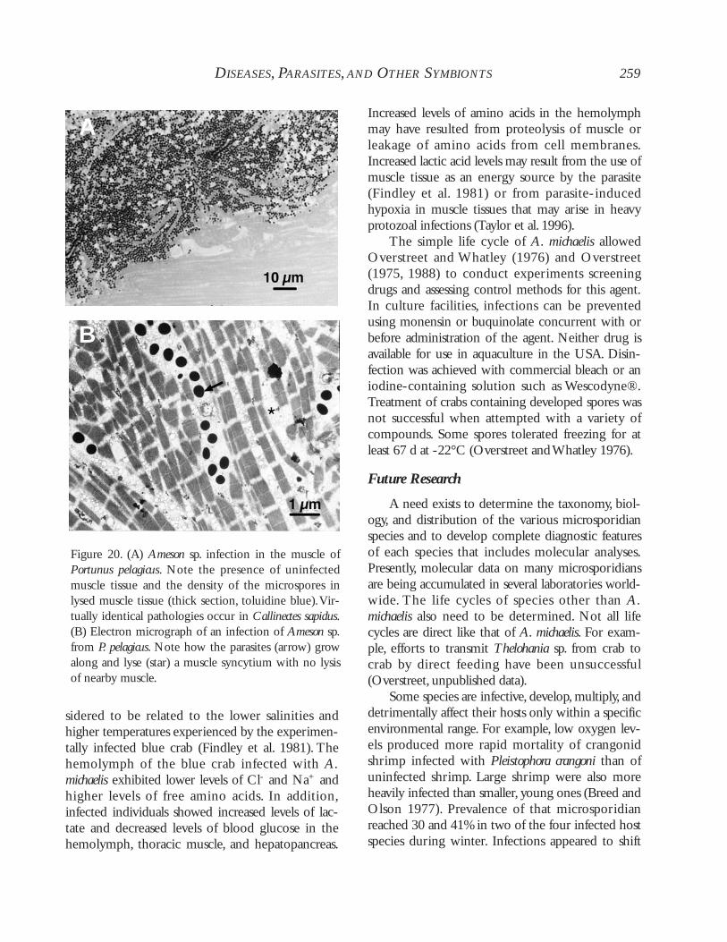
infects another.A “facultative symbiont” is not phys-iologically dependent on a host but can establish arelationship with it when the opportunity occurs. Itcontrasts with an “obligate symbiont,” which has aphysiological dependency on its host. The terms“infection” and “infestation” refer to internal andexternal invasion of the host by endo- and ecto-symbionts, respectively. “Commensalism” and“phoresy” are relationships where the symbiontderives benefit, but the host is not affected by theassociation. Commensalism results when the sym-biont shares nutritional resources or a living spacewith the host. Phoresy results when the symbiontuses the host for transportation. An “epibiont” is anorganism that lives on the external surface of thehost. In “mutualism,” both the symbiont and thehost benefit from the association. These definitionsrepresent a continuum that exists among symbioticassociations.
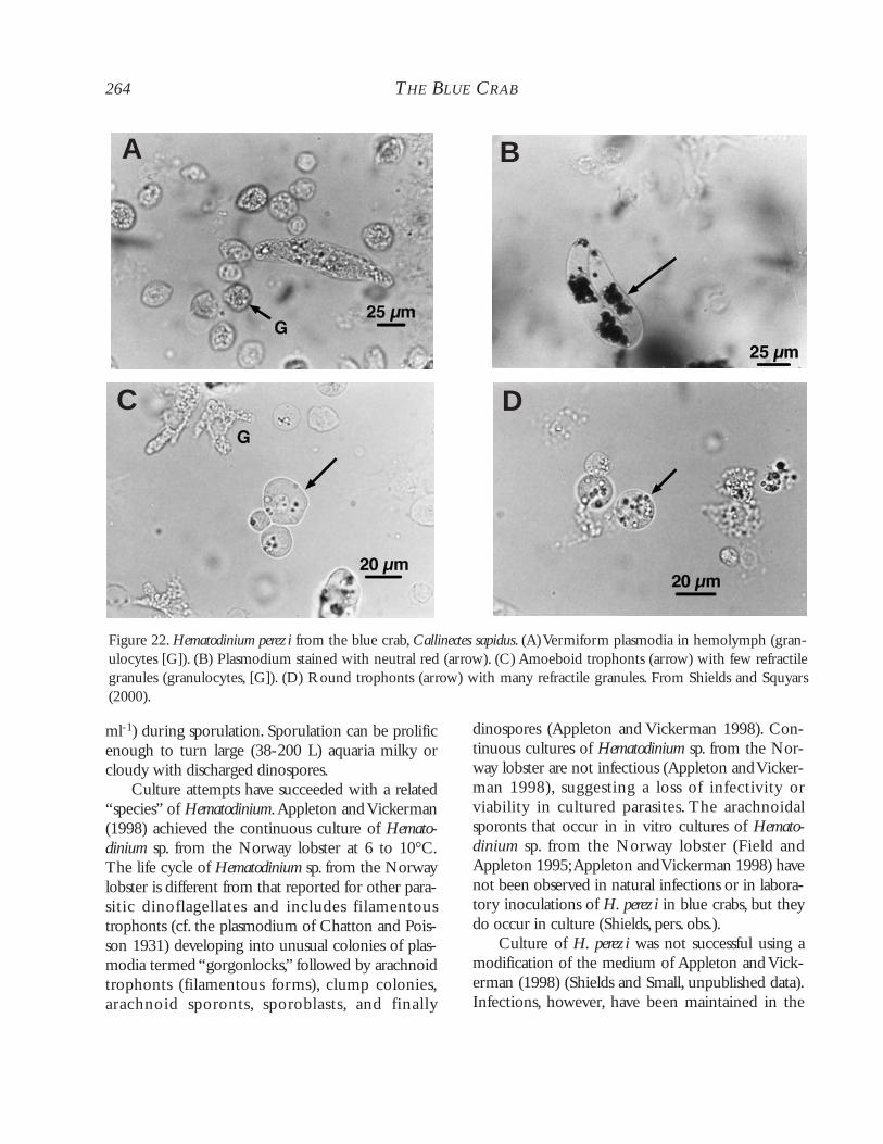
Standard parasitological terms were defined byMargolis et al. (1982) and updated by Bush et al.(1997). Briefly, “prevalence” is the number ofinfected hosts divided by the total number of hostsexamined, usually expressed as a percentage.“Inten-sity” is the number of parasites infecting a host, with“mean intensity” representing the mean number ofparasites per infected host. “Density” refers to thenumber of parasites per unit of host or habitat mea-sured in area, volume, or weight (e.g., parasites perml hemolymph), and is often used with bacterialand protozoal agents. An “epizootic,” or “epidemic”if related to people, is an outbreak of a disease, usu-ally expressed as a large increase in prevalence orintensity of infection in the host population.Whenoccurring over a wide geographic area, such an epi-zootic is referred to as a “panzootic.”An “enzootic,”or native, disease is one caused by factors consis-tently present in the affected host population, envi-ronment, or region.
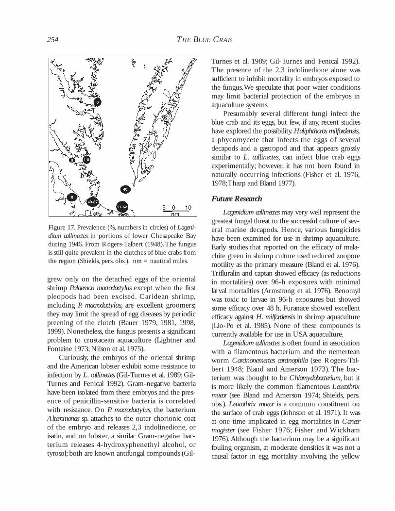
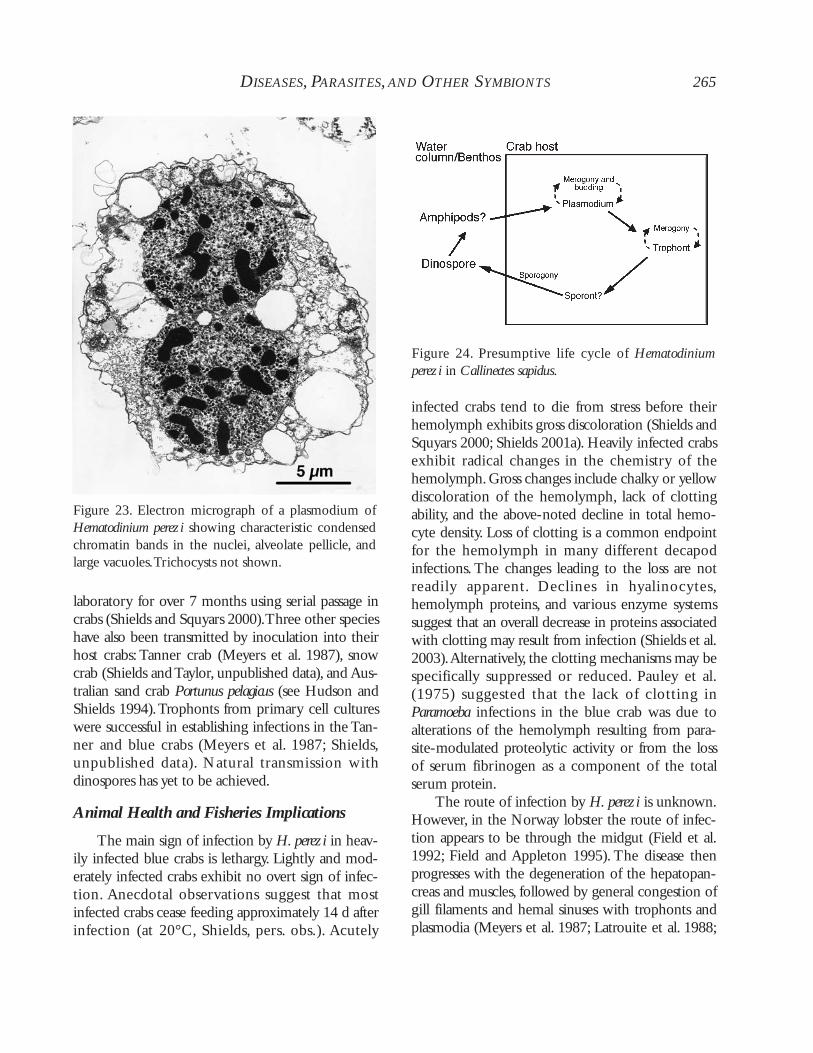
The first reported symbionts of the blue crabwere the rhizocephalan barnacle Loxothylacus texanusby Boschma (1933), the fungus Lagenidinium call-inectes by Couch (1942), and the nemertean wormCarcinonemertes carcinophila by Humes (1942). The
late 1960s and early 1970s saw the advent of scien-tific interest in diseases of the blue crab, especially ascrab fisheries became more fully exploited.With theexpansion of the softshell industry has come anincreased awareness of the role of diseases in theshort-term culture of the blue crab and their nega-tive effects on the fisheries. Blue crabs are nowknown to be infected by a large, disparate faunacomprised of viruses, bacteria, fungi, protozoa,helminths, and other crustaceans. Most of the para-sites and diseases are relatively benign and cause littlepathological alteration in the crab host. Several,however, cause considerable alteration and occasion-ally fulminate into epizootics, or outbreaks, resultingin crab mortalities. Unlike dead fish that float, deadcrabs generally sink, hence large mortalities often gounnoticed or underreported. The true influence ofseveral diseases, therefore, may be difficult to assesswithout intensive sampling in enzootic locations.
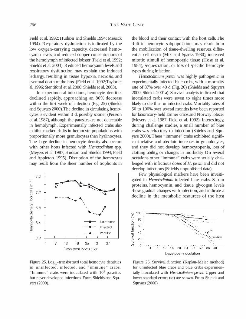
VIRAL INFECTIONS
Other than those from some penaeid shrimps,viral infections in the blue crab are some of the bet-ter known from a marine invertebrate host. Theseblue crab infections are known primarily fromdescriptive ultrastructural studies by Johnson (e.g.,1986a). There are seven or eight reported virusesinfective to C. sapidus along the Atlantic Coast, withat least two occurring in the Gulf of Mexico.Threespecies are lethal and two are found concurrentlywith other viruses, with strong experimental evi-dence indicating a synergistic pathogenic effect.Theidentification and characterization of the virusesinfecting crabs are less well established than thoseinfecting shrimps, perhaps because there is less eco-nomic incentive to culture crabs than to rearpenaeid shrimps. However, when one of the shrimpviruses is introduced to the blue crab, it can producean infection and induce mortality. In addition tohosting viruses that are apparently specific to theblue crab and its relatives, the blue crab can alsoaccumulate human enteric viruses.
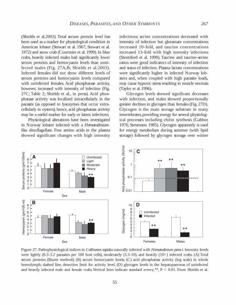
224 THE BLUE CRAB
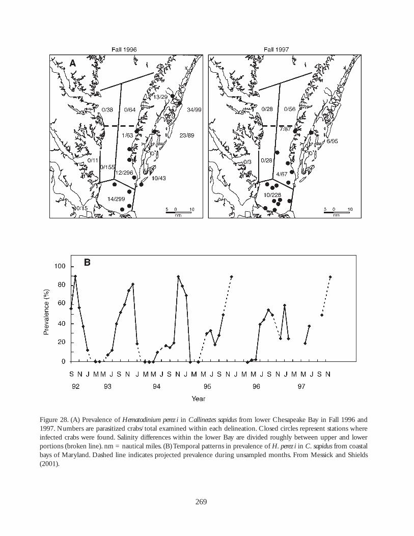
225
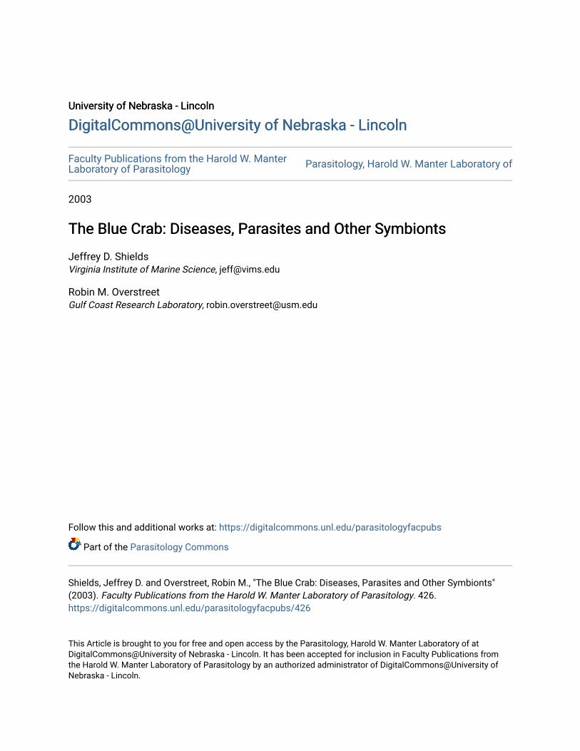
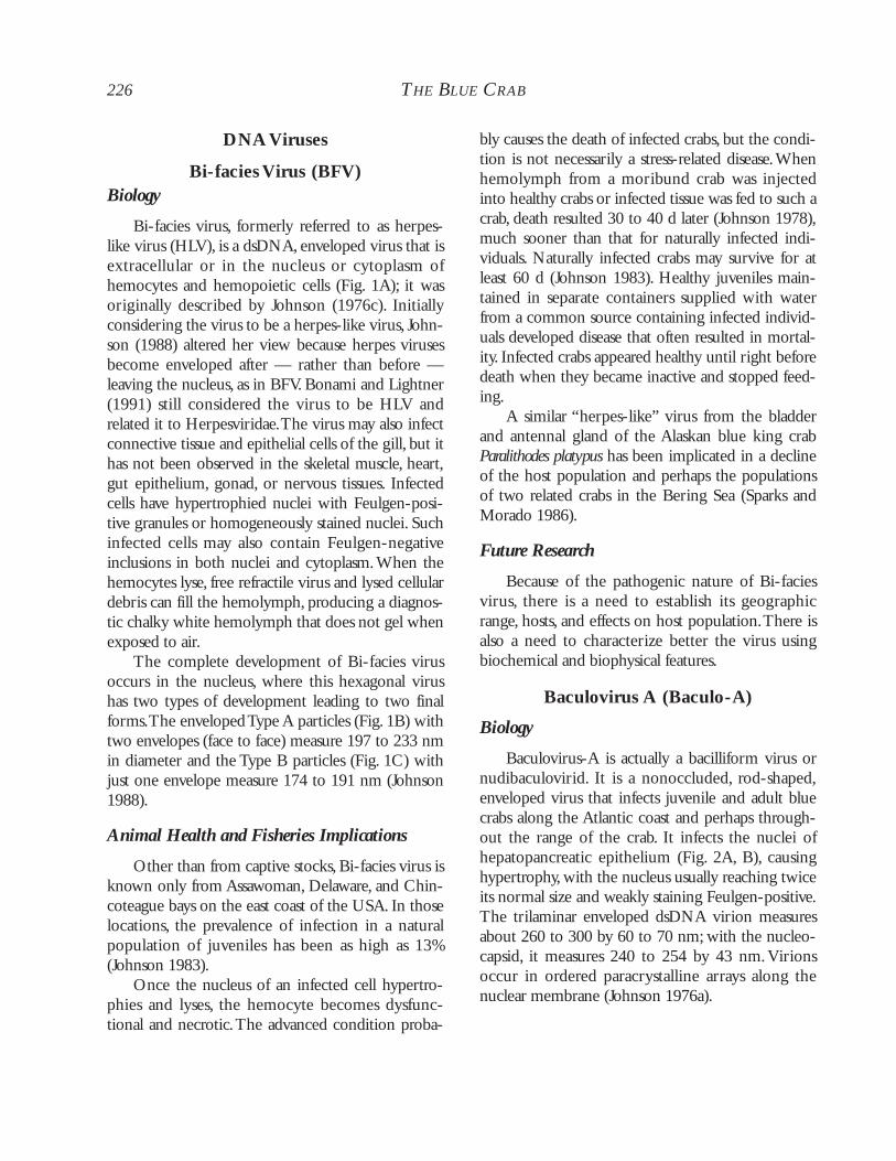
Figure 1. Bi-facies virus (BFV). (A) Infected hemocyte with large viral induced inclusions in both cytoplasm andnucleus. From Johnson (1988). (B) Type-A mature particle showing electron-dense core bound by electron-densesphere, which in turn is surrounded by an inner and outer membrane. From Overstreet (1978). (C) Type-B matureparticle showing single envelope. Note mass of rods (perhaps core of undeveloped BFV) in cytoplasmic material asso-ciated with fixed phagocyte. Note small rhabdo-like virus in same cell. From Johnson (1988).
A
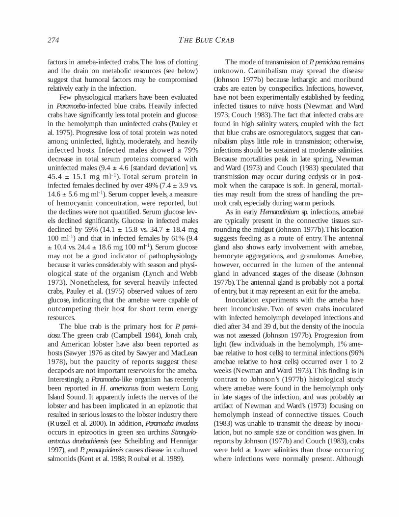
B C
DNA Viruses
Bi-facies Virus (BFV)Biology
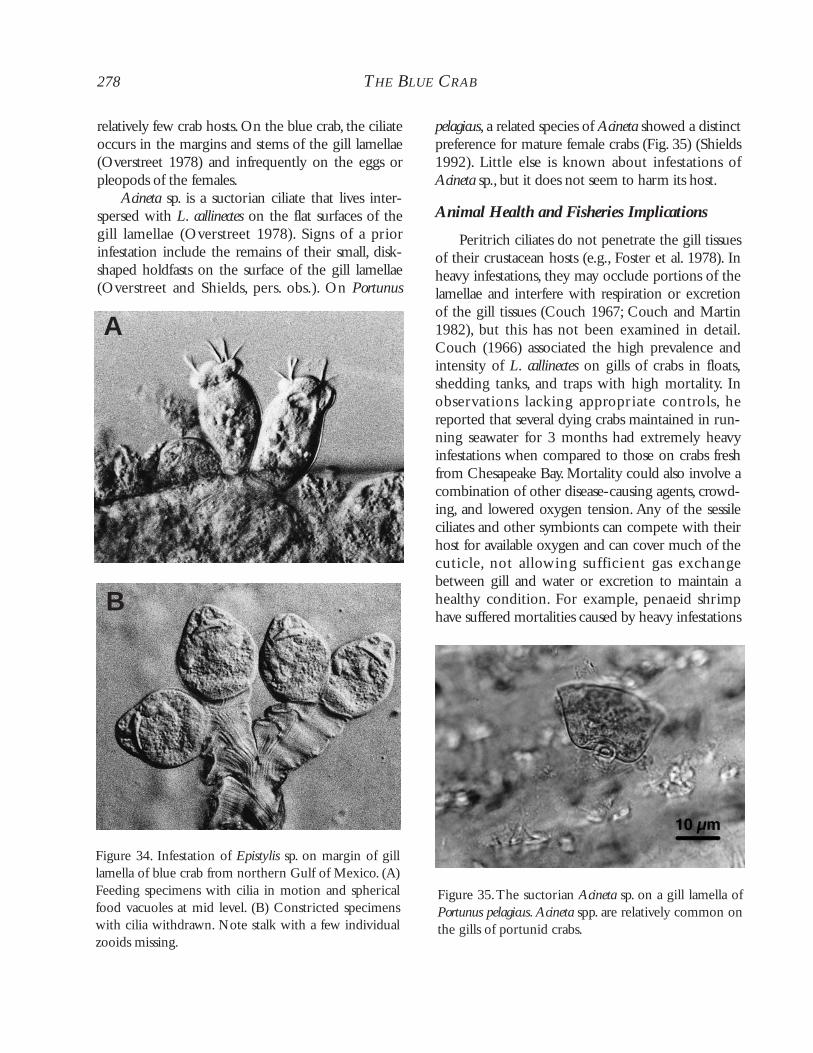
Bi-facies virus, formerly referred to as herpes-like virus (HLV), is a dsDNA, enveloped virus that isextracellular or in the nucleus or cytoplasm ofhemocytes and hemopoietic cells (Fig. 1A); it wasoriginally described by Johnson (1976c). Initiallyconsidering the virus to be a herpes-like virus, John-son (1988) altered her view because herpes virusesbecome enveloped after — rather than before —leaving the nucleus, as in BFV. Bonami and Lightner(1991) still considered the virus to be HLV andrelated it to Herpesviridae.The virus may also infectconnective tissue and epithelial cells of the gill, but ithas not been observed in the skeletal muscle, heart,gut epithelium, gonad, or nervous tissues. Infectedcells have hypertrophied nuclei with Feulgen-posi-tive granules or homogeneously stained nuclei. Suchinfected cells may also contain Feulgen-negativeinclusions in both nuclei and cytoplasm.When thehemocytes lyse, free refractile virus and lysed cellulardebris can fill the hemolymph, producing a diagnos-tic chalky white hemolymph that does not gel whenexposed to air.
The complete development of Bi-facies virusoccurs in the nucleus, where this hexagonal virushas two types of development leading to two finalforms.The enveloped Type A particles (Fig. 1B) withtwo envelopes (face to face) measure 197 to 233 nmin diameter and the Type B particles (Fig. 1C) withjust one envelope measure 174 to 191 nm (Johnson1988).
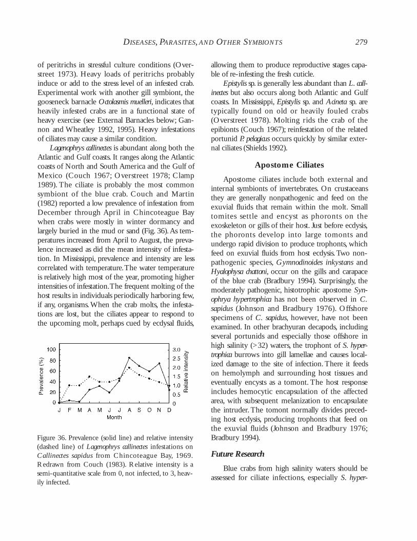
Animal Health and Fisheries Implications
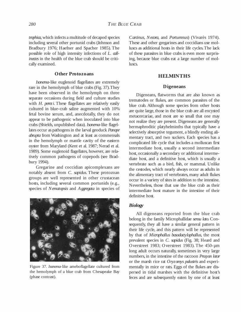
Other than from captive stocks, Bi-facies virus isknown only from Assawoman, Delaware, and Chin-coteague bays on the east coast of the USA. In thoselocations, the prevalence of infection in a naturalpopulation of juveniles has been as high as 13%(Johnson 1983).
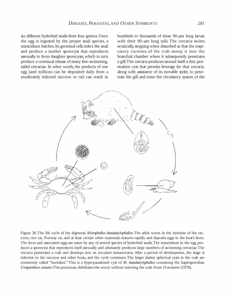
Once the nucleus of an infected cell hypertro-phies and lyses, the hemocyte becomes dysfunc-tional and necrotic.The advanced condition proba-
bly causes the death of infected crabs, but the condi-tion is not necessarily a stress-related disease.Whenhemolymph from a moribund crab was injectedinto healthy crabs or infected tissue was fed to such acrab, death resulted 30 to 40 d later (Johnson 1978),much sooner than that for naturally infected indi-viduals. Naturally infected crabs may survive for atleast 60 d (Johnson 1983). Healthy juveniles main-tained in separate containers supplied with waterfrom a common source containing infected individ-uals developed disease that often resulted in mortal-ity. Infected crabs appeared healthy until right beforedeath when they became inactive and stopped feed-ing.
A similar “herpes-like” virus from the bladderand antennal gland of the Alaskan blue king crabParalithodes platypus has been implicated in a declineof the host population and perhaps the populationsof two related crabs in the Bering Sea (Sparks andMorado 1986).
Future Research
Because of the pathogenic nature of Bi-faciesvirus, there is a need to establish its geographicrange, hosts, and effects on host population.There isalso a need to characterize better the virus usingbiochemical and biophysical features.
Baculovirus A (Baculo-A)
Biology
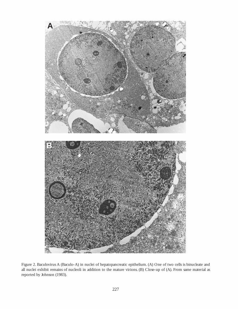
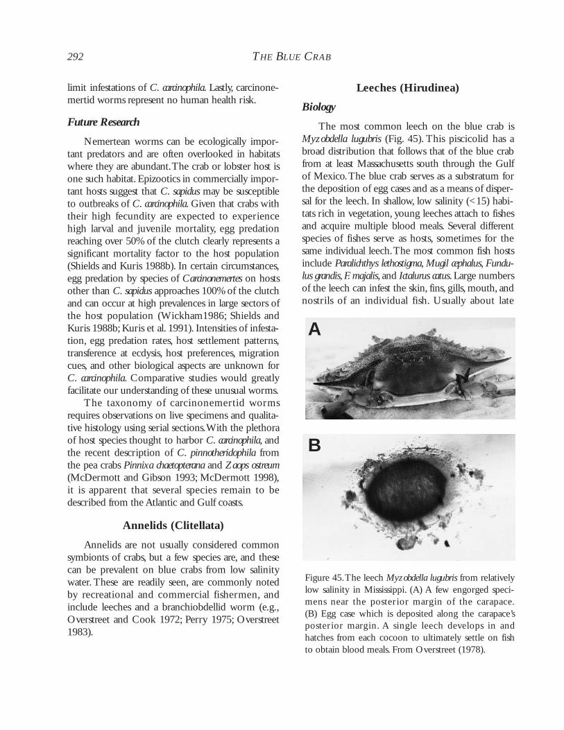
Baculovirus-A is actually a bacilliform virus ornudibaculovirid. It is a nonoccluded, rod-shaped,enveloped virus that infects juvenile and adult bluecrabs along the Atlantic coast and perhaps through-out the range of the crab. It infects the nuclei ofhepatopancreatic epithelium (Fig. 2A, B), causinghypertrophy, with the nucleus usually reaching twiceits normal size and weakly staining Feulgen-positive.The trilaminar enveloped dsDNA virion measuresabout 260 to 300 by 60 to 70 nm; with the nucleo-capsid, it measures 240 to 254 by 43 nm.Virionsoccur in ordered paracrystalline arrays along thenuclear membrane (Johnson 1976a).
226 THE BLUE CRAB
227
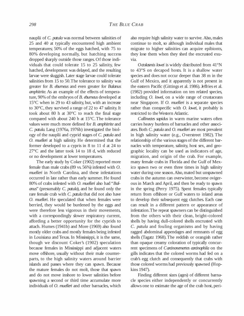
Figure 2. Baculovirus A (Baculo-A) in nuclei of hepatopancreatic epithelium. (A) One of two cells is binucleate andall nuclei exhibit remains of nucleoli in addition to the mature virions. (B) Close-up of (A). From same material asreported by Johnson (1983).
A
B
Animal Health and Fisheries Implications
Johnson (1983) thought that Baculo-A might bethe most ubiquitous of all the blue crab viruses. Itsprevalence typically ranged from 4 to 20% in allstages of the molt cycle of the blue crabs betweenLong Island Sound, Connecticut, and ChesapeakeBay, though Johnson (1983) reported 52% preva-lence in one collection from Chincoteague Bay,Vir-ginia. Johnson (1976a) found the agent in all collec-tions from crab populations in low to high salinitylocations.There, however, was no indication that anyinfected crab was harmfully affected. Nevertheless,microscopical signs of focal infection were observed;hypertrophied nuclei occurred most commonly inabsorptive cells (reserve cells, or R-cells) and lessoften in secretory (B-cells) and fibrillar (F-cells) cellsof infected crabs.
Future Research
Research might show that larval crabs areaffected or even killed by Baculo-A. For example, anoccluded baculovirus typically kills larval and post-larval penaeids but seldom older individuals (Over-street 1994).The apparently nonoccluded “tau” virusof the green crab Carcinus maenas, which may berelated to Baculo-A, kills its crab host (Bazin et al.1974). Feeding or injection can experimentallytransmit both the occluded and nonoccluded agents.Because Johnson (1983) thought Baculo-A mightbe the most ubiquitous of all the crab viruses, bluecrabs from the Gulf of Mexico surely should be sur-veyed for this as well as other viruses.
Baculovirus B (Baculo-B)
Biology
The nonoccluded Baculo-B virus exhibits simi-larities to Baculo-A, but it infects hemocytes andhematopoietic cells, often producing diagnostichyperchromatic areas in the center of the nucleus(Johnson 1983, 1986a). Nuclear and cellular hyper-trophy is not as marked as in cells infected with BFVor Baculo-A, but the cells stain more strongly withFeulgen.The infected hemopoietic cells and hemo-cytes exhibit pale, hypertrophied nuclei that can beeither homogeneous or rimmed with chromatin,
occasionally with hyperchromatic areas in the cen-ter.Virions mature after the nucleus becomes hyper-trophied.The enveloped virions appear ovoid, mea-suring about 100 by 335 nm with tapered androunded ends; developing virions are associated withintranuclear vesicles as has been observed in thehemocytes and some connective tissue cells of Carci-nus maenas (see Bazin et al. 1974), rather than longtubule-like structures as in Baculo-A.Virions occurin ordered arrays in the nucleoplasm.The cytoplasmbecomes a narrow rim around the nucleus with fewor no granules. Mature granulocytes are apparentlynot infected. Once a nucleus is infected, it rupturesand the virions invade the cytoplasm and then dis-perse into the extracellular space upon lysis of thecell.
Animal Health and Fisheries Implications
Infections occur at least in Chesapeake Bay,Maryland, and its tributaries and in ChincoteagueBay,Virginia. Experimentally infected crabs becamesick, but at least one was infected with other viruses(RhVA and EHV) (Johnson 1983). Infections arenot known to harm naturally-infected crabs.
Future Research
The pathogenic effect of the virus on the bluecrab, especially on larvae and young juveniles, needsto be determined. As with Baculo-A, biochemicaland biophysical data are needed.The lack of infec-tion in granulocytes is intriguing.The specificity ofBaculo-B for certain hemocyte-types should be fur-ther examined.
RNA Viruses
Reo-like Virus (RLV)
Biology
Reo-like virus, a nonoccluded member of theType I Reoviridae, infects primarily hemocytes,hemopoietic tissues, and glial cells (Fig. 3A, B).Vari-ous other ectodermally and mesodermally derivedtissues such as epidermis, gill, bladder, blood vesselendothelium,Y-organ, and connective tissue cells,including fixed phagocytes, can also be infected
228 THE BLUE CRAB
DISEASES, PARASITES,AND OTHER SYMBIONTS 229
229
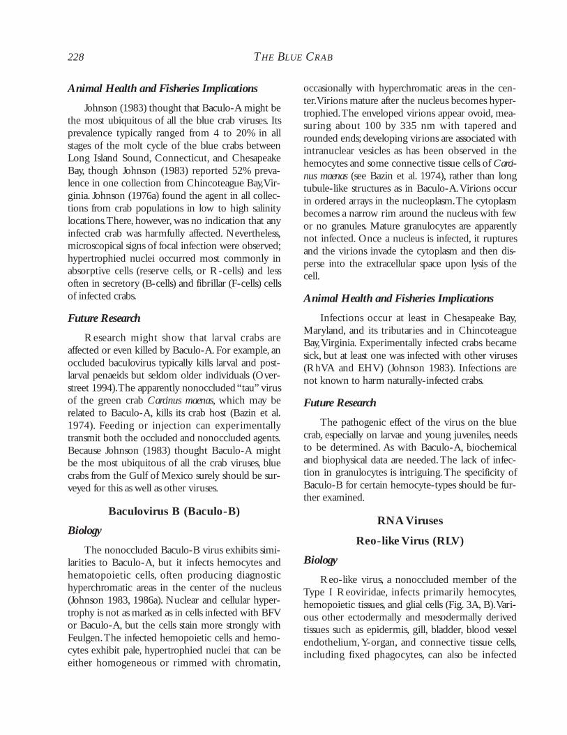
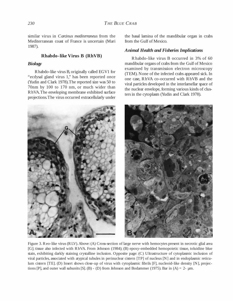
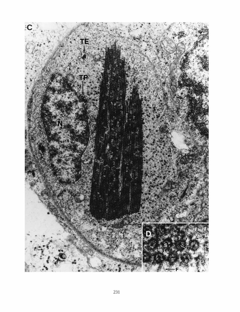
(Johnson 1983). Originally placed incorrectly inPicornaviridae, this reovirus has icosahedral virions.The virion is a nonenveloped dsRNA that measures55 to 60 nm in cross-section and occurs in the cyto-plasm, producing Feulgen-negative inclusions andincreased cytoplasmic volume. These inclusions,basophilic and angulate to rounded in shape, consti-tute paracrystalline arrays of virus particles as well assinuous proteinaceous filaments 20 to 30 nm indiameter (Fig. 3C, D). Infected hemocytes invadethe glia of the brain and thoracic ganglia, whichbecome necrotic. Because of this tissue destruction,the crab becomes sluggish and exhibits tremor andultimately paralysis (Johnson 1983).
Based on what is known about the virus, it maybe the same as that found in the harbor crab Liocarci-nus depurator (as Macropipus depurator) from theMediterranean Sea (Hukuhara and Bonami 1991).
Animal Health and Fisheries Implications
Reo-like virus has been found commonly injuvenile and adult crabs from Chincoteague andChesapeake bays, where it was associated with mor-talities (Johnson and Bodammer 1975; Johnson1983, 1984). Infected crabs occurred in high andlow salinity habitats.
Also present in RLV-infected crabs wasRhabdo-like virus A (RhVA),which seemed to pro-duce a synergistic response (Fig. 4) in the resultingglial necrosis and paralysis (Johnson 1983, 1984).Other viruses can also be present in RLV-infectedcrabs, such as another rhabdo-like virus, Baculo-A,and Baculo-B. The association between RLV andeach of those viruses requires investigation. Whenhemolymph infected with RLV plus RhVA wasinjected into healthy naïve crabs, those crabs died inas little as 3 to 4 d for pre- or postmolt individualsand 11 d for intermolt individuals.When crabs wereadministered infected tissues orally, it took 12 to 32d for the intermolt crabs to die (Johnson 1978,1983, 1986a). The virus can probably also enter byother routes, and the resulting infection represents apotential threat to crabs in shedding-tank systems.Diagnosis usually consists of examination of hemo-cytes or hemopoietic tissues of sluggish crabs pos-sessing hemolymph that either will not clot or that
exhibits a reduced clotting rate. In many cases, theexoskeleton becomes discolored, and the gills of thecrab turn a reddish to brownish color.
Future Research
Reo-like virus infects juveniles and adults inculture, but its prevalence in nature has not beenestablished. The virus needs to be bettercharacterized.
Rhabdo-like Virus A (RhVA)
Biology
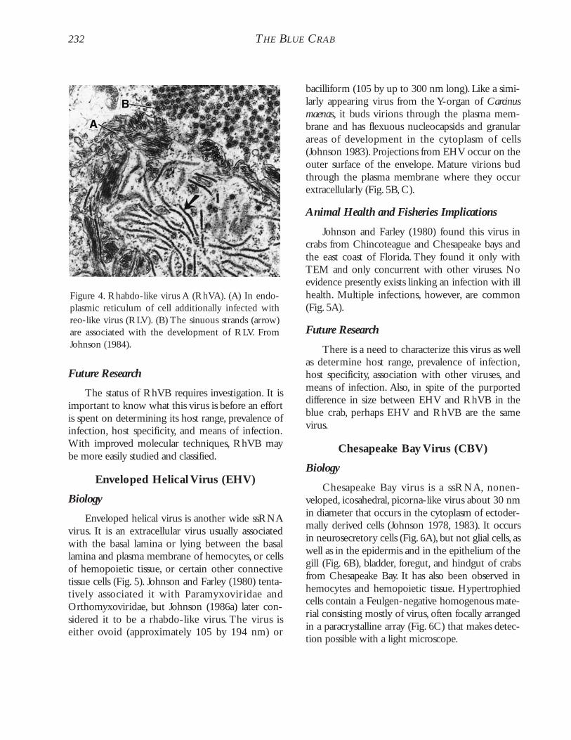
Rhabdo-like virus A probably occurs as a ubiquitousvirus in the blue crab along both the Atlantic andGulf of Mexico coasts.This small virus measures 20to 30 nm by 110 to 170 nm in bacilliform stageswith rounded ends or 20 to 30 nm by up to 600 nmlong in a filamentous flexuous stage (Jahromi 1977).It buds into the endoplasmic reticulum, infectingglial cells of ganglia and large nerves as well ashemocytes, hemopoietic tissue, connective tissues,and epithelium other than that of the alimentarytract and antennal gland. Rhabdo-like virus A infectssimilar sites as Reo-like virus. It does not infectaxons or striated muscle (Johnson 1978, 1983), but itdoes infect the mandibular organ. Initially, the viruswas incorrectly reported from the ecdysal gland, andconsequently the virus was originally called EGV2(Yudin and Clark 1978, 1979).
Animal Health and Fisheries Implications
Rhabdo-like virus A may exemplify a virusassociated with host stress. Infected crabs (Figs. 1C,3A) usually exhibited disease when they had beenmaintained under stressful laboratory conditions orwere infected with other viruses (RLV, EHV, CBV,BaculoB, or HLV) (Johnson 1983). Those notinfected with other viruses or not under stressapparently do not exhibit disease. Infection byRhVA produced no pathological signs visible withthe light microscope. As indicated above, sick crabsinfected with RLV had a mixed infection withRhVA (Figs. 3A, 4). Experimentally, an injectedinoculum of both of those viruses can kill a crab inas little as 3 d. The taxonomic relationship with a
230 THE BLUE CRAB
similar virus in Carcinus mediterraneus from theMediterranean coast of France is uncertain (Mari1987).
Rhabdo-like Virus B (RhVB)
Biology
Rhabdo-like virus B, originally called EGV1 for“ecdysal gland virus 1,” has been reported once(Yudin and Clark 1978).The reported size was 50 to70nm by 100 to 170 nm, or much wider thanRhVA.The enveloping membrane exhibited surfaceprojections.The virus occurred extracellularly under
the basal lamina of the mandibular organ in crabsfrom the Gulf of Mexico.
Animal Health and Fisheries Implications
Rhabdo-like virus B occurred in 3% of 60mandibular organs of crabs from the Gulf of Mexicoexamined by transmission electron microscopy(TEM). None of the infected crabs appeared sick. Inone case, RhVA co-occurred with RhVB and theviral particles developed in the interlamellar space ofthe nuclear envelope, forming various kinds of clus-ters in the cytoplasm (Yudin and Clark 1978).
Figure 3. Reo-like virus (RLV).Above: (A) Cross-section of large nerve with hemocytes present in necrotic glial area[G]; tissue also infected with RhVA. From Johnson (1984); (B) epoxy-embedded hemopoietic tissue, toluidine bluestain, exhibiting darkly staining crystalline inclusion. Opposite page: (C) Ultrastructure of cytoplasmic inclusion ofviral particles, associated with atypical tubules in perinuclear cistern [TP] of nucleus [N] and in endoplasmic reticu-lum cistern [TE]. (D) Insert shows close-up of virus with cytoplasmic fibrils [F], nucleoid-like density [N], projec-tions [P], and outer wall subunits [S]. (B) - (D) from Johnson and Bodammer (1975). Bar in (A) = 2- µm.
A B
231
C
D
232 THE BLUE CRAB
Future Research
The status of RhVB requires investigation. It isimportant to know what this virus is before an effortis spent on determining its host range, prevalence ofinfection, host specificity, and means of infection.With improved molecular techniques, RhVB maybe more easily studied and classified.
Enveloped Helical Virus (EHV)
Biology
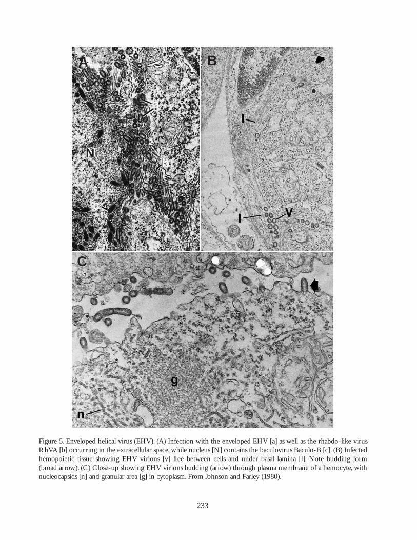
Enveloped helical virus is another wide ssRNAvirus. It is an extracellular virus usually associatedwith the basal lamina or lying between the basallamina and plasma membrane of hemocytes, or cellsof hemopoietic tissue, or certain other connectivetissue cells (Fig. 5). Johnson and Farley (1980) tenta-tively associated it with Paramyxoviridae andOrthomyxoviridae, but Johnson (1986a) later con-sidered it to be a rhabdo-like virus. The virus iseither ovoid (approximately 105 by 194 nm) or
bacilliform (105 by up to 300 nm long). Like a simi-larly appearing virus from the Y-organ of Carcinusmaenas, it buds virions through the plasma mem-brane and has flexuous nucleocapsids and granularareas of development in the cytoplasm of cells(Johnson 1983). Projections from EHV occur on theouter surface of the envelope. Mature virions budthrough the plasma membrane where they occurextracellularly (Fig. 5B, C).
Animal Health and Fisheries Implications
Johnson and Farley (1980) found this virus incrabs from Chincoteague and Chesapeake bays andthe east coast of Florida. They found it only withTEM and only concurrent with other viruses. Noevidence presently exists linking an infection with illhealth. Multiple infections, however, are common(Fig. 5A).
Future Research
There is a need to characterize this virus as wellas determine host range, prevalence of infection,host specificity, association with other viruses, andmeans of infection. Also, in spite of the purporteddifference in size between EHV and RhVB in theblue crab, perhaps EHV and RhVB are the samevirus.
Chesapeake Bay Virus (CBV)
Biology
Chesapeake Bay virus is a ssRNA, nonen-veloped, icosahedral, picorna-like virus about 30 nmin diameter that occurs in the cytoplasm of ectoder-mally derived cells (Johnson 1978, 1983). It occursin neurosecretory cells (Fig. 6A), but not glial cells, aswell as in the epidermis and in the epithelium of thegill (Fig. 6B), bladder, foregut, and hindgut of crabsfrom Chesapeake Bay. It has also been observed inhemocytes and hemopoietic tissue. Hypertrophiedcells contain a Feulgen-negative homogenous mate-rial consisting mostly of virus, often focally arrangedin a paracrystalline array (Fig. 6C) that makes detec-tion possible with a light microscope.
Figure 4. Rhabdo-like virus A (RhVA). (A) In endo-plasmic reticulum of cell additionally infected withreo-like virus (RLV). (B) The sinuous strands (arrow)are associated with the development of RLV. FromJohnson (1984).
233
Figure 5. Enveloped helical virus (EHV). (A) Infection with the enveloped EHV [a] as well as the rhabdo-like virusRhVA [b] occurring in the extracellular space, while nucleus [N] contains the baculovirus Baculo-B [c]. (B) Infectedhemopoietic tissue showing EHV virions [v] free between cells and under basal lamina [l]. Note budding form(broad arrow). (C) Close-up showing EHV virions budding (arrow) through plasma membrane of a hemocyte, withnucleocapsids [n] and granular area [g] in cytoplasm. From Johnson and Farley (1980).
C
BAA
N
Animal Health and Fisheries Implications
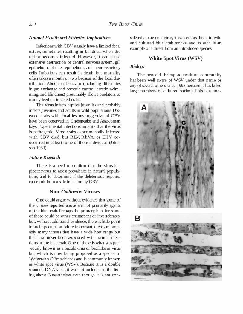
Infections with CBV usually have a limited focalnature, sometimes resulting in blindness when theretina becomes infected. However, it can causeextensive destruction of central nervous system, gillepithelium, bladder epithelium, and neurosecretorycells. Infections can result in death, but mortalityoften takes a month or two because of the focal dis-tribution. Abnormal behavior (including difficultiesin gas exchange and osmotic control, erratic swim-ming, and blindness) presumably allows predators toreadily feed on infected crabs.
The virus infects captive juveniles and probablyinfects juveniles and adults in wild populations. Dis-eased crabs with focal lesions suggestive of CBVhave been observed in Chesapeake and Assawomanbays. Experimental infections indicate that the virusis pathogenic. Most crabs experimentally infectedwith CBV died, but RLV, RhVA, or EHV co-occurred in at least some of those individuals (John-son 1983).
Future Research
There is a need to confirm that the virus is apicornavirus, to assess prevalence in natural popula-tions, and to determine if the deleterious responsecan result from a sole infection by CBV.
Non-Callinectes Viruses
One could argue without evidence that some ofthe viruses reported above are not primarily agentsof the blue crab. Perhaps the primary host for someof those could be other crustaceans or invertebrates,but, without additional evidence, there is little pointin such speculation. More important, there are prob-ably many viruses that have a wide host range butthat have never been associated with natural infec-tions in the blue crab. One of these is what was pre-viously known as a baculovirus or bacilliform virusbut which is now being proposed as a species ofWhispovirus (Nimaviridae) and is commonly knownas white spot virus (WSV). Because it is a doublestranded DNA virus, it was not included in the list-ing above. Nevertheless, even though it is not con-
sidered a blue crab virus, it is a serious threat to wildand cultured blue crab stocks, and as such is anexample of a threat from an introduced species.
White Spot Virus (WSV)
Biology
The penaeid shrimp aquaculture communityhas been well aware of WSV under that name orany of several others since 1993 because it has killedlarge numbers of cultured shrimp. This is a non-
234 THE BLUE CRAB
B
A
235
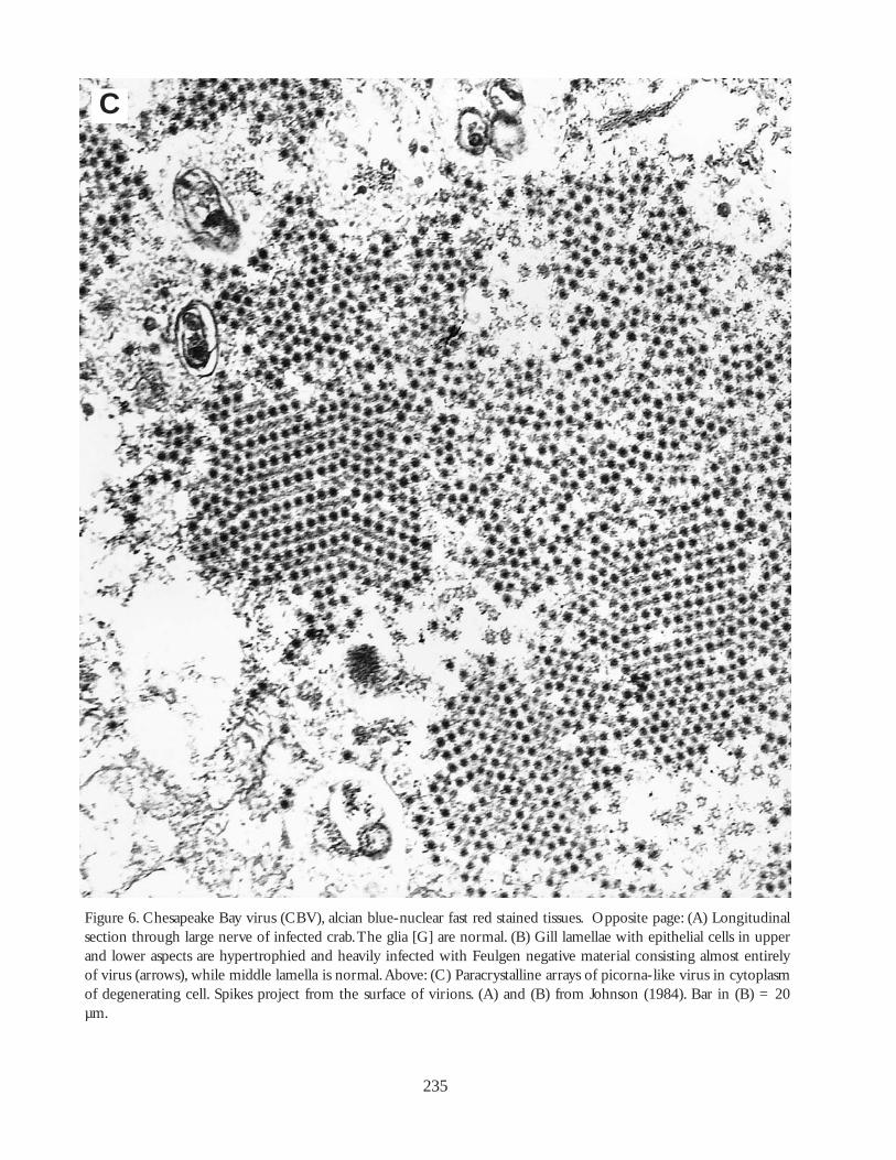
Figure 6. Chesapeake Bay virus (CBV), alcian blue-nuclear fast red stained tissues. Opposite page: (A) Longitudinalsection through large nerve of infected crab.The glia [G] are normal. (B) Gill lamellae with epithelial cells in upperand lower aspects are hypertrophied and heavily infected with Feulgen negative material consisting almost entirelyof virus (arrows), while middle lamella is normal.Above: (C) Paracrystalline arrays of picorna-like virus in cytoplasmof degenerating cell. Spikes project from the surface of virions. (A) and (B) from Johnson (1984). Bar in (B) = 20µm.
C
occluded rod-shaped particle with an apical enve-lope extension.The nucleocapsid is cylindrical withasymmetric ends and a superficially segmentedappearance (Durand et al. 1997).Virions measure 70to 150 nm by 275 to 380 nm. Infected cells can bediagnosed histologically by prominent eosinophilicto pale basophilic (with H&E staining), Feulgen-positive intranuclear inclusion bodies in hypertro-phied nuclei of cuticular epithelial (Fig. 7) and con-nective tissue cells. It can also be detected in theantennal gland epithelium, lymphoid organ sheathcells, hematopoietic tissues, and fixed phagocytes ofthe heart (Lightner 1996). Because of the interest inthis disease in aquaculture, gene probes and PCRdetection methods have been developed to quicklydetect an infection. Confirmation of bioassays isusually made with light microscopy (LM), TEM,PCR, and in situ DNA hybridization. In addition toinfecting several different penaeid shrimps, the viruscan infect a variety of other crustaceans, includingthe blue crab (Flowers et al. 2000; Krol 2002).
Animal Health and Fisheries Implications
White spot virus has been prevalent in penaeidaquaculture facilities in Asia and the Indo-Pacificwhere it has caused enormous losses to commercialshrimp farms.After Hurricane Georges in 1998, thevirus was introduced into South, Central, and NorthAmerica, where it caused major losses to penaeids.To assess for potential reservoir and carrier hosts,experimental work has been conducted on severalmarine and estuarine organisms. In Asia, the portu-nid sand crab Portunus pelagicus and mud or man-grove crab Scylla serrata as well as Acetes sp. wereexposed experimentally to WSV (Supamattaya et al.1998). All exposed specimens of Acetes sp. died in 3or 5 d following injection or immersion, respec-tively. All of the injected sand crabs died by day 8but only 20% of the mud crabs had died by day 9.Neither species of crab died when fed the virus, butboth demonstrated an infection histologically andcertainly can serve as reservoir hosts. On the otherhand, all naïve specimens of the injected blue crabfrom Mississippi and 66% of those fed infected tissuedied, and the infection in the bioassay animals wasconfirmed with PCR, TEM and LM histology
(Flowers et al. 2000; Krol 2002). White spot virusdemonstrates the potential for a virus common inshrimp aquaculture to have a serious influence as apathogen on wild or cultured stocks of the bluecrab.
Future Research
As indicated,WSV is an example of the abilityof the blue crab to serve as a host to a virus that isextremely pathogenic to members of another crus-tacean group. Regarding WSV, we need to knowwhat environmental, host, and viral conditions itwould take to transform an infected individual orstock into a panzootic with heavy mortalities. Thesame approach can be directed to other viruses thatcould be introduced into a habitat in which the bluecrab occurs.
Public Health Implicationsof Viruses
None of the crab viruses discussed above isharmful to humans or pose any public health threat.However, although the blue crab is not a filter feederlike the eastern oyster and other bivalves that con-centrate large numbers of human pathogenicviruses, it can readily accumulate human entericviruses. Hejkal and Gerba (1981) experimentallydetermined that poliovirus and other viruses in highconcentrations in water surrounding the crab wererapidly (within 2 h) acquired throughout the tissuesof the crab. Highest levels occurred in the digestivetract and hemolymph.Virus uptake was not affectedmuch by salinity, but the levels were clearly influ-enced by temperature. Some virus survived 6 d inthe hemolymph at 15°C, but at 25°C the rates ofboth uptake and removal were significantlyincreased, with none detected after 20 h. Conse-quently, especially in cool water, the blue crab canaccumulate (but not concentrate) harmful virusesfrom the surrounding water or from contaminatedfood in a polluted location, and then migrate to anuncontaminated location. Moreover, all or nearly allthe virus accumulated in crabs originating fromareas contaminated with municipal sludge and otherdumped wastes. Hejkal and Gerba (1981) also
236 THE BLUE CRAB
237
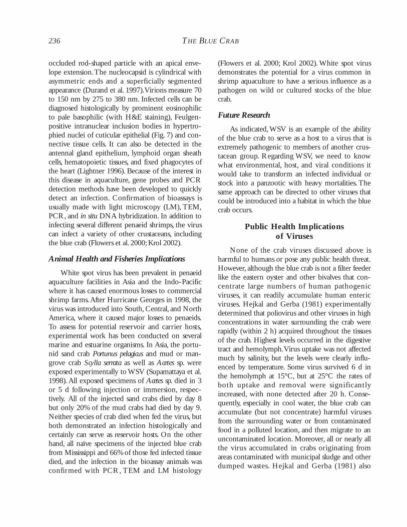
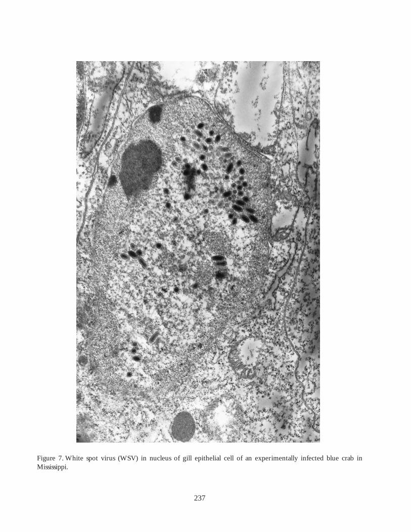
Figure 7. White spot virus (WSV) in nucleus of gill epithelial cell of an experimentally infected blue crab in Mississippi.
showed that although boiling a crab for 8 min(internal T of 70°C) inactivated 99.9% of the virus,in rare cases some active virus was still detected inswimming muscle after 16 min (internal T of 94°C).
Rotavirus and enteroviruses can be detectedsimply by separating the virus from tissuehomogenates at pH 9.5, concentrating by absorp-tion to protein precipitates at pH 3.5, followed byelution from the precipitates at pH 9.2 (Seidel et al.1983). Recovery effectiveness averaged 52% withpoliovirus and others when using the polyelectrolytecat-floc precipitation to remove toxic factors fromcell cultures without loss in virus recovery.Withoutsuch removal, the final elute had a toxic effect on thecells used for the assay.
General Future of Viral Research
Research possibilities dealing with blue crabviruses are begging for attention.As an example,TheCrustacean Society meeting in May 1999 (Lafayette,Louisiana) included a 16-paper symposium entitled“Blue Crab Mortality Symposium” that did notinclude a single paper that mentioned viral infec-tions even though at least RLV, BFV, and CBV canbe fatal and potentially serious pathogens to crabstocks. Increased experience with penaeid shrimpviruses has demonstrated how devastating a fewagents can be to wild and cultured stocks. Some ofthe matters that require future attention have beenindicated above under the separate viruses. Thereare, however, general approaches that should beaddressed. A basic need exists to fully characterizeeach known virus and to determine its host range,prevalence of infection, host specificity, and means oftransmission.We speculate that many viruses, underthe appropriate conditions, can have a devastatingeffect on cultured and wild crabs. Considering theassumed potential for catastrophes, we think thereexists a plethora of avenues to investigate. Mostinformation about viruses in the blue crab comesfrom descriptive studies by Phyllis Johnson and col-leagues. One must assume that the crab, an appar-ently good host for viruses, could or does harbornumerous others. Any of these could be eitherhighly specific to the blue crab or infective to a widerange of crustacean hosts. Both the geographic range
and the specificity (host range) of the known virusesrequire additional documentation. Prevalence ofinfection from a few locations should be docu-mented even though values for a given virus aredependent on environmental conditions and willdiffer yearly, seasonally, and geographically.Assumingthat the viruses all act differently, similar to what onefinds in penaeid viruses (e.g., Overstreet 1994;Lightner 1996), we think the conditions necessaryfor infection and inactivation for these should beestablished.The primary question is what conditionsor interactions are necessary to shift a harmlessinfection in equilibrium into a disease state and massmortality?
Based on the lack of critical examination forviruses along the range of the crab, at least some ofthe agents other than RhVA and RhVB can be pre-dicted to occur in the Gulf of Mexico and someprobably also in the Caribbean Sea. Considering theimportation of crabs from the Gulf of Mexico to theChesapeake Bay area, we think perhaps someChesapeake cases could have originated from theGulf. Perhaps the agent for some of these diseaseshas established an equilibrium with its host in thewild, including in the Gulf of Mexico. In any event,given the high population densities of blue crabs andshort-term culture of soft-shell crabs,we suggest thathigh mortalities in shedding facilities may fomentspread of viral pathogens. Given the fishing practicesinvolved in soft-shell culture, the movement of dis-eased crabs and introduction of pathogens to newareas is highly likely, especially in Chesapeake Baywhere molting crabs are shed in different watershedsfrom whence they came. For these reasons, we cau-tion against importation of soft-shell and hard crabsinto the Chesapeake area or the Gulf of Mexico.
As already indicated, the wealth of informationon viral infections in the blue crab has resulted fromultrastructural studies by Johnson, including experi-mental infections, stressing animals in confined sys-tems, and surveys. This work should be followedwith molecular and other techniques that are con-tinually being updated for viruses in penaeidshrimps (e.g., Lightner 1996). With additionalresearch, infections could be detected and distin-guished by genetic probes, PCR, in situ hybridiza-
238 THE BLUE CRAB
tion, dot blot hybridization, antibody tests (e.g.,ELISA and fluorescent), direct fresh microscopicalevaluation, and histopathological criteria as well asultrastructure of the viruses. Unfortunately, there isno well-established, continuous cell line developedfrom any crustacean. Hence, viral culture and plaquetests cannot be conducted as they are for virusesfrom most other animals.
Multiple infections often infect the same indi-vidual, and the pathogenic relationship among thedifferent species needs to be assessed. In some cases,an infection can become patent or an outbreak ofdisease can occur when crabs are in culture,crowded, or in a stressed environment (Johnson1978, 1984, 1986;Yudin and Clark 1979; Messickand Kennedy 1990). Consequently, in addition toproducing mortalities in cultured products, viralinfections may provide a good indication of envi-ronmental health in the natural environment.Knowledge of these agents suggests the need forincreased funding opportunities, if the crab is to becultured on an economically successful basis.
BACTERIAL DISEASES
Bacteria are ubiquitous in the marine environ-ment, and, not surprisingly, they are ubiquitous inthe blue crab. Although most bacteria are relativelybenign, several species of Vibrio have been impli-cated in several crab mortalities. Other bacteria suchas Listeria monocytogenes and Clostridium botulinumrepresent significant concerns to food safety(Williams-Walls 1968; Rawles et al. 1995; Petersonet al. 1997). Bacterial diseases of blue crabs sparkedsome debate in the 1970s. At that time, thehemolymph of blue crabs was thought to be sterilelike that of vertebrates and many other invertebrates(Bang 1970). Colwell et al. (1975) and Tubiash et al.(1975) found high prevalences of bacteria in thehemolymph of healthy blue crabs. Johnson (1976d),however, posited that bacterial infections were sim-ply acquired through wounds received by the roughhandling of crabs en route to markets. In 1982,Davisand Sizemore definitively showed that species of Vib-rio were present at low to moderate levels in thehemolymph and various other tissues of freshlycaught, unstressed crabs. Mean intensities were
found to range from 103 to 105 bacteria per ml, alevel too low in some cases to detect withmicroscopy (Davis and Sizemore 1982).
Stress plays a major role in the etiology andprognosis of bacterial infections in crustaceans (seeBrock and Lightner 1990). Blue crabs suffer consid-erable stress as a result of capture, handling, crowd-ing, transport, temperature, wounding, and poorwater quality, especially in poorly managed recircu-lating systems (Johnson 1976d). Given the back-ground of normal bacterial loads in healthy crabs,some stressed hosts will develop rampant, lethalinfections of species of Vibrio and other bacteria.Mortalities of crabs affecting the fishery are periodi-cally reported in the spring and summer, seasonswhen water temperatures and handling stress cantrigger outbreaks of bacterial disease.
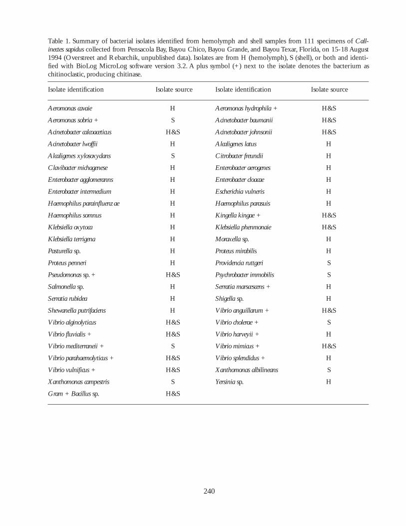
Blue crabs have a diverse fauna of opportunisticbacterial invaders. Using an elegant numericalapproach, Sizemore et al. (1975) and Colwell et al.(1975) identified several genera of bacteria from thehemolymph of market-bought, and freshly caught,“hardshell” blue crabs. The following species werecultured from C. sapidus in Maryland: Aeromonasspp., Pseudomonas spp., Vibrio spp., Bacillus spp., Acine-tobacter spp., Flavobacterium spp., and coliform bacteriasimilar to Escherischia coli; several isolates could notbe identified. Babinchack et al. (1982) isolatedEscherichia coli, Enterobacter aerogenes and Vibrio spp. onthe gills of blue crabs from South Carolina. Marshallet al. (1996) reported the presence of streptomycin-resistant Plesiomonas shigelloides from blue crabs fromMississippi. They implied that antibiotic resistancewas due to contamination of estuarine areas bywastewaters.There are other reports, including somein the grey literature. For example, Overstreet andRebarchik (unpublished) found 49 different bacter-ial isolates from blue crabs collected near Pensacola,Florida (Table 1). Forty-one of these were isolatedfrom the hemolymph. Sterile hemolymph wasnoted in only 24.3% of the 111 crabs examined.
Bacterial surveys have also been undertaken inother species of blue crabs. Rivera et al. (1999) cul-tured 23 different bacter ial isolates from thehemolymph of six specimens of C. boucourti fromthe eutrophic Mandry Channel, Puerto Rico.They
DISEASES, PARASITES,AND OTHER SYMBIONTS 239
240
Table 1. Summary of bacterial isolates identified from hemolymph and shell samples from 111 specimens of Call-inectes sapidus collected from Pensacola Bay, Bayou Chico, Bayou Grande, and Bayou Texar, Florida, on 15-18 August1994 (Overstreet and Rebarchik, unpublished data). Isolates are from H (hemolymph), S (shell), or both and identi-fied with BioLog MicroLog software version 3.2. A plus symbol (+) next to the isolate denotes the bacterium aschitinoclastic, producing chitinase.
Clavibacter michagenese H Enterobacter aerogenes H
Enterobacter agglomeranns H Enterobacter cloacae H
Enterobacter intermedium H Escherichia vulneris H
Haemophilus parainfluenzae H Haemophilus parasuis H
Haemophilus somnus H Kingella kingae + H&S
Klebsiella oxytoca H Klebsiella phenmonaie H&S
Klebsiella terrigena H Moraxella sp. H
Pasturella sp. H Proteus mirabilis H
Proteus penneri H Providencia ruttgeri S
Pseudomonas sp. + H&S Psychrobacter immobilis S
Salmonella sp. H Serratia marscescens + H
Serratia rubidea H Shigella sp. H
Shewanella putrifaciens H Vibrio anguillarum + H&S
Vibrio alginolyticus H&S Vibrio cholerae + S
Vibrio fluvialis + H&S Vibrio harveyii + H
Vibrio mediterraneii + S Vibrio mimicus + H&S
Vibrio parahaemolyticus + H&S Vibrio splendidus + H
Vibrio vulnificus + H&S Xanthomonas albilineans S
Xanthomonas campestris S Yersinia sp. H
Gram + Bacillus sp. H&S
found several human pathogens including Aeromonashydrophila, Pasteurella multocida, Pseudomonas mallei, P.cepacia, P. putrefasciens, Salmonella sp., Shigella flexeri, V.cholerae and Yersinia pseudotuberculosis, but, surpris-ingly, not V. parahaemolyticus.
Vibrio and Related BacterialInfections
Vibrio spp. are aerobic, heterotrophic, straight- orcomma-shaped, Gram-negative rods. Strains of V.parahaemolyticus exhibit lipase and lecithinase activity,liquefy gelatin, and hydrolyze casein (Krantz et al.1969). Such biochemical features may aid in theirinvasiveness (Krantz et al. 1969).At least three otherpathogens, V. vulnificus, V. cholerae, and V. alginolyticus,are also found in blue crabs (Colwell et al. 1975;Tubiash et al. 1975; Davis and Sizemore 1982). Vibriospp. make up the largest portion of bacterial speciespresent in blue crabs. Indeed, virtually pure culturesof Vibrio spp. were isolated directly from two heavilyinfected crabs (Davis and Sizemore 1982).
Biology
Vibrio parahaemolyticus, V. vulnificus, and V. choleraehave been isolated from the carapace, hemolymph,and digestive tract of the blue crab (see also Table 1).Vibrio parahaemolyticus is the most common speciesof bacteria isolated from crab hemolymph (Size-more et al. 1975; Davis and Sizemore 1982). Davisand Sizemore (1982) identified V. parahaemolyticus, V.vulnificus, and V. cholerae from the hemolymph of 23,7, and 2% and externally on 8, 2 and 1.5% of 140crabs, respectively. Vibrio cholerae was isolated fromfive crabs; none of the isolates consisted of thehuman pathogen 01 serovar, but non-01 serovars canalso be pathogenic (Aldova et al. 1968). Vibriocholerae may represent a significant public healththreat as infectious doses (104 to 108 organisms) arepossible from eating infected crabs (Davis and Size-more 1982).
Isolations of vibrios are typically made on thio-sulfate citrate bile salts (TCBS) agar followed by cul-turing on selective media to confirm physiologicaland biochemical characteristics. The TCBS agar ishighly selective for vibrios, but species in a few other
bacterial genera such as Photobacterium and otherswill grow on it. Formulations for TCBS are inex-pensive and simple to prepare.The difficulty in iden-tifying vibrios lies in the multitude of species andthe large number of strains within each species.Growth characteristics on selective media, immuno-probes with various antigens, primers for polymerasechain reactions, and DNA probes have all been usedto identify the multitude of species. At present, theleading method for identification uses variations ingene sequences from the small-subunit 16S riboso-mal region analyzed with the maximum-likelihoodand maximum-parsimony methods (e.g., Lambert etal. 1998; Farto et al. 1999). Strain variation is ana-lyzed by ribotyping using restriction enzymes andrestriction fragment length polymorphisms (Farto etal. 1999). Specific serovars of Vibrio cholerae are asso-ciated with disease, so ribotyping to identify thepathogenic forms is extremely important for properdiagnosis.
Animal Health and Fisheries Implications
The portal of bacterial entry into the crab maybe through wounding, limb autotomy, or roughhandling at time of capture (Tubiash et al. 1975;Johnson 1976d). The prevalence and communitycharacteristics of the bacterial flora on the externalsurfaces, however, is quite different from thatreported internally (Davis and Sizemore 1982). Inva-sion through the stomach appeared to provide theprimary avenue of entrance because the flora wasmore representative of that found in thehemolymph. Later, Sizemore and Davis (1985) con-cluded that the source of infections was from thecarapace, and crabs were likely to become infectedfrom injury or molting. Babinchak et al. (1982)equated the dark brown coloration of the gills withincreased densities of Vibrio spp. and fecal coliformbacteria that were assumed to be acquired from thesediments.They did not examine internal infectionsin the crab.
Injured crabs generally demonstrate heavierinfections than intact crabs. Tubiash et al. (1975)found that crabs injured during fishing had heavyinfections (> 6,600 MPN [most probable number],2.71 x 103 bacteria ml-1), while intact crabs had light
DISEASES, PARASITES,AND OTHER SYMBIONTS 241
infections (4.6 - 240 MPN, 1.67 x 10³ bacteria ml-1). The difference in prevalences between injuredand intact crabs was not discussed, but it was alsosignificant (86.6% vs. 77%, Chi-square, P<0.05). Incontrast,Davis and Sizemore (1982) reported no dif-ference in the intensity of infection between injuredand intact crabs. Welsh and Sizemore (1985), how-ever, found no difference in prevalence but didobserve a significant difference in intensity of bacte-rial infections between injured and intact crabs fromlightly stressed and highly stressed groups. Samplesize in the unstressed population was too small toassess any relationship with injury. They concludedthat although injured or stressed crabs were morelikely to suffer bacterial infections, bacteria were alsopresent in the healthy population.
Crabs trapped in cages during periods of rapidsalinity or other environmental changes can diefrom rapidly developing bacterial infections. Forexample, Overstreet (unpublished data) noted highlevels of bacteria (predominantly V. parahaemolyticus)in a large number of crabs not exhibiting conspicu-ously high levels of shell lesions but dying in traps inan area of Mississippi Sound where salinity hadrecently decreased. Similarly caged crabs were notdying in nearby areas that did not experiencedecreased salinity.
There are few outward signs that crabs possessbacterial infections. Heavily infected crabs becomeabnormally weak (Krantz et al. 1969), sluggish, andmoribund (Welsh and Sizemore 1985).These signs,however, are also common to infections with otherdisease agents (i.e., viruses, Hematodinium perezi, Ame-son michaelis, Paramoeba perniciosa, Mesanophrys chesa-peakensis). Upon dissection, moribund crabs showcharacteristic and extensive “anterior” acellular clotsin the anterodorsal and frontal blood sinuses andincomplete clotting of the hemolymph (Johnson1976d).“Cloudy” aggregations of hemocytes are fre-quently visible in the translucent regions of the gilllamellae and the 5th walking leg (Johnson 1976d).The gross signs of infection may change in the fall,presumably because of decreasing water tempera-tures. At that time, few crabs exhibit cellular aggre-gations and “anterior” clotting.
Histological observations showed a general
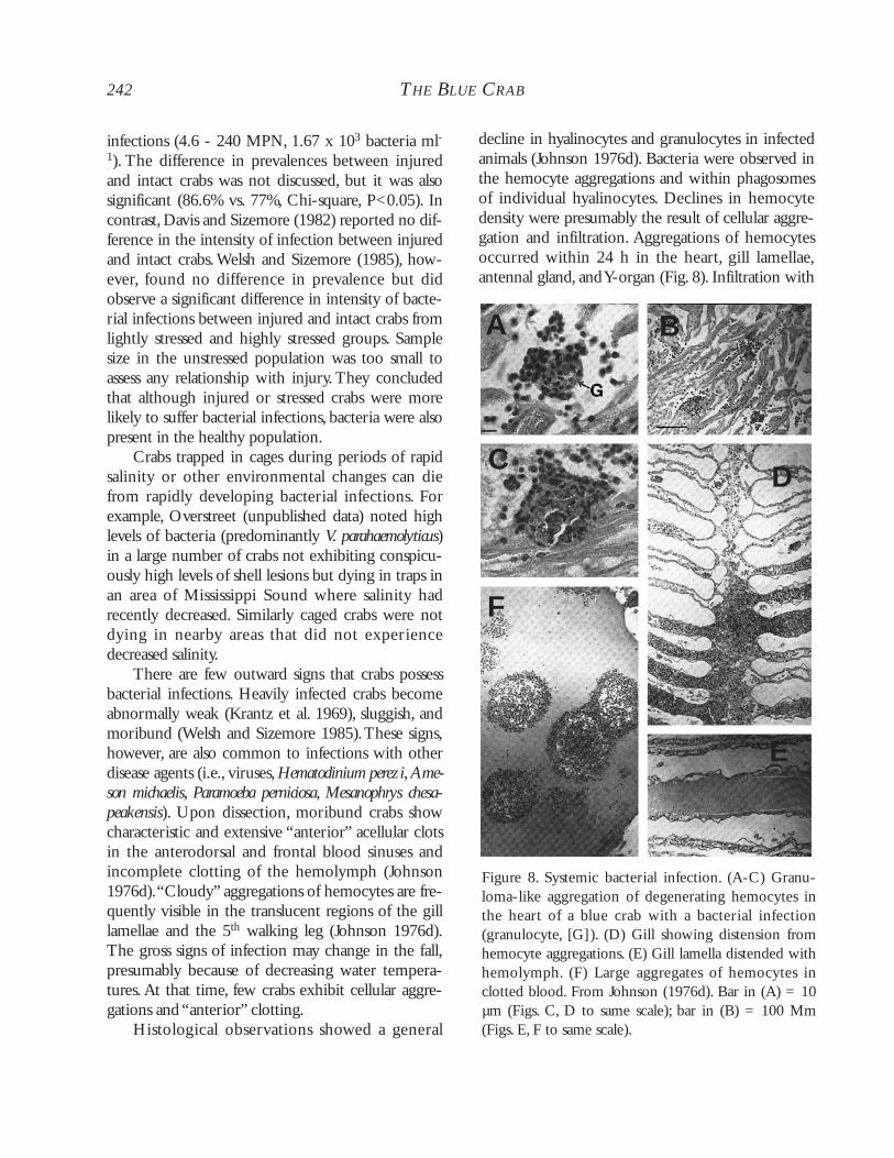
decline in hyalinocytes and granulocytes in infectedanimals (Johnson 1976d). Bacteria were observed inthe hemocyte aggregations and within phagosomesof individual hyalinocytes. Declines in hemocytedensity were presumably the result of cellular aggre-gation and infiltration. Aggregations of hemocytesoccurred within 24 h in the heart, gill lamellae,antennal gland, and Y-organ (Fig. 8). Infiltration with
242 THE BLUE CRAB
Figure 8. Systemic bacterial infection. (A-C) Granu-loma-like aggregation of degenerating hemocytes inthe heart of a blue crab with a bacterial infection(granulocyte, [G]). (D) Gill showing distension fromhemocyte aggregations. (E) Gill lamella distended withhemolymph. (F) Large aggregates of hemocytes inclotted blood. From Johnson (1976d). Bar in (A) = 10µm (Figs. C, D to same scale); bar in (B) = 100 Mm(Figs. E, F to same scale).
A B
CD
F
E
marked encapsulation was not apparent. Throm-boemboli formed as the aggregates were sloughedfrom the gill lamellae. Such emboli apparentlycaused ischemia through hemolymph stasis and fur-ther clot formation (Johnson 1976d). As the infec-tion progressed, nodules occurred less frequently inthe heart and blood sinuses, but more frequently inthe other organs. Large aggregations of hemocytesembolized and led to extensive focal necrosis anddegeneration in heavy infections. The hepatopan-creas showed significant involvement in infectedcrabs. The acinar epithelium exhibited massivesloughing and few mitotic figures, while karyolysisoccurred in affected cells (Johnson 1976d). Fixedhemocytes demonstrated karyorrhexis, pycnosis, andkaryolysis, possibly as a result of their phagocytosis ofthe virulent bacteria (Johnson 1976d).
Pathological effects occur quickly in bacterialinfections with mortalities occurring in as few as 2to 3 d (Johnson 1976d). Bacterial populations aretypically controlled by cellular and humoral defenses(i.e., phagocytosis, lectins, and callinectins; seeDefensive Responses below), but proliferation in thehemolymph may occur quickly in relation to watertemperature, handling, or other stressors (Davis andSizemore 1982).
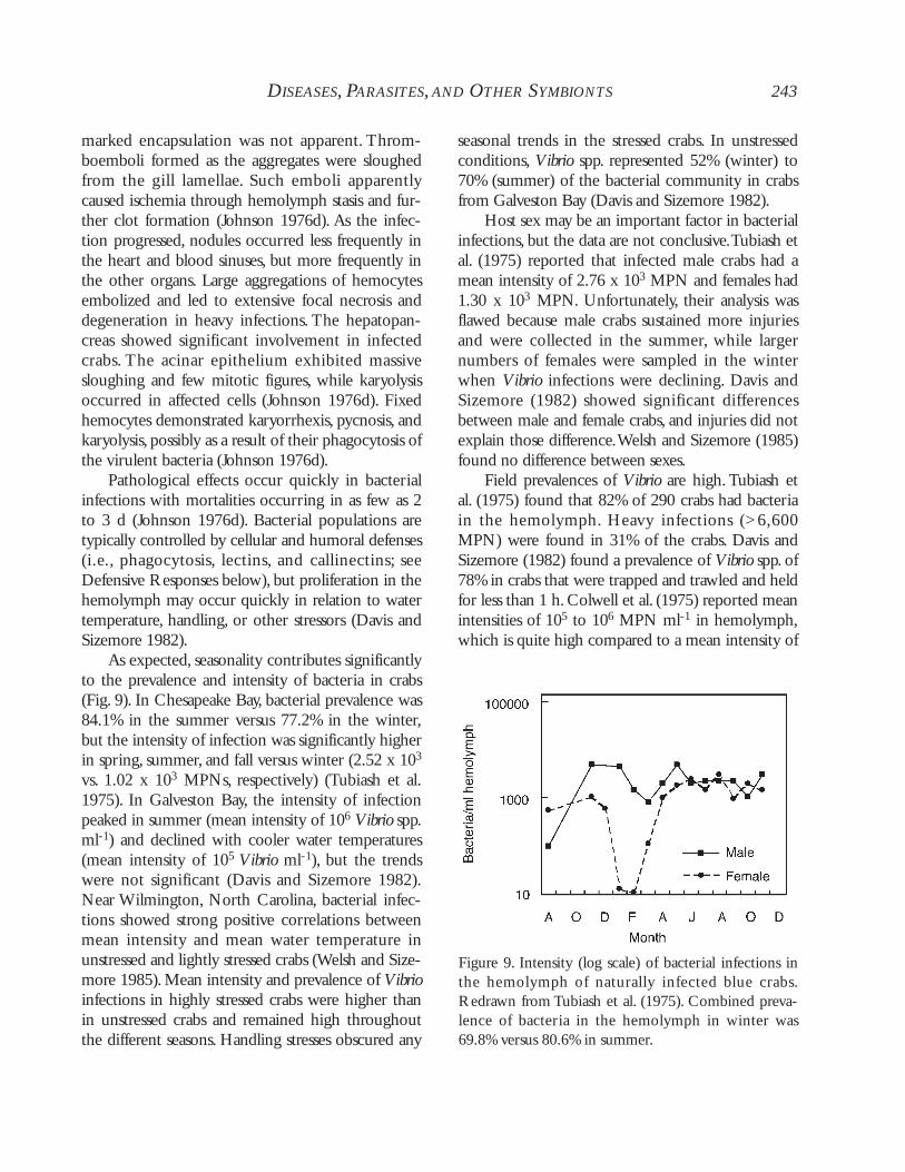
As expected, seasonality contributes significantlyto the prevalence and intensity of bacteria in crabs(Fig. 9). In Chesapeake Bay, bacterial prevalence was84.1% in the summer versus 77.2% in the winter,but the intensity of infection was significantly higherin spring, summer, and fall versus winter (2.52 x 10³
vs. 1.02 x 10³ MPNs, respectively) (Tubiash et al.1975). In Galveston Bay, the intensity of infectionpeaked in summer (mean intensity of 106 Vibrio spp.ml-1) and declined with cooler water temperatures(mean intensity of 105 Vibrio ml-1), but the trendswere not significant (Davis and Sizemore 1982).Near Wilmington, North Carolina, bacterial infec-tions showed strong positive correlations betweenmean intensity and mean water temperature inunstressed and lightly stressed crabs (Welsh and Size-more 1985). Mean intensity and prevalence of Vibrioinfections in highly stressed crabs were higher thanin unstressed crabs and remained high throughoutthe different seasons. Handling stresses obscured any
seasonal trends in the stressed crabs. In unstressedconditions, Vibrio spp. represented 52% (winter) to70% (summer) of the bacterial community in crabsfrom Galveston Bay (Davis and Sizemore 1982).
Host sex may be an important factor in bacterialinfections, but the data are not conclusive.Tubiash etal. (1975) reported that infected male crabs had amean intensity of 2.76 x 10³ MPN and females had1.30 x 103 MPN. Unfortunately, their analysis wasflawed because male crabs sustained more injuriesand were collected in the summer, while largernumbers of females were sampled in the winterwhen Vibrio infections were declining. Davis andSizemore (1982) showed significant differencesbetween male and female crabs, and injuries did notexplain those difference.Welsh and Sizemore (1985)found no difference between sexes.
Field prevalences of Vibrio are high. Tubiash etal. (1975) found that 82% of 290 crabs had bacteriain the hemolymph. Heavy infections (>6,600MPN) were found in 31% of the crabs. Davis andSizemore (1982) found a prevalence of Vibrio spp. of78% in crabs that were trapped and trawled and heldfor less than 1 h. Colwell et al. (1975) reported meanintensities of 105 to 106 MPN ml-1 in hemolymph,which is quite high compared to a mean intensity of
DISEASES, PARASITES,AND OTHER SYMBIONTS 243
Figure 9. Intensity (log scale) of bacterial infections inthe hemolymph of naturally infected blue crabs.Redrawn from Tubiash et al. (1975). Combined preva-lence of bacteria in the hemolymph in winter was69.8% versus 80.6% in summer.
2.4 x 10⁴ colony forming units (CFU) ml-1 (Davisand Sizemore 1982) and 1.8 x 10³ CFU ml-1 (Welshand Sizemore 1985). Welsh and Sizemore (1985)speculated that the absence of crabs with infectionsgreater than 104 CFU ml-1 might have been due tomortality or to moribund crabs not entering traps.Rivera et al. (1999), however, reported densities of2.9 x 107 CFU ml-1 in C. boucourti, with directcounts of 3.53 x 10⁹ to 4.64 x 10¹¹ bacteria ml-1.
Johnson (1976d) suggested that crabs acquiredbacterial infections from the stress of capture andhandling. Her results supported Bang’s (1970) viewthat the hemolymph of blue crabs was sterile andthat infections resulted from stress and trauma fromhandling. She did not, however, attempt to isolateand culture bacteria from crabs nor did she quantifyinfections that were observable with the light micro-scope (i.e., infections ≥ 106 bacteria ml-1) (Johnsonet al. 1981). Microscopic and histological analyseswere conducted for crabs that were roughly handled(commercially trapped, transported, and held out ofwater from 4 to 8 h) and for those that were trawledby research personnel (held out of water for < 6 h).The commercially caught crabs suffered 80% mor-tality over 12 d compared to 23% for the trawledcrabs. Bacterial infections were diagnosed histologi-cally in 85% of the mortalities from commerciallyfished crabs versus 45% in carefully handled crabs.Mortalities declined after 9 d, but the observationsmay have been confounded by a significant declinein water temperature. Mortality to bacterial infec-tions was further reduced in animals that were col-lected and handled gently (< 2 h exposure time).
In a rigorously controlled assessment of thestress issue,Welsh and Sizemore (1985) showed thatthe prevalence of bacteria in the spring and summerwas high (75%) in unstressed, freshly caught (pyra-mid traps) crabs. Lightly stressed crabs from pots(research collections using pots, with crabs held insitu for up to 24 h), and highly stressed crabs (pur-chased live from the market) showed somewhathigher prevalences (81 and 91%, respectively). Theintensity of infection was significantly lower in theunstressed crabs than in both of the stressed groups,and the lightly stressed crabs had significantly lowerintensities than the highly stressed group (mean
intensities of 14 vs. 19 vs. 46 CFU ml-1, respec-tively). Vibrio spp. comprised mean percentages of27, 26, and 44% of the bacterial community, respec-tively,with the heaviest infections represented almostsolely by Vibrio spp. From the stressed groups, onecan infer that heavy infections develop quickly fromlightly infected crabs.
Bacterial mortalities are common in sheddingfacilities. Messick and Kennedy (1990) used a split-plot design to examine host mortality and preva-lence of bacterial and viral infections in relation tothe type of holding system (flow-through vs. recir-culating) and crab density. Moribund crabs wereexamined histologically but not with isolation andculture techniques.Although there was no differencein the mean number of mortalities between systems,the total number of mortalities was higher in therecirculating systems (separately by month and intotal). Most of the mortalities could be attributed tobacterial and viral infections. Mortality rates werehighest in the recirculating system in June and Julyand declined in August. Interestingly, bacterial infec-tions were common in crabs from the flow-throughsystem. Messick and Kennedy’s (1990) findings sug-gest that flow-through systems may place less stresson crabs because mortalities were lower, eventhough bacteria were prevalent in the systems.Thestudy confirmed the importance of careful handlingof crabs to reduce mortalities in shedding systems.
Public Health Implications
Infections of Vibrio spp. in crabs warrant somepublic health concern. Raw or poorly preparedcrabmeat may be contaminated with pathogenicforms. Fortunately, neither the Southeast Asian deli-cacy of drunken crab (live crab marinated in winebefore eating) nor the habit of flavoring dishes withraw crab juices is popular in the USA. Thus, thereare few cases in the USA of bacterial poisoning fromeating crabs. Nonetheless, blue crabs should becooked thoroughly and eaten immediately or storedproperly before eating (Overstreet 1978). In addi-tion, V. cholerae shows a predilection for chitin (Huqet al. 1983; see Pruzzo et al. 1996), and attaches tothe chitin in the hindgut of blue crabs (Huq et al.1986). Because the bacterium attaches to chitin, it
244 THE BLUE CRAB
may potentially be transmitted by contaminatedcopepods (Huq et al. 1983, 1984; Chowdhury et al.1997; Montanari et al. 1999). Food handlers shouldbe aware of the potential for exposure to cholera,but at present, the possibility appears negligible inthe USA (Blake et al. 1980). Other species such as V.parahaemolyticus are often transmitted to cooled,cooked crabs from contact with live crabs or fromthe juices of uncooked crabs. In any case, live or rawcrabs should not be in contact with, be stored above,or otherwise contaminate cooked foods (Overstreet1978).
Future Research
Periodically the soft-shell industry experiencescrab mortalities from species of Vibrio. Blue crabmortalities are often localized regionally and can sig-nificantly reduce the short-term production of soft-shell crabs. In most cases, poor water quality, highstocking density, and other factors, such as tempera-ture, influence the level of stress in the host. Bacteriaare ubiquitous, and stress results in an increase inbacterial intensity. Nevertheless, given the impor-tance of crab mortalities in the soft-shell industry, itis remarkable that no one has published conclusiveexperiments to fulfill Koch’s postulates as has beendone with infections in lobster (Bowser et al. 1981).Because bacteria can be inoculated into crabs andrecovered (Shields, pers. obs.) and because bacterialclearance occurs quickly in some species (White andRatcliffe 1982; Martin et al. 1993), Koch’s postulatesshould be relatively easy to fulfill. Control groups ofuninfected crabs must be established with prior sam-pling. Injection studies, well-controlled mortalitystudies, and research to show better the associationsamong foci of infection, water quality, and otherstressors should be considered priorities.
There is at present no therapeutic treatment forsymptomatic crabs.As with many bacterial problemsin aquaculture, good culture practices and handlingtechniques are the best prophylaxis against bacteri-ally induced mortalities in shedding facilities. Lastly,crabs and other shellfish should be further assessed asindicators of pathogenic strains of Vibrio including V.parahaemolyticus, V. vulnificus, and V. cholerae in moni-toring programs for public health. Bacterial contam-
ination of crabs and other shellfish should not beignored.
Shell Disease (ChitinoclasticBacteria)
Shell disease is typically a non-fatal external bac-terial infection of the blue crab and other crus-taceans that have been subjected to stress. Injuriessustained from high stocking densities, long-termconfinement, molting, and environmental pollutantshave been implicated as stressors inducing shell dis-ease in many decapods (Rosen 1967; Iversen andBeardsley 1976; Overstreet 1978; Johnson 1983;Getchell 1989; Sindermann 1989; Smolowitz et al.1992). Chitinoclastic bacteria are a part of the nor-mal fauna found on crustaceans. Although bacteriaare clearly involved in the etiology of the disease,pollutants (i.e., sewage sludge, dredge spoils, heavymetals, organic debris) and other symbionts can playa significant role in the syndrome (Young and Pearce1975; Couch 1983; Morado et al. 1988; Gemperlineet al. 1992; Weinstein et al. 1992; Ziskowski et al.1996;Andersen et al. 2000).
Biology
Shell disease was first described from the Ameri-can lobster Homarus americanus (see Hess 1937). Asimilar disease was observed in freshwater crayfish inthe 1880s, but it was later determined to be a fungus(krebspest, or burn-spot disease caused byAphanomyces astaci).While fungal infections of crus-taceans also cause shell lesions, few fungi have beenisolated from the characteristic lesions of shell dis-ease (Rosen 1967). Chitinoclastic bacteria are iso-lated by streaking infected shell onto difco-marineagar with precipitated chitin (Skerman medium)(Cook and Lofton 1973) or by swabbing the lesionwith a sterile loop and inoculating into enrichmentbroth with chitin (Malloy 1978).
Shell disease in blue crabs is typically caused bysmall, chitinoclastic, gram-negative rods (Rosen1967).The genera of bacteria have been tentativelyidentified as Vibrio, Beneckea (now Vibrio), andPseudomonas (Cook and Lofton 1973). As noted inTable 1, 14 of the 49 bacteria collected from nearPensacola, Florida, produced chitinase, an enzyme
DISEASES, PARASITES,AND OTHER SYMBIONTS 245
allowing the bacteria to break down the crab’s chiti-nous exoskeleton (Overstreet and Rebarchik,unpublished). The bacteria belonged in the generaVibrio, Aeromonas, Pseudomonas, Kingella, and Serratia.As in other studies, V. parahaemolyticus was the pre-dominant species. Kingella kingae, also common inthe crab and also found in the eastern oyster andshrimps in the Gulf of Mexico, is a non-motilegram-negative, straight rod that has not beenregarded before now as a chitinoclastic bacterium. Itis known from the human respiratory system anddoes not grow in media with NaCl concentrationsof 4% and higher; perhaps it was introduced into theecosystem following a heavy rainfall and is not atypical part of the normal microbial flora of the bluecrab. Other bacteria (Photobacterium sp., V. anguil-larum, and Vibrio spp.) have been isolated fromlesions on Chionoecetes bairdi (Baross et al. 1978).
The crustacean exoskeleton is comprised ofthree layers: the epicuticle, the exocuticle, and themembranous layer overlying the living epidermis(Skinner 1962, 1985; Green and Neff 1972; O’Brienet al. 1991). Polyphenolic substances contained inthe epicuticle provide resistance to microbial degra-dation (Dennell 1960), but a wide range of bacteriacolonizes the surface of the epicuticle, and slowdegradation of the cuticle over the molt cycle of thehost may allow penetration of chitinoclastic bacteria(Baross et al. 1978). Damage to the exoskeleton pro-vides a portal of entry for chitinoclastic bacteria(Cook and Lofton 1973; Malloy 1978), and thedeveloping lesions represent a portal of entry andmedia for other infectious agents.Trauma, fungi, andother events may also effect portals of entry(Getchell 1989). In shrimp, lipolytic enzymes mayinitiate the lesion,with chitinase, lipase, and proteasesimportant to lesion development (Cipriani et al.1980). Lipases may be important in initiating inva-sion through the waxy epicuticle, with chitinasesand proteases facilitating expansion into the chitin-rich exocuticle and membranous layer.
Animal Health and Fisheries Implications
Early stages of shell disease initiate as numerous,small, brown puncture- or crater-like marks on the
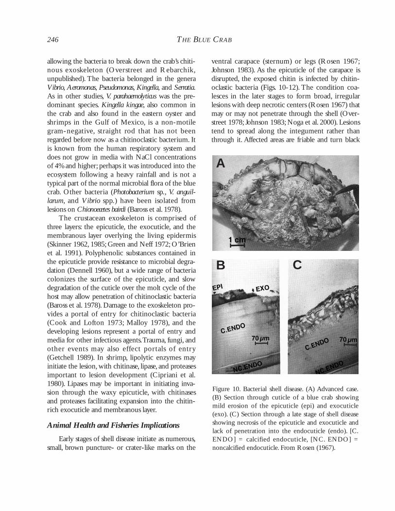
ventral carapace (sternum) or legs (Rosen 1967;Johnson 1983). As the epicuticle of the carapace isdisrupted, the exposed chitin is infected by chitin-oclastic bacteria (Figs. 10-12). The condition coa-lesces in the later stages to form broad, irregularlesions with deep necrotic centers (Rosen 1967) thatmay or may not penetrate through the shell (Over-street 1978; Johnson 1983; Noga et al. 2000). Lesionstend to spread along the integument rather thanthrough it. Affected areas are friable and turn black
246 THE BLUE CRAB
Figure 10. Bacterial shell disease. (A) Advanced case.(B) Section through cuticle of a blue crab showingmild erosion of the epicuticle (epi) and exocuticle(exo). (C) Section through a late stage of shell diseaseshowing necrosis of the epicuticle and exocuticle andlack of penetration into the endocuticle (endo). [C.ENDO] = calcified endocuticle, [NC. ENDO] =noncalcified endocuticle. From Rosen (1967).
A
B C
or blue from melanin deposition (Johnson 1983).Rosen (1970) viewed the necrotic pits as miniaturecommunities of bacterial colonizers, includingchitinoclastic and non-chitinoclastic forms.
In advanced cases, the lesion penetrates into thenoncalcified membranous layer, and limbs and spinesmay become necrotic and are lost (Rosen 1967).The gills can also be attacked (Johnson 1983).Infected American lobsters show varying stages ofhost response ranging from cellular infiltration, epi-cuticle deposition, and melanization to pseudomem-brane formation (Smolowitz et al. 1992). Lightly andmoderately infected individuals can overcome thedisease by molting (Rosen 1967), but the area of thelesions may not reflect the severity of the disease(Noga et al. 2000). Newly molted crabs are usuallyfree of shell disease, but in advanced cases, the newinstar dies from an inability to cast off the old molt(Sandifer and Eldridge 1974; Fisher et al. 1976;Overstreet 1978; Johnson 1983). Older blue crabs,which molt less frequently, are most affected by thedisease (Sandifer and Eldridge 1974). Heavilyinfected crabs are lethargic, weak, and die whenstressed.
Cook and Lofton (1973) inoculated cultures ofbacteria directly onto sterile, rasped, or scraped sur-faces of crab exoskeletons. After a few weeks, shellnecrosis was observed on all of the rasped surfaces.Inoculated but undamaged areas did not obtain dis-
DISEASES, PARASITES,AND OTHER SYMBIONTS 247
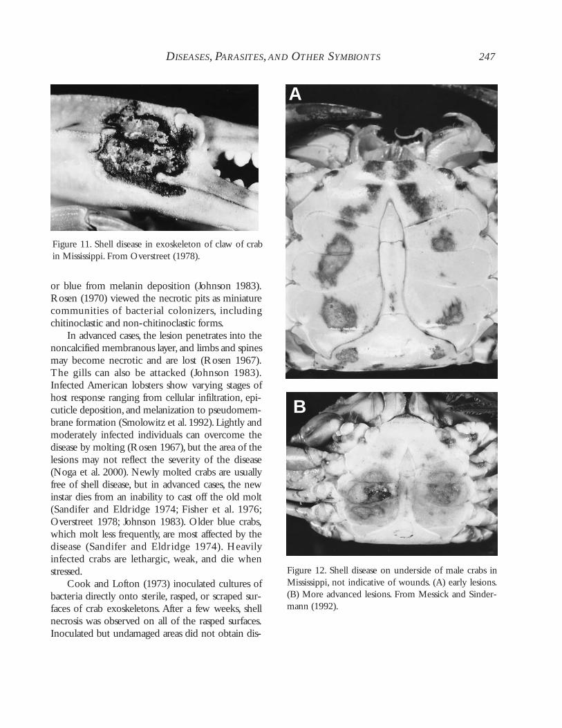
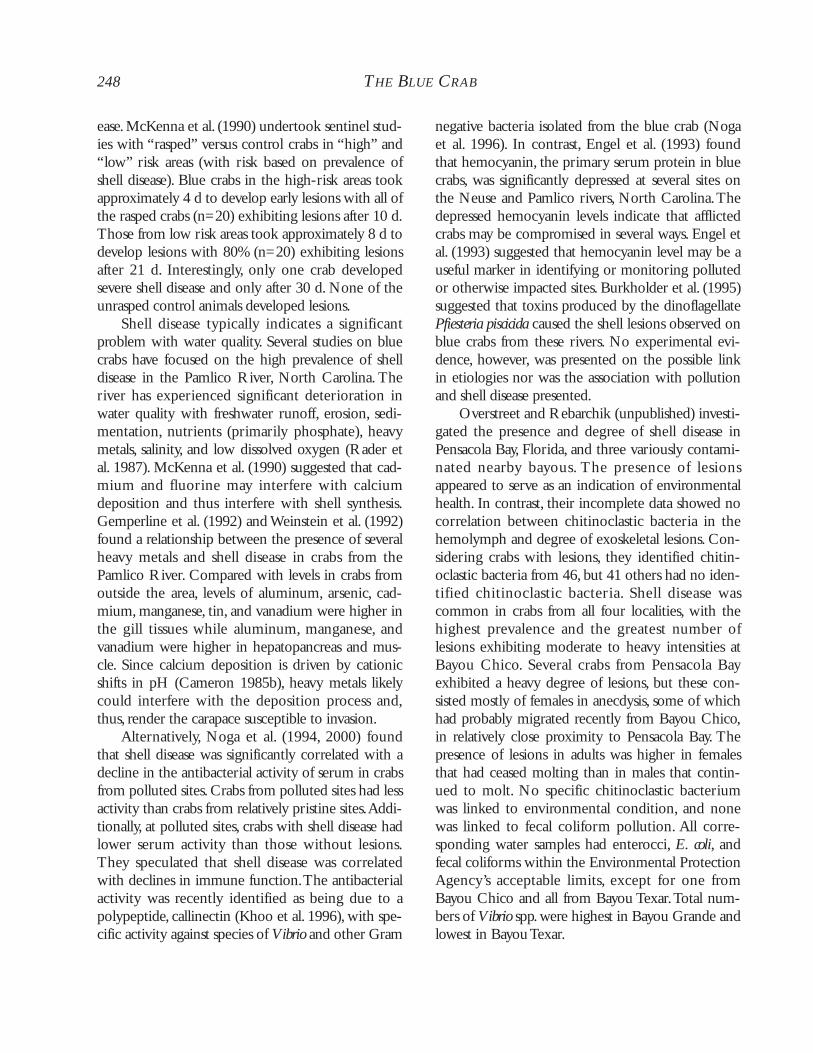
Figure 11. Shell disease in exoskeleton of claw of crabin Mississippi. From Overstreet (1978).
Figure 12. Shell disease on underside of male crabs inMississippi, not indicative of wounds. (A) early lesions.(B) More advanced lesions. From Messick and Sinder-mann (1992).
A
B
ease.McKenna et al. (1990) undertook sentinel stud-ies with “rasped” versus control crabs in “high” and“low” risk areas (with risk based on prevalence ofshell disease). Blue crabs in the high-risk areas tookapproximately 4 d to develop early lesions with all ofthe rasped crabs (n=20) exhibiting lesions after 10 d.Those from low risk areas took approximately 8 d todevelop lesions with 80% (n=20) exhibiting lesionsafter 21 d. Interestingly, only one crab developedsevere shell disease and only after 30 d. None of theunrasped control animals developed lesions.
Shell disease typically indicates a significantproblem with water quality. Several studies on bluecrabs have focused on the high prevalence of shelldisease in the Pamlico River, North Carolina. Theriver has experienced significant deterioration inwater quality with freshwater runoff, erosion, sedi-mentation, nutrients (primarily phosphate), heavymetals, salinity, and low dissolved oxygen (Rader etal. 1987). McKenna et al. (1990) suggested that cad-mium and fluorine may interfere with calciumdeposition and thus interfere with shell synthesis.Gemperline et al. (1992) and Weinstein et al. (1992)found a relationship between the presence of severalheavy metals and shell disease in crabs from thePamlico River. Compared with levels in crabs fromoutside the area, levels of aluminum, arsenic, cad-mium, manganese, tin, and vanadium were higher inthe gill tissues while aluminum, manganese, andvanadium were higher in hepatopancreas and mus-cle. Since calcium deposition is driven by cationicshifts in pH (Cameron 1985b), heavy metals likelycould interfere with the deposition process and,thus, render the carapace susceptible to invasion.
Alternatively, Noga et al. (1994, 2000) foundthat shell disease was significantly correlated with adecline in the antibacterial activity of serum in crabsfrom polluted sites. Crabs from polluted sites had lessactivity than crabs from relatively pristine sites.Addi-tionally, at polluted sites, crabs with shell disease hadlower serum activity than those without lesions.They speculated that shell disease was correlatedwith declines in immune function.The antibacterialactivity was recently identified as being due to apolypeptide, callinectin (Khoo et al. 1996), with spe-cific activity against species of Vibrio and other Gram
negative bacteria isolated from the blue crab (Nogaet al. 1996). In contrast, Engel et al. (1993) foundthat hemocyanin, the primary serum protein in bluecrabs, was significantly depressed at several sites onthe Neuse and Pamlico rivers, North Carolina.Thedepressed hemocyanin levels indicate that afflictedcrabs may be compromised in several ways. Engel etal. (1993) suggested that hemocyanin level may be auseful marker in identifying or monitoring pollutedor otherwise impacted sites. Burkholder et al. (1995)suggested that toxins produced by the dinoflagellatePfiesteria piscicida caused the shell lesions observed onblue crabs from these rivers. No experimental evi-dence, however, was presented on the possible linkin etiologies nor was the association with pollutionand shell disease presented.
Overstreet and Rebarchik (unpublished) investi-gated the presence and degree of shell disease inPensacola Bay, Florida, and three variously contami-nated nearby bayous. The presence of lesionsappeared to serve as an indication of environmentalhealth. In contrast, their incomplete data showed nocorrelation between chitinoclastic bacteria in thehemolymph and degree of exoskeletal lesions. Con-sidering crabs with lesions, they identified chitin-oclastic bacteria from 46, but 41 others had no iden-tified chitinoclastic bacteria. Shell disease wascommon in crabs from all four localities, with thehighest prevalence and the greatest number oflesions exhibiting moderate to heavy intensities atBayou Chico. Several crabs from Pensacola Bayexhibited a heavy degree of lesions, but these con-sisted mostly of females in anecdysis, some of whichhad probably migrated recently from Bayou Chico,in relatively close proximity to Pensacola Bay. Thepresence of lesions in adults was higher in femalesthat had ceased molting than in males that contin-ued to molt. No specific chitinoclastic bacteriumwas linked to environmental condition, and nonewas linked to fecal coliform pollution. All corre-sponding water samples had enterocci, E. coli, andfecal coliforms within the Environmental ProtectionAgency’s acceptable limits, except for one fromBayou Chico and all from Bayou Texar.Total num-bers of Vibrio spp.were highest in Bayou Grande andlowest in Bayou Texar.
248 THE BLUE CRAB
DISEASES, PARASITES,AND OTHER SYMBIONTS 249
The benthic life style of the blue crab con-tributes to the transmission of shell disease. Sedi-ments foster high densities of chitinoclastic bacteria(Seki 1965; Cook and Lofton 1973; Hood andMeyers 1977); hence, burying activity places crabs indirect contact with the highest densities of chiti-nolytic forms.Vogan et al. (1999) found a higherprevalence of lesions on the posterodorsal carapace,and the ventral surfaces of the legs of Cancer pagurus.They suggested that lesions develop from sand abra-sion with subsequent infection resulting from theburying activity of the crab. Young and Pearce(1975) showed that lesions developed in lobstersheld in aquaria containing sludge from a dumpsite.McKenna et al. (1990) found that lesions on bluecrabs were more frequent on the anterodorsal cara-pace and on the posterodorsal carapace than else-where on the carapace or limbs, but they did notrecord lesions on the sternum, one of the morecommon areas for initial infections (Rosen 1967;Iversen and Beardsley 1976; Overstreet 1978; John-son 1983; Getchell 1989). Dredged crabs fromChesapeake Bay exhibit the characteristic pinpointlesions on the sterna and limbs in late winter pre-sumably from their residence in the sediments(Shields, pers. obs.). Although Hood and Meyers(1974) found peak populations of chitinoclastic bac-teria in the spring and summer, shell disease in bluecrabs was highest in fall and winter (Sandifer andEldridge 1974). Thus, shell disease on blue crabsarises from abrasions acquired from their buryingactivities, from other wounds to the epicuticle, andfrom stress due to poor water quality.
Shell disease is contagious, especially in long-term, crowded conditions such as those found atAmerican lobster holding facilities (Rosen 1970;Sandifer and Eldridge 1974). Mortality can be highin lobster facilities, but less so with blue crabs whereculture conditions are generally of short duration(e.g., soft-shell production). Brock and Lightner(1990) note that shell disease is often associated withstress and that the underlying causes of stress must bedetermined in a differential diagnosis.Wound avoid-ance, proper attention to hygiene, and proper hus-bandry lessen the prevalence of shell disease in lob-ster culture systems (Stewart 1980).
Heavily diseased animals can be difficult to treat.Dips of malachite green have been used for lobsters(Fisher et al. 1978), and antibiotic baths (penicillin-streptomycin, furanace, erythromycin, oxolinic acid)and malachite green and formalin have been usedfor shrimp (Tareen 1982; Brock 1983; El-Gamal etal. 1986). Disinfection of aquaria can be achievedwith bleach solutions. Advanced cases should bedestroyed to prevent further spread and to avoidexcessive trauma.
In general, shell disease is not a significant factorin mortality of wild stocks of crustaceans. Highprevalences of it, however, may indicate significantissues involving water quality or stress in residentcrustacean populations. Shell disease does have asmall economic impact in that afflicted animals arenot aesthetically pleasing to eat; thus there may be alower grading of meat value (Rosen 1970; Getchell1989). Because blue crabs are not held for long peri-ods, shell disease in culture systems is not a signifi-cant issue. Rosen (1967) recorded 3% prevalence inthree shedding houses in Maryland. Sandifer andEldridge (1974) reported monthly prevalences infield and commercial samples of 15,000 crabs col-lected from four locations in South Carolina. Preva-lences ranged from 0.0 to 53.1%, with field sampleshaving a higher overall prevalence (9.2%) comparedwith that in commercial samples (3.4%). Males wereslightly more susceptible than females, but the datawere not consistent. McKenna et al. (1990) did anextensive survey of shell disease in the PamlicoRiver (trawl, pot, and sentinel studies). In July 1987,5% of 1459 trawled crabs had shell disease. Maleshad a prevalence of 5.1%, females, 16.2%, and imma-ture females, 2.5%.Although more crabs were foundin shallower waters, there was no associationbetween shell disease and depth (5.1 vs. 4.4% preva-lence, 0-1.82 m vs. 1.83-3.65 m depth, respectively,Chi-square).
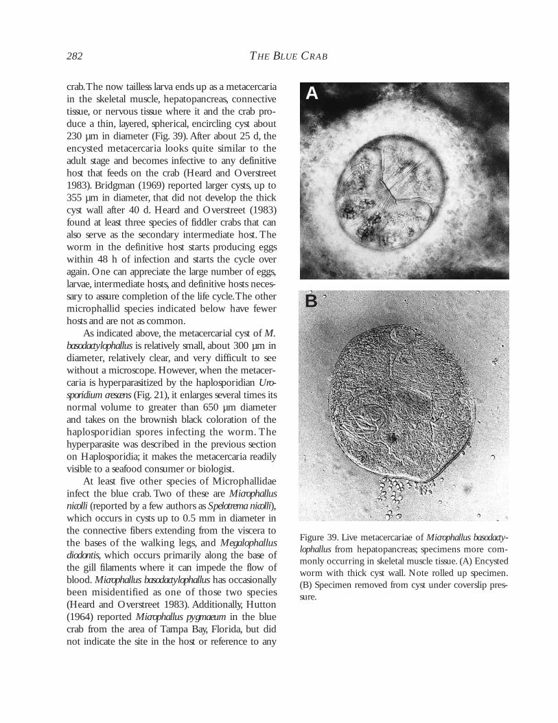
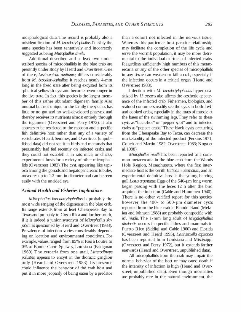
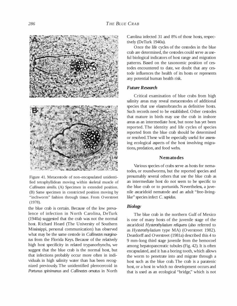
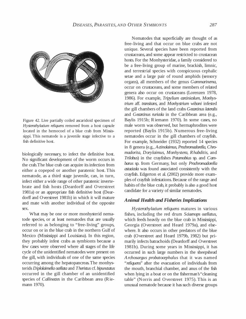
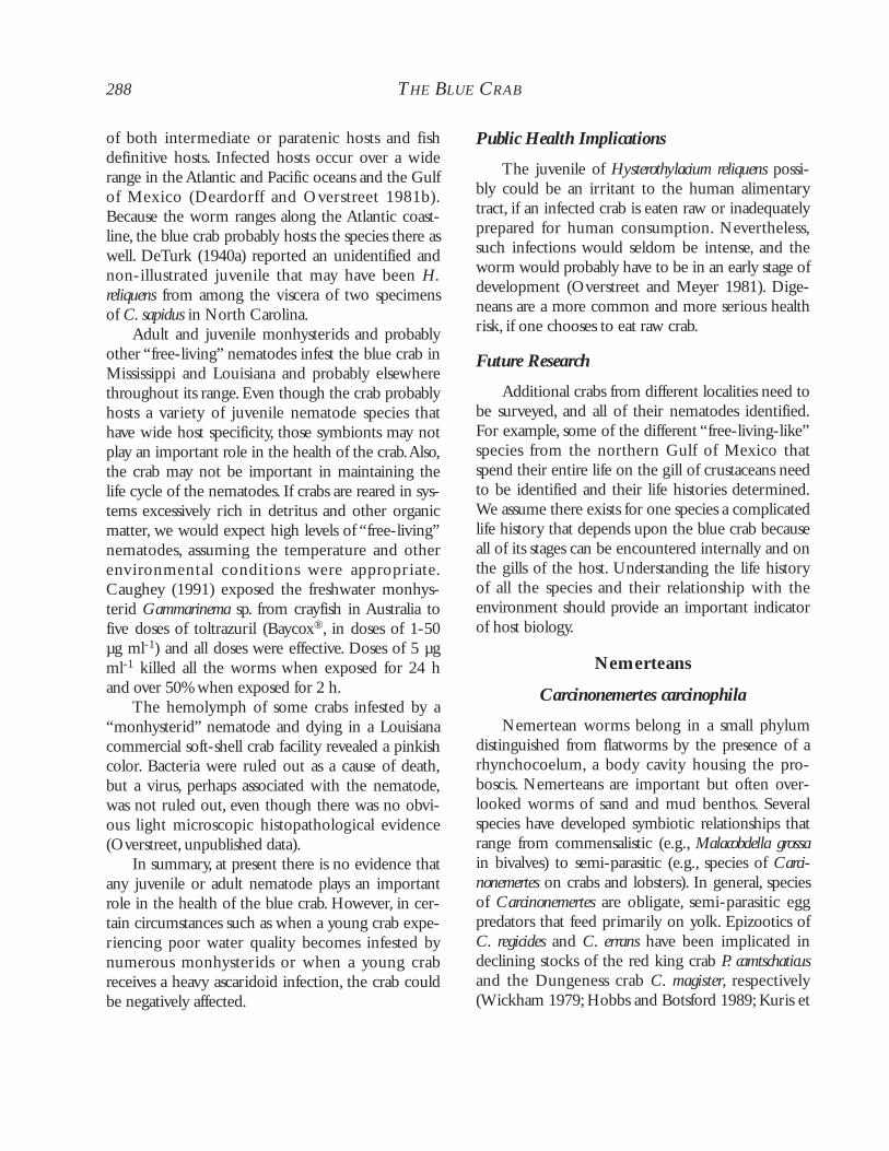
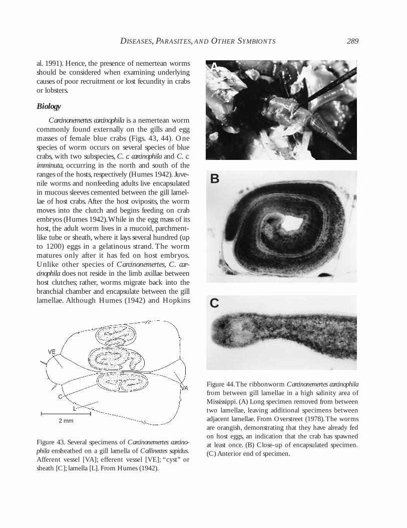
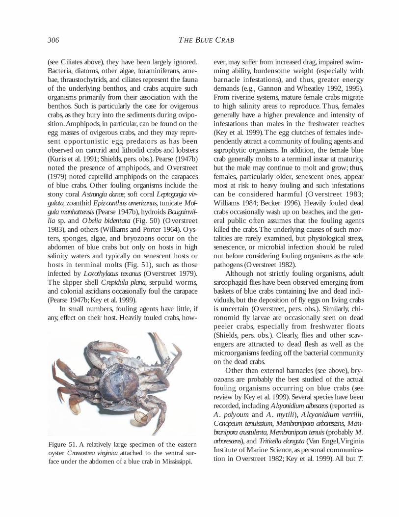
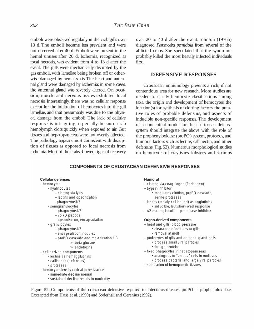
Future Research
Shell disease is relatively innocuous in bluecrabs.However, significant research questions can beaddressed through study of the disease. Stress is a sig-nificant issue in the onset of numerous diseases and
conditions in invertebrates. Further investigation ofthe association between different stressors and theonset of shell disease will provide a useful model tostudy the effects of stress on crustaceans and otherinvertebrates. Indeed, the decrease in immune func-tion and decline in hemocyanin in diseased crabsfrom polluted waters (e.g., Engel et al. 1993; Noga etal. 1994, 1996) highlights the need for just suchstudies. In addition, the prevalence of shell diseasemay be an excellent indicator of water quality (e.g.,pollution, nutrient enrichment). Thus, monitoringfor shell disease may provide an inexpensive earlywarning tool for pollutants or other stressors.
Other Bacteria
Other bacterial infections have been reportedfrom the blue crab.A rare, Gram-negative bacteriumhas been observed in the midgut and hepatopan-creas (Johnson 1983). It was associated with focalnecrosis of the hepatic tubules.An unusual filamen-tous, non-septate Gram-negative bacter ium(“strand-like” organism - Johnson 1983; Messick1998) was also observed attached to the tubules ofthe hepatopancreas. It was reported from 2% of crabsfrom the Atlantic and Gulf coasts (Johnson 1983).The prevalence of the bacterium was lower in flow-through (12%) versus recirculating seawater (31%)systems in the summer (Messick and Kennedy1990).The bacterium exhibited a peak in prevalencein the summer (up to 16% in Maryland), and signifi-cantly higher prevalences in juveniles than adults(Messick 1998). The localized effect of the bac-terium included subtle changes in the epithelial cellswith the formation of syncytia. The strand-likeorganisms are of doubtful pathogenicity to their crabhost as the epithelial cells of the hepatopancreas arebeing replaced continuously (Johnson 1983; Messick1998).
Rickettsiales-like organisms (RLOs) are typi-cally considered obligate intracellular bacterial para-sites.They are rarely reported from crustaceans. Massmortalities of shrimp, however, have been associatedwith RLOs (Krol et al. 1991; Lightner et al. 1992;Loy et al. 1996). In the blue crab, the prevalence of
RLOs from field samples is low, probably because ofthe few histological analyses available. Rickettsiales-like organisms had a prevalence of 2.3% in a Mary-land shedding facility, but heavy infections were notfatal (Messick and Kennedy 1990). In a later study, asingle infected crab exhibited a focal infection of thehepatopancreas, with moderate increases in hemo-cyte numbers in the adjacent hemal spaces (Messick1998). Given the emerging importance and abun-dance of RLOs in invertebrates, especially molluscs,it is surprising that so few have been described inCrustacea.
Leucothrix mucor is an ubiquitious filamentousbacterium found on the external surfaces, gills, andeggs of crabs, algae, and various other surfaces (John-son et al. 1971; Bland and Brock 1973). It is com-mon in the egg masses and occasionally on the gillsof the blue crab (Bland and Amerson 1974; Shields,pers. obs.). Bacterial fouling of the egg masses occurswith several crustaceans. Leucothrix mucor was sus-pected of contributing to egg mortalities in Cancerspp. (Fisher et al. 1976), but its contribution to mor-talities was negligible; rather, nemertean worms wereshown to cause significantly more egg mortalitythrough predation (Shields and Kuris 1988a). Thepresence of many strains of L. mucor and relatedspecies typically develop on hosts in the presence ofexcess nutrients, such as in natural areas contami-nated with domestic sewage or in overfed aquacul-ture facilities. Solangi et al. (1979) tested a variety oftreatments on infestations on the brine shrimp. Bac-terial mats and associated debris sloughed as a singleunit, most effectively with 100 ppm Terramycin(®)(an oxytetracycline formulation). In the field, somecrustaceans preen themselves of infestations withtheir third maxillipeds and, thus, control their infes-tations (Bauer 1979).
Lastly, bdellovibrios occur on the external sur-faces of the carapace and gills of the blue crab (Kel-ley and Williams 1992). Bdellovibrios are predatorybacteria that are ubiquitous in the marine environ-ment.They are not pathogenic and apparently feedon other bacteria.They should be considered foul-ing organisms.
250 THE BLUE CRAB
DISEASES, PARASITES,AND OTHER SYMBIONTS 251
FUNGI
Historically, the oomycetes (water molds) weredescribed as fungi. With the advent of TEM andadvanced molecular techniques, they have beenremoved from the fungi and placed in their ownphylum. For the sake of simplicity, we refer to theoomycetes and other “lower fungi” as fungi. Fungalinfections in crustaceans range from the benign tothe severe. Epizootic outbreaks have affected severalcrustaceans including copepods, crayfishes, tannercrab, penaeid shrimps, and American lobster. One ofthe most notorious and disastrous is krebspest,Aphanomyces astaci. Introduced into Europe with theAmerican signal crayfish Pacifastacus leniusculus in the1880s, it has since wiped out most stocks of theEuropean crayfish Astacus astacus (Unestam 1973).Fungal diseases in crustaceans have been reviewedby Unestam (1973), Alderman (1976), Lightner(1981), and Johnson (1983). In aquaculture, infec-tions of Lagenidium callinectes, Haliphthoros milfordensis,and Fusarium solani are relatively common inpenaeid shrimp and American lobster embryos,imposing significant threats to the culture of thesecrustaceans (Lightner 1981). The embryos of theblue crab can be experimentally infected with H.milfordensis, but natural infections have not beenreported in that host (Tharp and Bland 1977).
Lagenidium callinectes
Fortunately, there are few serious fungal infec-tions reported from the blue crab. A significantpathogen, Lagenidium callinectes was first isolated fromthe embryos of the blue crab (Couch 1942). It hassince been reported from the embryos and larvae ofseveral decapod crustaceans and algae.
Biology
Lagenidium callinectes is a holocarpic oomycetefungus-like protist that attacks and kills crabembryos. The fungal thallus consists of coenocytic,intramatrical hyphae within a crab embryo, andextramatrical hyphae that function in sporogenesisand spore discharge (Fig. 13).The life cycle of L. call-inectes was elucidated by Bland and Amerson (1973)
with additional work on an algal isolate by Gotelli(1974a, b). Sporangia start discharging spores after 12to 15 h in sterile sea water, with continued releaseextending over 48 h.The pyriform zoospores, 10 by13 µm, have two flagella arising from a groove thatspans the length of the spore. Polycomplexes indica-tive of meiosis have been observed in the encystingspores (Amerson and Bland 1973). Cysts, 9 to 11 µmin diameter, germinate a single germ tube that pene-trates the egg and grows rapidly into vegetativehyphae that in turn ramify throughout the embryo.Young hyphae have few septa. Septa usually delimitsporangia or separate older sections of the hyphae(Gotelli 1974b).
Upon death of the embryo, several holocarpicsporangia form at the end of discharge tubes (Fig.14) and release monoplanetic zoospores. Sporogene-sis begins at the tips of the extramatrical hyphaewith the formation of septa to differentiate the spo-rangium (Gotelli 1974a). After the discharge tubedevelops, cytoplasm is discharged (5-30 min) into agelatinous vesicle. Flagellar formation precedescleavage and the flagella can be observed activelybeating inside the sporangium (Gotelli 1974a).Cleavage is rapid and spore release occurs within 10min of sporogenesis. From 20 to 200 zoospores areproduced by a single sporangium. Zoospores swimto, encyst on, and infect new embryos, but extrama-tr ical hyphae also grow into and infect newembryos.
Figure 13. Lagenidium callinectes. Sporangial develop-ment in a modified Vishniac medium.
252 THE BLUE CRAB
Several strains of L. callinectes have been isolatedfrom decapod embryos and algae. Couch (1942)described one with short extramatrical hyphae, andBland and Amerson (1973) described another withelongate extramatrical hyphae. Bahnweg and Bland(1980) indicated the need for a review of the taxon-omy of the group based on biochemical attributesbetween different isolates. In a statistical analysisusing morphological and physiological parameters,Crisp et al. (1989) showed that strain differences arepart of intraspecific variation. Molecular taxonomywould no doubt improve our understanding of thistaxon.
Nutrient requirements vary significantly amongstrains of Lagenidium callinectes. Fungal growth israpid,with hyphal tips recognizable after 12 h in sea-water PYG-agar (Bland and Amerson 1973). An
algal strain was grown on a defined medium con-taining glutamate, glucose, vitamin B constituents,and trace metal mix (Gotelli 1974a,b).Vitamin B1was required for the isolate from the blue crab (L-1),and cultures grew better on simple sugars (e.g., fruc-tose, glucose) than on complex carbohydrates andpolysaccharides (Bahnweg and Bland 1980). Moststrains are obligate marine forms, but isolates fromthe American lobster and the Dungeness crab Cancermagister do not require NaCl (Bahnweg and Gotelli1980). Isolate L-1 is strongly proteolytic, and chitinis not used for growth (Bahnweg and Bland 1980).Chitin is not present in hyphal walls, but beta-glu-cans and both 1-3 and 1-6 glucosamines are abun-dant (Bertke and Aronson 1992). The nitrogensources and simple energy requirements of L. call-inectes appear representative of a marine saprophyte,not a “fastidious” parasite (Bahnweg and Bland1980); this is not surprising when one considers therich “medium” of an undifferentiated crab embryo.
Animal Health and Fisheries Implications
In the blue crab, Lagenidium callinectes naturallyinfects embryos. It has not been observed in zoeae,albeit experimentally infected larvae quickly lose theability to swim; hence, larvae sink and their mortal-ity may be difficult to document in the field(Rogers-Talbert 1948). The presence of the funguscan be observed as brown or gray patches in theclutch. Dead eggs are opaque and smaller thanhealthy ones (Couch 1942; Rogers-Talbert 1948).The fungus is usually restricted to the periphery ofthe clutch, penetrating rarely more than 3 mm intoit (Fig. 15). Diseased eggs die before hatching. Inheavy infections, the fungus may kill approximately25% of the clutch. Older clutches are attacked moreheavily than recently laid ones (Fig. 16).
Transmission to new hosts is rapid. Zoeae ofCancer magister acquire infections within 48 h ofmolting, and uninfected females can develop infec-tions within 2 to 3 d of being housed with infectedfemales or exposed to water that contains infectedclutches (Armstrong et al. 1976). Infections establishin the visceral organs of the larvae and then ramifythroughout the body and tissues (Armstrong et al.
Figure 14. Lagenidium callinectes. Dead crab egg showingextramatrical hyphae with sporangia containing devel-oping zoospores (arrow). Note the opacity of the egg.
1976). Zoeal stages may become infected during themolt (Armstrong et al. 1976), but because the ger-mination tube can penetrate the chorion of the egg,the tube can probably penetrate through the thincuticle of the larva. In larval specimens of C. magister,the fungus was highly pathogenic, with 40% mortal-ity over 7 d.Transmission can be difficult to controland may involve other crustaceans such as brineshrimp that are often used as food (Ho and Lightner,personal communication in Armstrong et al. 1976).
Ovigerous blue crabs experience a high preva-lence of the fungus throughout the summer inChesapeake Bay (Fig. 16) (Rogers-Talbert 1948).There is an apparent lag period in May when crabsare not infected. In North Carolina, the fungus has ahigh prevalence from May through June (95%) withprevalence in July (after the main ovigerous period)dropping to 30%, and to zero thereafter (Bland andAmerson 1973, 1974).Anecdotally, the prevalence ofthe fungus increased with the density of female crabs(Bland 1974).
Although the fungus has a wide salinity toler-ance, in Chesapeake Bay it is primarily restricted tothe lower regions of the Bay where salinity is high(Fig. 17; Rogers-Talbert 1948; Bahnweg and Bland1980). In the 1940s, the fungus occurred at highprevalences (40-62%) in the Virginia MarineResources Commission’s Crab Sanctuary (Rogers-Talbert 1948) and was moderate to high in YorkRiver,Virginia, in the 1960s (Scott 1962).
Natural infections have been reported from theembryos of the blue crab (Sandoz et al. 1944;Rogers-Talbert 1948), the barnacle Chelonibia patula(Johnson and Bonner 1960), the zoeae of Cancermagister (Armstrong et al. 1976), and algae (Fuller etal. 1964; Gotelli 1974a,b).The embryos of the crabsDyspanopeus texana, Panopeus herbstii, and Pinnotheresostreum but not those of Libinia emarginata, Menippemercenaria, or Sesarma cinereum are susceptible toinfection (Rogers-Talbert 1948; Bland and Amerson1974).The reason for such differences in susceptibil-ity has not been resolved, though the depth of thechorion or its resiliency may be factors (but seebelow).
In an elegant study of bacterial and fungal pa-thogenesis, Fisher (1983) showed that L. callinectes
DISEASES, PARASITES,AND OTHER SYMBIONTS 253
Figure 15. (A) Dead blue crab egg with fungal hyphae.(B) Live crab egg for compariso. (C) Representativecross-section through a healthy clutch of a blue crab.(D) Cross-section through a clutch infected withLagenidium callinectes showing the peripheral location(arrow) of most infected eggs. From Rogers-Talbert(1948).
Pre
vale
nce
(%)
JuneJulyAugust
100
80
60
40
20
0Yellow Black
Color of clutchBrown
Figure 16. Prevalence of Lagenidium callinectes inclutches of blue crabs during summer months.Yellow(early), brown (mid) and black (late) refer to the rela-tive age of the clutch. Data from Rogers-Talbert(1948).
grew only on the detached eggs of the orientalshrimp Palaemon macrodactylus except when the firstpleopods had been excised. Caridean shrimp,including P. macrodactylus, are excellent groomers;they may limit the spread of egg diseases by periodicpreening of the clutch (Bauer 1979, 1981, 1998,1999). Nonetheless, the fungus presents a significantproblem to crustacean aquaculture (Lightner andFontaine 1973; Nilson et al. 1975).
Curiously, the embryos of the oriental shrimpand the American lobster exhibit some resistance toinfection by L. callinectes (Gil-Turnes et al. 1989; Gil-Turnes and Fenical 1992). Gram-negative bacteriahave been isolated from these embryos and the pres-ence of penicillin-sensitive bacteria is correlatedwith resistance. On P. macrodactylus, the bacteriumAlteromonas sp. attaches to the outer chorionic coatof the embryo and releases 2,3 indolinedione, orisatin, and on lobster, a similar Gram-negative bac-terium releases 4-hydroxyphenethyl alcohol, ortyrosol; both are known antifungal compounds (Gil-
Turnes et al. 1989; Gil-Turnes and Fenical 1992).The presence of the 2,3 indolinedione alone wassufficient to inhibit mortality in embryos exposed tothe fungus.We speculate that poor water conditionsmay limit bacterial protection of the embryos inaquaculture systems.
Presumably several different fungi infect theblue crab and its eggs, but few, if any, recent studieshave explored the possibility. Haliphthoros milfordensis,a phycomycete that infects the eggs of severaldecapods and a gastropod and that appears grosslysimilar to L. callinectes, can infect blue crab eggsexperimentally; however, it has not been found innaturally occurring infections (Fisher et al. 1976,1978;Tharp and Bland 1977).
Future Research
Lagenidium callinectes may very well represent thegreatest fungal threat to the successful culture of sev-eral marine decapods. Hence, various fungicideshave been examined for use in shrimp aquaculture.Early studies that reported on the efficacy of mala-chite green in shrimp culture used reduced zooporemotility as the primary measure (Bland et al. 1976).Trifluralin and captan showed efficacy (as reductionsin mortalities) over 96-h exposures with minimallarval mortalities (Armstrong et al. 1976). Benomylwas toxic to larvae in 96-h exposures but showedsome efficacy over 48 h. Furanace showed excellentefficacy against H. milfordensis in shrimp aquaculture(Lio-Po et al. 1985). None of these compounds iscurrently available for use in USA aquaculture.
Lagenidium callinectes is often found in associationwith a filamentous bacterium and the nemerteanworm Carcinonemertes carcinophila (see Rogers-Tal-bert 1948; Bland and Amerson 1973). The bac-terium was thought to be Chlamydobacterium, but itis more likely the common filamentous Leucothrixmucor (see Bland and Amerson 1974; Shields, pers.obs.). Leucothrix mucor is a common constituent onthe surface of crab eggs (Johnson et al. 1971). It wasat one time implicated in egg mortalities in Cancermagister (see Fisher 1976; Fisher and Wickham1976).Although the bacterium may be a significantfouling organism, at moderate densities it was not acausal factor in egg mortality involving the yellow
254 THE BLUE CRAB
Figure 17. Prevalence (%, numbers in circles) of Lageni-dium callinectes in portions of lower Chesapeake Bayduring 1946. From Rogers-Talbert (1948).The fungusis still quite prevalent in the clutches of blue crabs fromthe region (Shields, pers. obs.). nm = nautical miles.
rock crab Cancer anthonyi (see Shields and Kuris1988a).The cumulative effect of bacterial and fungalagents and predatory worms may cause significantmortality to the clutches of crabs (Wickham 1986;Shields and Kuris 1988a; Kuris et al. 1991). One canspeculate on the adaptive significance of egg preda-tion by the worm inside the clutch versus fungalmortality (and abrasion) on the periphery.
Other Fungi
Leptolegnia marina is a saprolegnid oomycete thatwas first reported in ova, embryos, and body organsof the pea crab Pinnotheres pisum by Atkins (1954).While seemingly rare, it has been reported from anumber of invertebrate eggs, including those ofother crabs Pinnotheres pisum, P. pinnotheres, and Call-inectes sapidus and the bivalves Barnea candida andCardium echinatum (Atkins 1954; Johnson and Pin-schmidt 1963). It or a related species also occurs as afacultative parasite on cultured salmonid eggs(Kitancharoen et al. 1997).The fungus has profuselybranched, intramatrical and extramatrical hyphae,but often the latter appear as the “apical portions ofthe sporangia” (Johnson and Penschmidt 1963).Sporangia are similar to hyphae in appearance withan apical or lateral discharge pore. Spores form intwo to three rows within a sporangium.The diplan-etic, biflagellate spores encyst and excyst as reniformbiflagellate spores 8 to 14 µm long by 3 to 4 µmwide. Oogonia are 6 to 40 µm in breadth, generallywith hypogynous antheridia or lacking antheridia,with potential oospores developing in the form of alarge oogonial mass. The saprolegnids on salmonideggs have been successfully treated with ozone(Benoit and Matlin 1966).
Another fungus-like protist, a thraustochytrid,also occurs on the egg masses of C. sapidus (Rogers-Talbert 1948). Most thraustochytrids are saprophytic,but some are serious pathogens to molluscs (QPX)(Maas et al. 1997) and sea grasses (wasting disease)(Short et al. 1987). In the milieu of the egg mass,several fungus-like protists, including thraustochy-trids and oomycetes, are attracted to dying and deadeggs, which make an excellent medium for benthicsaprobes (Shields 1990).
Yeast infections are rare in crustaceans. None hasbeen reported from C. sapidus. However, yeast infec-tions have been observed in the hemolymph of bluecrabs from Virginia and Mississippi (Overstreet andShields, pers. obs.), but their prevalence and role indisease in nature have not been investigated.
PROTOZOANS
Microspora
The blue crab hosts a variety of microsporidiansthat are in many cases destructive to the crab.Microspora is a phylum containing strictly intracel-lular parasitic species that produce small (usually < 6µm) unicellular spores with an imperforate wall.Thespores lack mitochondria, but contain a sporoplasmand a hatching apparatus, including an extrusiblehollow polar tube that injects the sporoplasm intothe host cell. In species of many genera, including“Pleistophora” and Thelohania parasitic in crabs, sporesdevelop inside a membranous structure termed thesporophorous vesicle (SPV, previously termed pans-poroblast). In members of other genera, such as Ame-son, the spores do not develop in an SPV.The phylo-genetic position of the phylum is uncertain (Müller1997). It has been considered by some as one of theearliest diverging eukaryotes, related to blue-greenalgae (e.g., Sogin et al. 1989). In contrast, other evi-dence involving the loss of mitochondria (Germotet al. 1997; Hirt et al. 1997) and the presence ofcytokeratin filaments and desmosomal analoguessuggests the group may be more recently derived, atleast in the sense of acquiring the latter features froma vertebrate host (Weidner et al. 1990). Severalrecent studies also have suggested a close relationshipbetween Microspora and fungi (e.g., Keeling andMcFadden 1998). All microsporidians are parasitic,with some infecting vertebrates and others infectinginvertebrates; some have a direct life cycle notrequiring an intermediate or additional host, butothers need another host or life stage.The life cyclefor most species is not known.Whereas morpholog-ical features seen under the light microscope are stilluseful to identify and classify species, other charac-teristics involving developmental, ultrastructural,
DISEASES, PARASITES,AND OTHER SYMBIONTS 255
biochemical, and molecular features are now beingused in the systematics of the phylum and are con-tinually being updated (e.g., Larsson 1986, 1999;Sprague et al. 1992; Pomport-Castillon et al. 1997).
Biology
Most well-known and probably most commonof the species infecting the blue crab is Amesonmichaelis (Fig. 18), previously known as Nosemamichaelis (see Sprague 1965, 1970). It will serve as arepresentative for this group of parasites (Fig. 19), butother known species will be discussed.The life cycleof A. michaelis was first described by Weidner (1970),who demonstrated a direct cycle not requiringanother host.The fresh spore is relatively small andovoid; its size varies. For example, in crabs fromMaryland, spores measured 2.2 µm long by 1.7 µmwide, with a polar tube about 40 µm long (Sprague1977). In crabs from Louisiana, spores were 1.2 to3.5 µm long by 0.9 to 2.0 µm wide, averaging 1.3 by1.0 µm (Weidner 1970). In Louisiana and Missis-sippi, spores averaged 1.9 by 1.5 µm (Overstreet1988). When infected crab tissues or spores areingested by an uninfected crab, the spore everts itscoiled polar tube by means of its lamellar polaroplastand rapidly injects the sporoplasm into an epithelial
cell lining the lumen of the mid-gut.The vegetativecell then invades, develops, and multiplies in thehemocytes in the adjacent submucosal connectivetissue of the midgut. When the hemocytes reachskeletal muscles, the parasite in these cells undergoesfurther development in the myofibrils, first formingchains of eight meronts (merogony) that separateinto pairs of cells that finally result in isolated maturespores (sporogony) (Weidner 1970; Weidner andOverstreet, unpublished data).
Aspects of the nutrition, invasion into the hostcell, and other features of A. michaelis are consideredrepresentative of the phylum in general (e.g.,Weid-ner 1972, 1976). One should appreciate the fact thatif the sporoplasm is injected into a general culturemedia such as medium 199 or blue crab gut extract,rather than being injected into a host cell, it will dis-integrate within minutes (Weidner 1972). Whenadenosine triphosphate, but not various other com-pounds, is added to the culture medium, the sporo-plasm maintains its structural integrity for at least 6 h(Weidner and Trager 1973).
At least five species in addition to A. michaelisinfect the blue crab. One is a species of Thelohaniathat is presently being described (Weidner et al.1990; Overstreet and Weidner, in preparation). Thelo-hania sp. has eight spores developing in an SPV. Thisvesicle has a highly persistent membrane. Thespecies, which infects skeletal muscles as do theother species known to infect blue crabs, hascytoskeletal features, as indicated above, that suggestmicrosporidians are highly evolved and that Theloha-nia sp. is especially complex (Weidner et al. 1990).Nosema sapidi (referred to as Ameson sapidus byCouch and Martin [1982], Noga et al. [1998], andothers) has single spores massed in the skeletal mus-cle that measure 3.6 µm long by 2.1 µm wide. It hasbeen reported in the blue crab from North Carolinaonly (DeTurk 1940a).What was originally describedas Nosema sapidi in an unpublished thesis (DeTurk1940a) actually consisted of both A. michaelis and N.sapidi (see Sprague 1977). Pleistophora cargoi has anSPV containing 32 to >100 ellipsoidal, mononucle-ate mature spores measuring approximately 5.1 µmlong by 3.3 µm wide (4-6 by 3-4 µm) when live(Sprague 1977). The extruded polar tube is usually
256 THE BLUE CRAB
Figure 18. Oil emersion micrograph of fresh skeletalmuscle tissue exhibiting the microsporan Amesonmichaelis in Louisiana. From Overstreet (1978).
unevenly thick along most of its 80-µm lengthexcept where it narrows abruptly near the distal end.When additional material is examined, P. cargoi willmost likely be transferred to another genus becausespecies of Pleistophora sensu stricto do not infect crus-taceans.A cooked crab from Mississippi contained aspecies of “Pleistophora,” but it was too altered toidentify (Overstreet, unpublished data). A relatedspecies, Pleistophora sp. of Johnson (1972) fromNorth Carolina is reported to have smaller sporesthan P. cargoi (see Sprague 1977). Finally, a hyperpara-site identified as Nosema sp. infects an unidentifiedmicrophallid metacercaria in C. sapidus, rather than
infecting crab tissue directly (Sprague and Couch1971).
Animal Health and Fisheries Implications
Ranges for the different species are not known,but at a minimum they extend from at leastDelaware and Chesapeake bays to Louisiana andprobably farther south for A. michaelis (see Over-street and Whatley 1976; Overstreet 1988). In theLake Pontchartrain area, commercial crabbers esti-mated prevalence to be less than 1% year-around,with more than 1% in relatively warm areas, lagoons,and close to shore (Overstreet and Whatley 1976),
DISEASES, PARASITES,AND OTHER SYMBIONTS 257
Figure 19. Life cycle of Ameson michaelis.This direct life cycle proceeds without an intermediate host.The maturespore is acquired when an uninfected crab feeds on infected crab tissue or on spores ultimately released from suchtissue. The polar tube everts from the ingested spore and the sporoplasm passes through the tube and apparentlyinfects a crab hemocyte. Once reaching muscle tissue, the intracellular parasite undergoes development first in astring of eight cells and then of two cells before producing single spores, which occur in large numbers within themuscle.The muscle becomes chalky and weakened from this vegetative multiplication. From Overstreet (1978).
where infections occurred in crabs measuring 2 to13 cm in carapace width. Prevalences of up to 10%occur in restricted locations from Chesapeake Bayto Georgia (Messick 2000).
Of the other species infecting the blue crab inthe Gulf of Mexico, the species of Thelohaniapresently being described by Overstreet and Weid-ner (in preparation; Weidner et al. 1990) occurs incrabs from at least Mississippi and Central Florida.What may be the same species occurs in Chesa-peake Bay (Shields, unpublished data). In the GulfCoast locations, infections are associated with mor-bidity and mortalities (Steele and Overstreet, unpub-lished data). At least one other species of Thelohaniaexists in the Gulf, but nothing is known about it. Inregard to the other species reported from the bluecrab along the Atlantic coast, Nosema sapidi occurs inBeaufort, North Carolina, as does A. michaelis (seeSprague 1977). Only 3 of 120 crabs examined byDeTurk (1940a) exhibited microsporidian infec-tions. Pleistophora cargoi was originally described fromone crab in the Patuxent River, Maryland (Sprague1966), and we are unaware of any other reports.Pleistophora sp. from North Carolina with smallerspores than P. cargoi infected 2% of a population incoastal waters of salinity less than 171, but not inhigher salinity water (Johnson 1972).The hyperpar-asite identified as Nosema sp. occurred in Pry Coveon the Eastern Shore of Chesapeake Bay, Maryland.What may be the same microsporidian occurredalso in North Carolina, South Carolina, and Georgia(Sprague and Couch 1971).
All species of microsporidians from the bluecrab are relatively rare. Infected crabs become inac-tive and the occasional “outbreaks” may be artifactscaused by the departure of uninfected crabs fromshallow water habitats during seasonal migrations.Findley et al. (1981) demonstrated lactate concentra-tions six to seven times higher in the muscle tissueand hemolymph of infected crabs compared withthose in uninfected ones. Commercial fishers com-
monly collect crabs infected by A. michaelis near oramong the vegetation along the shorelines ofLouisiana lakes. Perhaps infected crabs have abuildup of lactic acid that inhibits their innatemigration and spawning behaviors (Overstreet andWhatley 1976; Overstreet 1988). Because of the par-asite’s direct life cycle, dead or weakened crabs serveas a source of the disease for their cannibalisticcohorts, resulting in deleterious effects on stocksboth in culture or in confined habitats in the wild.Infected crabs, including dead individuals andspoiled tissues, should not be returned to the waterbecause the infection could spread (Overstreet 1978,1988).
Crabs infected by A. michaelis in ChesapeakeBay and Louisiana are considered to have “sick crabdisease,” or “cotton crab disease.” Experimentallyinfected crabs are clearly weakened by the agent,with at least some dying (Weidner 1970; Overstreetand Whatley 1976); but in the natural environment,infected individuals may seem to be healthy. Theparasite lyses infected muscle and adjacent tissues(Fig. 20).The actin and myosin filaments of the hostdisassemble in the presence of the sporoblasts (Weid-ner 1970; Overstreet and Weidner 1974), and a cell-free extract of infected tissue can produce lysis ofnormal blue crab muscle tissue (Vernick andSprague 1970). Grossly, the infected muscles appearchalky white through joints of the appendages, andthe abdomen may appear greyish (Overstreet 1988).This species does not have an SPV, also called a“pansporoblast wall,” like species in some other gen-era such as Inodosporus spraguei in the grass shrimpPalaemonetes pugio and P. kadiakensis. The SPV in I.spraguei was suggested by Overstreet and Weidner(1974) to function as a sink for toxic byproducts ofparasitic metabolism. Such a sink could provide pro-tection for the host shrimp, and would be unavail-able in infections of A. michaelis in the blue crab.
Ameson michaelis also promotes an internal bio-chemical imbalance in the host. Findley et al. (1981)found significant changes in hemolymph proteinand ion concentrations in infected blue crabs. Thisimbalance is in marked contrast to the condition ininfections of Thelohania maenadis in Carcinus maenas(see Vivarès and Cuq 1981).The difference was con-
258 THE BLUE CRAB
1 Salinity is presented as a pure ratio with no dimensions or
units, according to the Practical Salinity Scale (UNESCO
1985).
sidered to be related to the lower salinities andhigher temperatures experienced by the experimen-tally infected blue crab (Findley et al. 1981). Thehemolymph of the blue crab infected with A.michaelis exhibited lower levels of Cl- and Na+ andhigher levels of free amino acids. In addition,infected individuals showed increased levels of lac-tate and decreased levels of blood glucose in thehemolymph, thoracic muscle, and hepatopancreas.
Increased levels of amino acids in the hemolymphmay have resulted from proteolysis of muscle orleakage of amino acids from cell membranes.Increased lactic acid levels may result from the use ofmuscle tissue as an energy source by the parasite(Findley et al. 1981) or from parasite-inducedhypoxia in muscle tissues that may arise in heavyprotozoal infections (Taylor et al. 1996).
The simple life cycle of A. michaelis allowedOverstreet and Whatley (1976) and Overstreet(1975, 1988) to conduct experiments screeningdrugs and assessing control methods for this agent.In culture facilities, infections can be preventedusing monensin or buquinolate concurrent with orbefore administration of the agent. Neither drug isavailable for use in aquaculture in the USA. Disin-fection was achieved with commercial bleach or aniodine-containing solution such as Wescodyne®.Treatment of crabs containing developed spores wasnot successful when attempted with a variety ofcompounds. Some spores tolerated freezing for atleast 67 d at -22°C (Overstreet and Whatley 1976).
Future Research
A need exists to determine the taxonomy, biol-ogy, and distribution of the various microsporidianspecies and to develop complete diagnostic featuresof each species that includes molecular analyses.Presently, molecular data on many microsporidiansare being accumulated in several laboratories world-wide. The life cycles of species other than A.michaelis also need to be determined. Not all lifecycles are direct like that of A. michaelis. For exam-ple, efforts to transmit Thelohania sp. from crab tocrab by direct feeding have been unsuccessful(Overstreet, unpublished data).
Some species are infective, develop, multiply, anddetrimentally affect their hosts only within a specificenvironmental range. For example, low oxygen lev-els produced more rapid mortality of crangonidshrimp infected with Pleistophora crangoni than ofuninfected shrimp. Large shrimp were also moreheavily infected than smaller, young ones (Breed andOlson 1977). Prevalence of that microsporidianreached 30 and 41% in two of the four infected hostspecies during winter. Infections appeared to shift
DISEASES, PARASITES,AND OTHER SYMBIONTS 259
Figure 20. (A) Ameson sp. infection in the muscle ofPortunus pelagicus. Note the presence of uninfectedmuscle tissue and the density of the microspores inlysed muscle tissue (thick section, toluidine blue).Vir-tually identical pathologies occur in Callinectes sapidus.(B) Electron micrograph of an infection of Ameson sp.from P. pelagicus. Note how the parasites (arrow) growalong and lyse (star) a muscle syncytium with no lysisof nearby muscle.
A
B
the sex ratio in favor of females as well as effectivelycastrating those females. The relationship betweenenvironmental factors and infection by A. michaelisalso needs critical investigation. Apparently, A.michaelis infects the blue crab most readily in areas oflow salinity, high temperature, and reduced oxygen(Findley et al. 1981). Alternatively, these conditionsmay represent those where infected crabs survivelonger (Overstreet and Whatley 1976).
The effect of infections on the crab populationmay be more pronounced in juveniles than inmature crabs (Overstreet and Whatley 1976). Exper-imental infections of A. michaelis clearly demonstratethat infected crabs are not as active or strong as non-infected ones, but no critical evaluation has beenconducted. In the case of Thelohania sp., infectionsmay weaken the blue crab host sufficiently for it tobe eaten by the next host in the life cycle of the par-asite.Vivarès (1975) indicated that Carcinus maenasinfected with Thelohania maenadis when comparedwith uninfected ones exhibited increasedexploratory activity and decreased aggressive reac-tions, agonistic reaction time, strength, and locomo-tive speed when out of the water. Most likely, bluecrabs infected with any microsporidian are morevulnerable prey than their uninfected counterparts.
Host resistance may explain the relatively lowprevalence of infection considering the potential forhigh rates of infection. Overstreet and Whatley(1976) collected A. michaelis-infected crabs inLouisiana (Lake Pontchartrain and Lake Borgne)and administered the spores per os to wild crabs col-lected from inshore waters nearby in Mississippiwhere infections had not been observed. Exposuresto several groups of crabs usually produced infec-tions in 40 to 65% of the individuals over a several-year period.Then, abruptly and continually for sev-eral attempts with additional groups of crabs, sporesthat had proven to be infective as well as additionalfresh spores from Louisiana crabs no longer pro-duced infections. At that time, two crabs infectedwith A. michaelis were collected in Back Bay ofBiloxi, Mississippi. Perhaps, weather conditions andcurrent flow had allowed crabs or the parasite fromLouisiana to enter the Mississippi estuary and with itpromote some type of resistance. Regardless, much
remains to be learned about factors that influencethe temporal and spatial dynamics of microsporidianinfections in the blue crab.
Haplosporidia
The Haplosporidia is a small phylum of spore-forming protozoans that have a multinucleatednaked plasmodial stage in their life cycle. Memberscontain uninucleated spores without extrudiblepolar tubules, but they contain mitochondria, char-acteristic haplosporosomes, and an anterior orificeor operculum. Most of the approximately 40 knownspecies occur in molluscs, but a few infect crus-taceans and other invertebrates. Little is knownabout them, and various unclassified spore-formingprotists have at one time or another been placedwithin the group (e.g., Sprague 1979).There is con-siderable attention directed to the group because ofthe severe panzootics caused by Haplosporidium nel-soni in the eastern oyster Crassostrea virginica alongthe Middle Atlantic USA and to a lesser extent otherspecies in wild oysters and in hatchery-producedspat. (See recent references by Burreson et al. [2000],who discussed increased virulence in introduced H.nelsoni, and by Hine and Thorne [1998, 2000], whodiscussed recent mortalities in Australia.) Problemsoccur in identifying and classifying members of theHaplosporidia sensu stricto. The group is consideredto be a phylum by some (Perkins 1990; Flores et al.1996) and a major component of the phylum Asce-tospora by others (e.g., Sprague 1979; Corliss 1994).
Presently, all or nearly all species are placed intothree genera, but authors disagree as to what featuresare diagnostic for each. Members of the genusUrosporidium, perhaps the basal genus of the extantgenera (Siddall et al. 1995; Flores et al. 1996), have anoval or spherical spore with an anterior orifice cov-ered by a tongue of wall material tucked inside theaperture.The several species in the genus are primar-ily hyperparasites of digeneans or nematodes andhave characteristic extensions of the epispore cyto-plasm. Members of the genera Haplosporidium andMinchinia have an oval spore with an operculumcovering the orifice. Differentiating species intothese two genera is contentious, especially because
260 THE BLUE CRAB
DISEASES, PARASITES,AND OTHER SYMBIONTS 261
what are considered species of Haplosporidium do notform a monophyletic assemblage (e.g., McGovernand Burreson 1990; Flores et al. 1996; Perkins 1996);nevertheless, many taxonomists temporarily considerthose of Haplosporidium to have a spore with fila-ments wrapped around it that do not form a promi-nent extension visible with the light microscope.Using that criterion, the genus includes severalfreshwater and mar ine species occurr ing indecapods and a variety of invertebrates, whereasMinchinia contains species parasitizing marine poly-chaetes and molluscs only. Members of genus Min-chinia have spores possessing prominent extensions,or “tails,” visible with the light microscope (Perkins2000). No life cycle of a species of haplosporidianhas been established, but an intermediate host in thecycle is suspected (e.g., Perkins 1996, 2000).
Biology
Most obvious of the haplosporidians in the bluecrab is Urosporidium crescens because it causes “peppercrabs,” crabs exhibiting “pepper-spot,” or “buck-shot,” in the skeletal muscles, visceral organs, and gills(Fig. 21, see also Fig. 38). The haplosporidian doesnot infect the actual crab tissue, but rather it hyper-parasitizes the encysted metacercaria of the digeneanMicrophallus basodactylophallus that infects the crab.When this parasite infects the fluke and undergoesextensive multiplication, its brownish colored sporesin the greatly enlarged worm create a black spotreadily visible to the naked eye. The ultrastructureand development of U. crescens was described byPerkins (1971). A hyperparasitized worm can be upto three or more times larger (up to 1 mm) than theuninfected 450-µm long worm within its 200-µmdiameter cyst.The infected worm has no functionalreproductive organs, can hardly move whenexcysted, and serves only as a culture medium for U.crescens. Couch (1974) described details of the histo-logical changes in the same species misidentified asMegalophallus sp. (see Heard and Overstreet 1983).
At least one other haplosporidian infects crabtissue directly. Newman et al. (1976) reported a Hap-losporidium-like agent in a few moribund crabs inNorth Carolina and Virginia. Affected crabs exhib-
ited opaque hemolymph with uninucleated cellscontaining perinuclear haplosporosomes and mito-chondria; interstitially, multinuclear plasmodial stagesof this parasite occupied much of the vascularspaces. As with members of the Haplosporidia, thenuclei of the plasmodial stage contained bundles ofmicrotubules and the cytoplasm of these cells con-tained no haplosporosome, but they did have emptymembrane-bound vesicles the same size as the hap-losporosomes (Newman et al. 1976). No spore wasobserved in those cases.
A focal haplosporidian infection with developedspores in the hepatopancreas of the blue crab was
Figure 21. (A) Pepper-spot disease (arrow) in the con-nective tissues surrounding the foregut of a blue crab.(B) Spores of Urosporidium crescens in the tissues of atrematode metacercaria infecting the blue crab (wetsmear). Note the dark coloration of the mature spores.Bar in (A) = 5 mm.
A
B
found in Mississippi (Overstreet, pers. obs.); thisspecies may or may not be the same as that foundalong the Middle Atlantic coast. The ultrastructureof the agent is presently being examined to deter-mine its identity and to find out whether it is con-specific with that from the blue crab in the Atlantic,conspecific with U. crescens from a concurrent dige-nean infection, conspecific with one from othercrabs or hosts in the area, or a unique species (Kroland Overstreet, pers. obs.).
Few species of Haplospor idia have beenreported from crustaceans, although Haplosporidiumlouisiana infects xanthid mud crabs from both theAtlantic seaboard and the Gulf of Mexico. Thepaucity of known species probably reflects the lackof attention given to investigation of this groupbecause we have seen species in majids, penaeids,and amphipods as well as molluscs (Heard, Over-street, and Shields, unpublished data).
Animal Health and Fisheries Implications
Reports, several of them unsubstantiated, of U.crescens exist from microphallids found north ofChesapeake Bay to as far south as at least Texas.Thishaplosporidian was originally described fromSpelotrema nicolli (probably a misidentification ofMicrophallus basodactylophallus) from North Carolina(DeTurk 1940b). Because the “pepper-spots” pro-duce an unappetizing appearance, the infected crabsare either unmarketable as a seafood product or havea lesser value and therefore negatively influence thecrab fishery (Perkins 1971; Couch and Martin 1982;Overstreet 1983; Noga et al. 1998). From a humanhealth perspective, U. crescens will not infect humans,but the hyperparasitized, hypertrophied, darkly-col-ored, infected, encysted metacerceria may keep theseafood consumer from eating the nearly invisible,opaque, co-occuring uninfected metacercaria of M.basodactylophallus, which may be infective and patho-genic to humans who eat the crab host uncooked(Heard and Overstreet 1983).
Sprague (1982) indicated that although U.crescens and other species of Urosporidium in trema-todes were not known to be of any practical signifi-cance as pathogens, the metacercaria infected with
U. crescens became hypertrophied and eventuallytransformed into a large bag of haplosporidianspores. Nevertheless he continued,“it remains appar-ently healthy and active.” We note that an infectedmetacercaria can hardly move and does not possessthe reproductive organs typical of the healthy speci-men; consequently, infected worms are neitherhealthy nor active, and the haplosporidian, whereasnot pathogenic to the crab, is highly pathogenic tothe worm.
The unidentified Haplosporidium-like agentreported by Newman et al. (1976) was suspected tocause morbidity in the crabs from ChincoteagueBay, Virginia, and coastal North Carolina. As indi-cated above, no spore stage was present. From Vir-ginia, more than 475 crabs examined throughoutthe year by Phyllis Johnson included only threeinfected with the haplosporidian; those cases were inMay and July (Newman et al. 1976). Unlike theapparent rare pathogenic infection along the Atlanticcoast, the one in Mississippi did not seem to severelyharm the host; it infected the alimentary tractepithelium and formed brownish spores.
Future Research
One of the most important problems concern-ing the Haplosporidia is to determine a representa-tive life cycle of a member of the group and ascer-tain if any additional host exists. The one or morehaplosporidian species within tissues of the blue crabshould be identified and the distributions deter-mined. More important, the question of whetherthe agent(s) causes disease and mortality in the hostneeds to be confirmed. If it does cause disease, whatis the relationship among salinity, temperature, size ofcrab, and parasite? A better understanding of thebiology of U. crescens may allow for a better under-standing of the biology of other crab- and oyster-infecting species.
Dinoflagellata/DinophyceaeHematodinium perezi
Hematodinium perezi is a parasitic dinoflagellatethat proliferates internally in the hemolymph andtissues of crustaceans. It infects the blue crab, where
262 THE BLUE CRAB
it is highly pathogenic, and in most cases kills itshost.Two species in the genus, H. perezi and H. aus-tralis, have been described, but several forms warrantspecies status. Hematodinium perezi, the type species,was originally described from the green crab Carci-nus maenas from France (Chatton and Poisson 1931).The parasite in the blue crab is morphologicallyidentical to H. perezi, and until there is convincingmolecular or ultrastructural data to the contrary, werefer to H. perezi sensu lato Chatton and Poisson1931 as the infectious form in the American bluecrab (see Newman and Johnson 1975; MacLean andRuddell 1978).
Species of Hematodinium or Hematodinium-likedinoflagellates have been identified from a widerange of host species from many geographic regions.On the eastern seaboard of the USA, H. pereziinfects the blue crab (Newman and Johnson 1975;Couch 1983), the rock crabs Cancer irroratus and C.borealis, and the lady crab Ovalipes ocellatus (seeMacLean and Ruddell 1978).A possibly related par-asite has been reported from several genera ofamphipods (Johnson 1986b).Well-studied but unde-scribed species of Hematodinium-like dinoflagellateshave caused significant damage to fisheries for theTanner crab Chionoecetes bairdi and snow crab C.opilio (see Meyers et al. 1987, 1990, 1996;Taylor andKhan 1995), the edible crab Cancer pagurus (seeLatrouite et al. 1988), the velvet crab Necora puber(see Wilhelm and Miahle 1996), the harbor crab Lio-carcinus depurator (see Wilhelm and Boulo 1988), andthe Norway lobster Nephrops norvegicus (see Field etal. 1992; Field and Appleton 1995). In Tanner andsnow crabs, the parasite causes “bitter crab disease,”resulting in unpalatable, unmarketable crabs. Infectedblue crabs do not taste bitter; perhaps they die beforeacquiring the vinegary, bitter flavor.
Biology
Infections with H. perezi can be difficult to diag-nose.The vermiform plasmodium is the most readilyidentifiable stage in the hemolymph (Fig. 22), butthe trophont or vegetative stage is that most fre-quently observed; to the layperson, it is often con-fused with a hemocyte. Previously, infections were
described as a neoplastic granulocytemia from theblue crab (Newman 1970). Neutral red is an excel-lent vital stain for the parasite in fresh hemolymph,because the dye is taken up in the Golgi apparatus ofthe actively synthesizing parasites; hemocytes do notgenerally acquire the stain. Electron micrographsreveal the distinctive dinokaryon, alveolate pellicle,and trichocysts that firmly establish affinities of theparasite to the dinoflagellates (Fig. 23).
At least four different stages can be observed inthe hemolymph.The vermiform, multinucleate plas-modia are motile and range in length from approxi-mately 15 to 100 µm.They are found in early infec-tions and probably ar ise from an infectiousdinospore. The ameboid trophont resembles animmature or atypical hemocyte ranging in size from9 to 22 µm. Trophonts occur in at least two mor-phologically different stages: an ameboid form withfew, small refractile granules and a large roundedform with many, large refractile granules.The lattermay represent a sporont because it is generallyobserved in later stages of infection. Dinospores arerarely observed in the hemolymph. Distinct macro-spores and microspores (cf. Meyers et al. 1987, 1990;Appleton and Vickerman 1998) occur in the bluecrab, but they have not been characterized (Shields,pers. obs.). In massive infections, few host hemocytesremain, and rounded forms of the parasite (presporeor effete stages) can be observed in the hemolymph.Effete stages of the parasite have many large vacuolesand are typically associated with large quantities ofcellular debris in the hemolymph.
Partial progression of the life cycle of H. perezihas been observed in primary cultures of the parasite(Shields, unpublished data). The multinucleate plas-modium initially reproduces by budding and furtherdevelops by merogony to produce the vegetativetrophont (Fig. 24).Ameboid trophonts undergo suc-cessive rounds of binary fission to produce prespores(rounded trophonts) or sporonts that undergo rapiddivision to produce dinospores. Sporulation is rapidand occurs over 2 to 4 d in blue crabs (Shields andSquyars 2000); the ephemeral sporont may developover a very brief period; it is rarely observed in pre-pared smears. Dinospore density in the hemolymphcan be extraordinarily high (1.6 x 10⁸ dinospores
DISEASES, PARASITES,AND OTHER SYMBIONTS 263
ml-1) during sporulation. Sporulation can be prolificenough to turn large (38-200 L) aquaria milky orcloudy with discharged dinospores.
Culture attempts have succeeded with a related“species” of Hematodinium.Appleton and Vickerman(1998) achieved the continuous culture of Hemato-dinium sp. from the Norway lobster at 6 to 10°C.The life cycle of Hematodinium sp. from the Norwaylobster is different from that reported for other para-sitic dinoflagellates and includes filamentoustrophonts (cf. the plasmodium of Chatton and Pois-son 1931) developing into unusual colonies of plas-modia termed “gorgonlocks,” followed by arachnoidtrophonts (filamentous forms), clump colonies,arachnoid sporonts, sporoblasts, and finally
dinospores (Appleton and Vickerman 1998). Con-tinuous cultures of Hematodinium sp. from the Nor-way lobster are not infectious (Appleton and Vicker-man 1998), suggesting a loss of infectivity orviability in cultured parasites. The arachnoidalsporonts that occur in in vitro cultures of Hemato-dinium sp. from the Norway lobster (Field andAppleton 1995;Appleton and Vickerman 1998) havenot been observed in natural infections or in labora-tory inoculations of H. perezi in blue crabs, but theydo occur in culture (Shields, pers. obs.).
Culture of H. perezi was not successful using amodification of the medium of Appleton and Vick-erman (1998) (Shields and Small, unpublished data).Infections, however, have been maintained in the
264 THE BLUE CRAB
Figure 22. Hematodinium perezi from the blue crab, Callinectes sapidus. (A) Vermiform plasmodia in hemolymph (gran-ulocytes [G]). (B) Plasmodium stained with neutral red (arrow). (C) Amoeboid trophonts (arrow) with few refractilegranules (granulocytes, [G]). (D) Round trophonts (arrow) with many refractile granules. From Shields and Squyars(2000).
A B
C D
laboratory for over 7 months using serial passage incrabs (Shields and Squyars 2000).Three other specieshave also been transmitted by inoculation into theirhost crabs: Tanner crab (Meyers et al. 1987), snowcrab (Shields and Taylor, unpublished data), and Aus-tralian sand crab Portunus pelagicus (see Hudson andShields 1994).Trophonts from primary cell cultureswere successful in establishing infections in the Tan-ner and blue crabs (Meyers et al. 1987; Shields,unpublished data). Natural transmission withdinospores has yet to be achieved.
Animal Health and Fisheries Implications
The main sign of infection by H. perezi in heav-ily infected blue crabs is lethargy. Lightly and mod-erately infected crabs exhibit no overt sign of infec-tion. Anecdotal observations suggest that mostinfected crabs cease feeding approximately 14 d afterinfection (at 20°C, Shields, pers. obs.). Acutely
infected crabs tend to die from stress before theirhemolymph exhibits gross discoloration (Shields andSquyars 2000; Shields 2001a). Heavily infected crabsexhibit radical changes in the chemistry of thehemolymph.Gross changes include chalky or yellowdiscoloration of the hemolymph, lack of clottingability, and the above-noted decline in total hemo-cyte density. Loss of clotting is a common endpointfor the hemolymph in many different decapodinfections. The changes leading to the loss are notreadily apparent. Declines in hyalinocytes,hemolymph proteins, and various enzyme systemssuggest that an overall decrease in proteins associatedwith clotting may result from infection (Shields et al.2003).Alternatively, the clotting mechanisms may bespecifically suppressed or reduced. Pauley et al.(1975) suggested that the lack of clotting inParamoeba infections in the blue crab was due toalterations of the hemolymph resulting from para-site-modulated proteolytic activity or from the lossof serum fibrinogen as a component of the totalserum protein.
The route of infection by H. perezi is unknown.However, in the Norway lobster the route of infec-tion appears to be through the midgut (Field et al.1992; Field and Appleton 1995). The disease thenprogresses with the degeneration of the hepatopan-creas and muscles, followed by general congestion ofgill filaments and hemal sinuses with trophonts andplasmodia (Meyers et al. 1987; Latrouite et al. 1988;
DISEASES, PARASITES,AND OTHER SYMBIONTS 265
Figure 24. Presumptive life cycle of Hematodiniumperezi in Callinectes sapidus.
Figure 23. Electron micrograph of a plasmodium ofHematodinium perezi showing characteristic condensedchromatin bands in the nuclei, alveolate pellicle, andlarge vacuoles.Trichocysts not shown.
Field et al. 1992; Hudson and Shields 1994; Messick1994). Respiratory dysfunction is indicated by thelow oxygen-carrying capacity, decreased hemo-cyanin levels, and reduced copper concentrations ofthe hemolymph of infected lobster (Field et al. 1992;Shields et al. 2003). Reduced hemocyanin levels andrespiratory dysfunction may explain the inducedlethargy, resulting in tissue hypoxia, necrosis, andeventual death of the host (Field et al. 1992;Taylor etal. 1996; Stentiford et al. 2000; Shields et al. 2003).
In experimental infections, hemocyte densitiesdeclined rapidly, approaching an 80% decreasewithin the first week of infection (Fig. 25) (Shieldsand Squyars 2000).The decline in circulating hemo-cytes is evident within 3 d, possibly sooner (Perssonet al. 1987), although the parasites are not detectablein hemolymph. Experimentally infected crabs alsoexhibit marked shifts in hemocyte populations withproportionally more granulocytes than hyalinocytes.The large decline in hemocyte density also occurswith other hosts infected with Hematodinium spp.(Meyers et al. 1987; Hudson and Shields 1994; Fieldand Appleton 1995). Disruption of the hemocytesmay result from the sheer number of trophonts in
the blood and their contact with the host cells.Theshift in hemocyte subpopulations may result fromthe mobilization of tissue-dwelling reserves, differ-ential cell death (Mix and Sparks 1980), increasedmitotic stimuli of hemopoetic tissue (Hose et al.1984), sequestration, or loss of specific hemocytetypes during infection.
Hematodinium perezi was highly pathogenic inexperimentally infected blue crabs, with a mortalityrate of 87% over 40 d (Fig. 26) (Shields and Squyars2000; Shields 2001a). Survival analysis indicated thatinoculated crabs were seven to eight times morelikely to die than uninfected crabs. Mortality rates of50 to 100% over several months have been reportedfor laboratory-held Tanner crabs and Norway lobster(Meyers et al. 1987; Field et al. 1992). Interestingly,during challenge studies, a small number of bluecrabs was refractory to infection (Shields and Squ-yars 2000).These “immune” crabs exhibited signifi-cant relative and absolute increases in granulocytes,and they did not develop hemocytopenia, loss ofclotting ability, or changes in morbidity. On severaloccasions other “immune” crabs were serially chal-lenged with infectious doses of H. perezi and did notdevelop infections (Shields, unpublished data).
Few physiological markers have been investi-gated in Hematodinium-infected blue crabs. Serumproteins, hemocyanin, and tissue glycogen levelsshow gradual changes with infection, and indicate adecline in the metabolic resources of the host
266 THE BLUE CRAB
Figure 26. Survival function (Kaplan-Meier method)for uninfected blue crabs and blue crabs experimen-tally inoculated with Hematodinium perezi. Upper andlower standard errors (se) are shown. From Shields andSquyars (2000).
Figure 25. Log10-transformed total hemocyte densitiesin uninfected, infected, and “immune” crabs.“Immune” crabs were inoculated with 105 parasitesbut never developed infections. From Shields and Squ-yars (2000).
(Shields et al.2003). Total serum protein level hasbeen used as a marker for physiological condition inAmerican lobster (Stewart et al. 1967; Stewart et al.1972) and snow crab (Courmier et al. 1999). In bluecrabs, heavily infected males had significantly lowerserum proteins and hemocyanin levels than unin-fected males (Fig. 27A,B; Shields et al.2003).Infected females did not show different levels ofserum proteins and hemocyanin levels comparedwith uninfected females. Acid phosphatase activity,however, increased with intensity of infection (Fig.27C; Table 2; Shields et al., in press). Acid phos-phatase activity was localized intracellularly in theparasite (as opposed to lysozymes that occur extra-cellularly in oysters); hence, acid phosphatase activitymay be a useful marker for early or latent infections.
Physiological alterations have been investigatedin Norway lobster infected with a Hematodinium-like dinoflagellate. Free amino acids in the plasmashowed significant changes with high intensity
infections; serine concentrations decreased withintensity of infection but glutamate concentrationsincreased 10-fold, and taurine concentrationsincreased 13-fold with high intensity infections(Stentiford et al. 1999). Taurine and taurine-serineratios were good indicators of intensity of infectionand status of infection. Plasma lactate concentrationswere significantly higher in infected Norway lob-sters and, when coupled with high parasite loads,may cause hypoxic stress resulting in muscle necrosis(Taylor et al. 1996).
Glycogen levels showed significant decreaseswith infection, and males showed proportionallygreater declines in glycogen than females (Fig. 27D).Glycogen is the main storage substrate in manyinvertebrates, providing energy for several physiolog-ical processes including chitin synthesis (Gabbot1976; Stevenson 1985). Glycogen apparently is usedfor energy metabolism during summer (with lipidstorage) followed by glycogen storage over winter
DISEASES, PARASITES,AND OTHER SYMBIONTS 267
UninfectedLightModerateHeavy
Female MaleSex
Female MaleSex
876543210
876543210
A
B
Ser
um p
rote
ins
(gm
/100
ml)
Hem
ocya
nin
(gm
/100
ml)
**
**
UninfectedInfected
10
1
0.1
0.01
109876543210
Aci
d ph
osph
atas
e ac
tivity
(S
U/m
l)G
lyco
gen
(mg/
ml)
Females Males
C
D
** ****
** **
Figure 27. Pathophysiological indices in Callinectes sapidus naturally infected with Hematodinium perezi. Intensity levelswere lightly (0.3-3.2 parasites per 100 host cells), moderately (3.3-10), and heavily (10+) infected crabs. (A) Totalserum proteins (Biuret method); (B) serum hemocyanin levels; (C) acid phosphatase activity (log scale) in wholehemolymph; dashed line, detection limit for activity level. (D) glycogen levels in the hepatopancreas of uninfectedand heavily infected male and female crabs.Vertical lines indicate standard errors; **, P < 0.01. From Shields et al.
55
268 THE BLUE CRAB
(with lipid metabolism) (Heath and Barnes 1970;Nery and Santos 1993). Large quantities of glycogencan be found in the epidermis and underlying con-nective tissues before ecdysis (Travis 1955; Johnson1980). The juvenile green crab C. carcinus storesglycogen in the hepatopancreas before moltingwhereas large, adult crabs (anecdysial) do not storelarge quantities (Heath and Barnes 1970).The cessa-tion of feeding in infected blue crabs may hasten thedepletion of glycogen, but lightly infected blue crabshave successfully molted in the laboratory (Shields,unpublished data). Because there is no difference inthe prevalence of H. perezi in postmolt, intermolt,and premolt crabs (Messick and Shields 2000), thereduction in glycogen may not occur before molt-ing in lightly infected blue crabs.
Hematodinium perezi is endemic to the highsalinity waters of the mid-Atlantic states (Messickand Shields 2000).The parasite occurs in blue crabsin high salinity (>11) waters from Delaware toFlorida and in the Gulf of Mexico (Newman andJohnson 1975; Messick and Sinderman 1992; Mes-sick and Shields 2000). It has occurred along theDelmarva Peninsula of Delaware, Maryland, and Vir-ginia at moderate prevalences for several years (Mes-sick and Shields 2000).The parasite occurs in smallfoci along the Peninsula throughout much of theyear, with small to moderate outbreaks occurring inthe mouth and southeastern portions of the main-stem of Chesapeake Bay during fall (Fig. 28). In
1975, 30% prevalence was reported in blue crabsfrom Florida, where it was thought to have a largeeffect on the crab population (Newman and John-son 1975). In 1991 and 1992, an epizootic of theparasite affected 70 to 100% of the juvenile bluecrabs in the seaside bays of Maryland and Virginia(Messick 1994). Commercial watermen reportedreduced catches, as well as lethargic, moribund, anddead crabs in pots and shedding facilities. The epi-zootic virtually shut down the blue crab fishery inseaside bays of the Delmarva Peninsula. In 1996 and1997, prevalence ranged from 10 to 40% on theeastern embayments of the Delmarva Peninsula and1 to 10% in the eastern portions of lower Chesa-peake Bay (Messick and Shields 2000).
Hematodinium perezi exhibits a strong peak inprevalence in fall and a rapid decline in winter (Fig.28B) (Messick and Shields 2000). Temperatureexperiments with infected crabs suggest that somecrabs either lose their infections or overwinter withlatent infections (Messick et al. 1999). Messick(1994) found that juvenile crabs had a higher preva-lence of infection than older, mature crabs (as highas 100 vs. 70%, respectively; prevalence in juvenileswas generally 20% higher than in adults). The fallpeak in prevalence in mature females and thepredilection of the disease for juvenile crabs indi-cates that during epizootics the disease may threatenreproduction in the fishing sanctuaries and possiblyhave a negative effect on survivorship of the nextseason’s crabs. Peak periods of mortality may kill alarge portion of the population, leaving only unin-fected and lightly infected crabs. Sudden mortalitiesmay explain the decline in mean intensity inNovember and prevalence in December, and thelow to moderate prevalences occurring throughspring. The possible effect of the disease on recruit-ment to the stock has not been modeled as has beendone for Cancer magister and the egg-predatornemertean worm Carcinonemertes errans (see Hobbsand Botsford 1989).
The smaller coastal fisheries appear at most riskto H. perezi because the infection occurs in highsalinity waters. Epizootics, however, tend to occurduring fall, at or near the end of the primary migra-
Table 2. Number of crabs naturally infected with Hema-todinium perezi with detectable levels of acid phos-phatase in their hemolymph. From Shields et al. 2003.SU = standard units.
Infectionlevel Below
(parasites per detection Above100 host cells) (0.1 SU/ml) detection
Uninfected (0) 16 3
Light (0.3-3.2) 4 4
Moderate (3.3-10) 5 11
Heavy (10+) 2 13
269
Figure 28. (A) Prevalence of Hematodinium perezi in Callinectes sapidus from lower Chesapeake Bay in Fall 1996 and1997. Numbers are parasitized crabs/total examined within each delineation. Closed circles represent stations whereinfected crabs were found. Salinity differences within the lower Bay are divided roughly between upper and lowerportions (broken line). nm = nautical miles. (B) Temporal patterns in prevalence of H. perezi in C. sapidus from coastalbays of Maryland. Dashed line indicates projected prevalence during unsampled months. From Messick and Shields(2001).
270 THE BLUE CRAB
270
tion and reproductive periods. In 1996, the parasitewas present at low prevalences during fall in themain spawning grounds of the blue crab in Chesa-peake Bay.The peak season for crab reproduction islate spring and summer (Van Engel 1958, 1987; Hillet al. 1989). Therefore, although crab reproductionoccurred during fall, the parasite was not presentduring the peak periods of reproduction. However,1996 exhibited unusually high rainfall/runoff andlower than average temperatures. The parasite doesnot occur in salinities below 11; therefore, it couldfeasibly infect and cause significant mortalities tojuvenile and adult crabs in much of the mainstem ofthe Bay.
The Delmarva Peninsula may be an ideal regionfor the growth and spread of parasitic diseases inblue crabs. Epizootics of both H. perezi andParamoeba perniciosa recur there.The region possessesseveral characters that may facilitate such epizootics,including relatively closed crab populations (in thiscase, little immigration and emigration of juvenilesand adults); relatively high salinity with the potentialfor entrainment of water within the backwaters andlagoonal systems; and stressful conditions such ashigh temperatures, seasonal hypoxia, seasonal fishing,and predation pressure (Shields 1994). Similar condi-tions exist in many small estuaries along the mid-Atlantic and southeastern USA.
Overfishing, density-dependent juvenile mortal-ity, and predation are considered overarching causesfor declining stocks. In stock assessment models, dis-ease is considered as natural mortality, and it is typi-cally given a low, stable, discrete value (e.g., Lipciusand Van Engel 1990; Abbe and Stagg 1996; Rugoloet al. 1998). Stock assessment models do not con-sider the potential for epizootics and resulting mor-talities caused by H. perezi. Differential models ofexploitation by region may be warranted, especiallyduring or after epizootics (Shields and Squyars2000). Given that all of the blue crabs in ChesapeakeBay migrate through high salinity waters, there ispotential for the parasite to have a large effect on thefishery.
Future Research
Clearly, the genus Hematodinium needs bettertaxonomic definition. Given the importance of thedisease to several commercial fisheries, high priorityshould be given to the taxonomy of the genus.Thetype species requires ultrastructural study to com-pare it with other forms. Unfortunately, the typespecies may be rare in the green crab, in that Chat-ton and Poisson (1931) found three infected indi-viduals out of 3500 crabs examined. Molecular andelectron microscopic studies using parasites from thetype host should be undertaken to resolve taxo-nomic questions to adequately diagnose specieswithin the taxon.
Molecular probes should be developed to deter-mine whether other crabs (Cancer spp., Ovalipes spp.)are alternate hosts for H. perezi, whether amphipodsare involved in the life cycle or transmission of theparasite, and whether low level infections persist incrabs through the winter and spring. For example,because Hematodinium-like infections occur inamphipods (Johnson 1986b) that are a significantcomponent of the diet of juvenile blue crabs, aDNA probe may be the best means of determiningif amphipods are reservoir hosts for the crab parasite.The internal transcribed spacer region of the riboso-mal RNA gene cluster from H. perezi has beendescr ibed (Hudson and Adlard 1994, 1996).Although their study did not include the typespecies (H. perezi from Carcinus maenas), Hudson andAdlard (1996) found substantial sequence variationamong the different species of Hematodinium fromthe Norway lobster, the Tanner crab, and the bluecrab.Thus, species-specific PCR primers or custom-made DNA probes should provide exquisitely sensi-tive tools to investigate transmission pathways in thelife cycle of the parasite.
Additional studies are needed to obtain H. pereziin continuous culture. Success with modified physi-ological saline will improve our efforts to study theparasite. Successful in vitro culture of the organismwill facilitate study of many comparative aspects ofits transmission and life history. For example, sporu-
DISEASES, PARASITES,AND OTHER SYMBIONTS 271
lation is relatively rapid and difficult to observe in H.perezi from blue crabs. The development and pro-duction of dinospores for transmission studies andthe determination if resting cysts occur in the lifecycle are two immediate goals for cultureexperiments.
Mortality and life cycle/transmission studiesshould be done with juveniles and adults to refinemodels for better estimates of natural, disease-induced mortality, and to identify important factorsin the host-parasite association. For example, experi-mental evidence indicates an innate resistance insome crab hosts. Such resistance needs to be docu-mented and more fully characterized.
RHIZOPODA
Paramoeba perniciosa
“Gray crab” disease is a relatively commonaffliction recognized by watermen along the Del-marva Peninsula (Delaware, Maryland, and Virginia).It is caused by a systemic infection of the pathogenicameba Paramoeba perniciosa.The ameba causes mor-talities in high salinity waters, and the disease isnamed for the darkly discolored sternum and ventralsurfaces of heavily infected and dead crabs (Spragueand Beckett 1966; Sprague et al. 1969).The diseasewas first thought to be viral in origin because thecausative agent resembled a semigranulocyte with aninclusion-like “Nebenkörper,” a secondary nucleus(Sprague and Beckett 1966; Sprague et al. 1969).Aswith Hematodinium perezi, there is anecdotal evi-dence that the ameba is limited to salinities over 25(Sawyer et al. 1970).
Biology
Paramoeba perniciosa is a lobose ameba with fewlinguifom lobopods and a distinct, intenselybasophilic nucleosome, or secondary nucleus (Fig.29) (Sprague et al. 1969).The Nebenkörper is Feul-gen positive, is siderophilic, and possesses twobasophilic polar caps (Sprague et al. 1969). Theorganelle is comprised of an eukaryotic nucleus situ-ated as a polar cap with finger-like extensions into a
prokaryotic-like nucleus, the whole enclosed in dis-crete cytoplasm (Fig. 30) (Perkins and Castagna1971). Phagocytosis occurs in the cytoplasm of theNebenkörper; the organelle is apparently a symbiontthat has established a mutualistic relationship withthe host ameba (Grell 1968; Perkins and Castagna1971).The formation of such a relationship has beenwell documented with a bacterial symbiont inAmoeba proteus and can occur over relatively shortperiods (see Jeon 1983).
The parasite has two forms that can both befound in the same individual (Fig. 30). The smallform is spherical and ranges from 3 to 7 µm long,
Figure 29. Representative forms of Paramoeba perniciosafrom Callinectes sapidus. From Sprague and Beckett(1966).
272 THE BLUE CRAB
but the large form is lobose and measures from 10 to25 µm (Sawyer 1969; Sprague et al. 1969; Johnson1977b; Couch 1983). The small form (Fig. 30) ismore commonly seen and can be observed in thehemolymph in the late stages of the disease (Johnson1977b).The large form generally occurs in the con-nective tissues of the antennal gland, the endotheliaof the blood vessels, and within the nervous system;it rarely occurs in the blood except in late-stage, ter-minal infections. Mitotic activity in the ameba wasobserved in 30 to 40% of the heavy infections and
generally in large organisms.The presence of abun-dant small forms, the paucity of mitotic figures, andthe distribution of the two forms indicate thatmitotic activity may be synchronous and of shortduration (Johnson 1977b).The ameba does not formcysts and does not form extensive pseudopodialextensions (Couch 1983).
Paramoeba perniciosa has not been grown in con-tinuous culture. Primary cultures in 10% calf serum-agar overlaid with sterile sea water showed the bestsurvival at 2 weeks (Sprague et al. 1969). Othermedia were also tested, including sterile seawatersupplemented with bacteria, yeast, algae, or diatoms;10% calf or crab serum in sterile sea water; andbiphasic media of serum-agar overlaid with serumor sea water. Neither cysts nor trophonts of theameba were found in cultured sediments obtainedfrom aquaria containing infected crabs (Sprague etal. 1969). Couch (1983) considered P. perniciosa anopportunistic invader because other members of thegenus are free-living. Because the parasite was notobtained in culture in standard media for nonpara-sitic forms, Sprague et al. (1969) considered theameba an obligate rather than a facultative parasite.
Animal Health and Fisheries Implications
Crabs with light and moderate infectionsexhibit no overt sign of disease. Heavily infectedcrabs, however, are sluggish and often die shortlyafter capture (Johnson 1977b). Infected “peelers” dieshortly after molting. Mortality in laboratory-heldanimals can be sporadic (Johnson 1977b). During anepizootic, Newman and Ward (1973) examined crabmortality in relation to the disease. In an unusualand uncontrolled mortality study, they estimated thatP. perniciosa caused a 30% loss to the population ofcrabs in Chincoteague Bay during June 1971.
In light infections, the ameba occurs in theantennal gland,Y-organ, and endothelial lining of theblood vessels. Connective tissues of the midgut arefrequently involved, containing numerous amebaeand infiltrating host hemocytes (Johnson 1977b).The hemal spaces of the gills and heart rarely haveamebae in light infections, but those in thehepatopancreas are occasionally invaded. As the
Figure 30. Paramoeba perniciosa in Callinectes sapidus. (A)Small (star) and large (arrow) forms of amebae in theheart. From Shields (pers. obs.). (B) Electron micro-graph of the Nebenkörper or secondary nucleus of P.perniciosa: cytoplasm of Nebenkörper [C]; polar caps ofNebenkörper [EN]; prokaryotic-like nucleoid ofNebenkörper [M]; host nucleus [N], Nebenkörper[Nb]; phagosomes [P] within cytoplasm of Nebenkör-per. From Perkins and Castagna (1971).
A
B
infection progresses, the hemal spaces, connectivetissues, muscles, and nerve tissues are infiltrated bythe amebae. In heavy infections, necrotic lesionsoccur in the hemopoietic tissues but never in thepericardial connective tissue (Johnson 1977b).
Hemolymph smears often reveal a large amountof lysed muscle (Sprague et al. 1969). Necrosis of theheart muscle and focal necrosis and lysis of skeletalmuscle occasionally occur in heavily infected crabs(Johnson 1977b). Lysis of the connective tissues,hemocytes, hemopoeitic tissues, and Y-organ can alsooccur in heavy infections. Because hypoxia cancause focal necrosis in the muscles of the blue crab(Johnson 1976b, d), and because hemocyanin isdepleted in heavily infected crabs (Pauley et al.1975), Johnson (1977b) speculated that necrosis ofthe muscles may result from hypoxia rather thandirectly from effects of the amebae.A similar patho-logical condition that includes the additional burdenof the parasite’s respiration and intensity of infectionhas been posited for Norway lobster infected withHematodinium sp. (Taylor et al. 1996).
In terminal infections, P. perniciosa is abundant inthe hemolymph. The sheer number of organismsmay cause extensive disruption of the connectivetissues, hemopoetic tissues, and Y-organ (Johnson1977b). The epidermis is also involved in heavyinfections, with a displacement or lysis of epithelialcells.The effect on the metabolism of the host is sig-nificant. In approximately half of the heavily infectedcrabs, the reserve cells (R cells) of the hepatopan-creas appeared to be depleted, and little fat wasstored (Johnson 1977b). Hosts were clearly depletedof metabolic reserves.
Hemolymph from heavily infected crabs iscloudy and does not clot (Sprague et al. 1969;Sawyer et al. 1970; Johnson 1977b).The lack of clot-ting may result from alterations of the hemolymphby the proteolytic activity modulated by the parasite,from the loss of serum fibrinogen as a component ofthe total serum protein (Pauley et al. 1975), or fromthe loss of hyalinocytes which carry clotting factors(see Defensive Responses). There is some evidencefor the destruction of the alpha subunits of hemo-cyanin (Pauley et al. 1975).The loss of hemocyaninindicates that death is due to a combination of
hypoxia and nutrient depletion (Pauley et al. 1975).Amebae do not generally occur in the
hemolymph until late in the course of the infection.In heavy infections, amebae can reach densities ashigh as 238,500 cells per mm3 (Sawyer et al. 1970).In such infections, the parasite virtually replaces thehemocytes and may thus contribute to the loss ofvital functions of the hemolymph, hemocytes, andmuscle (Couch 1983). Hemocyte densities werevariable in light and moderate infections, but inheavy infections, hemocytopenia was evident(Sawyer et al. 1970). The cause of the decline inhemocyte numbers is unknown, but lysis of phago-cytic cells, aggregations of hemocytes, and endocrinedisruption of the hemopoietic tissues have been pos-tulated (Johnson 1977b). Johnson (1977b) suggestedthat a study of the mitotic index of the hemopoietictissue during the molt cycle in relation to pathogen-esis of the infection may help determine the fate ofthe hemocytes.
Dead or degraded amebae occur in the lumenof the heart, even in light infections (Johnson1977b). Free and fixed hemocytes are capable ofphagocytizing amebae; however, the hemocytes areoften destroyed during the process (Johnson 1977b).Phagocytic hemocytes form aggregates in the hemalspaces of the antennal gland, not in the hepatopan-creas or gills. The phagocytic hemocytes are rarelynecrotic (Johnson 1977b). In contrast, in bacterialinfections, bacteria-laden hemocytes are depositedin the antennal gland,Y-organ, hepatopancreas, andgills, and such hemocytes frequently form largeaggregates of necrotic cells in the hemal spaces(Johnson 1976d). Granulocytes and hyalinocytesphagocytize bacter ia, but apparently onlyhyalinocytes phagocytize amebae (Johnson 1977b).
Encapsulation and nodule formation are less fre-quently observed in amebic infections than in bacte-rial infections (Johnson 1977b). Hemocytes infiltrateareas with amebae, especially the regions around themidgut and the antennal gland, but nodule forma-tion is uncommon. The fixed phagocytes of thehepatopancreas do not appear to play a role in com-batting the disease (Johnson 1977b).Whereas there isclearly a cellular defensive response, there has beenno study on the potential role of specific humoral
DISEASES, PARASITES,AND OTHER SYMBIONTS 273
274 THE BLUE CRAB
factors in ameba-infected crabs.The loss of clottingand the drain on metabolic resources (see below)suggest that humoral factors may be compromisedrelatively early in the infection.
Few physiological markers have been evaluatedin Paramoeba-infected blue crabs. Heavily infectedcrabs have significantly less total protein and glucosein the hemolymph than uninfected crabs (Pauley etal. 1975). Progressive loss of total protein was notedamong uninfected, lightly, moderately, and heavilyinfected hosts. Infected males showed a 79%decrease in total serum proteins compared withuninfected males (9.4 ± 4.6 [standard deviation] vs.45.4 ± 15.1 mg ml-1). Total serum protein ininfected females declined by over 49% (7.4 ± 3.9 vs.14.6 ± 5.6 mg ml-1). Serum copper levels, a measureof hemocyanin concentration, were reported, butthe declines were not quantified. Serum glucose lev-els declined significantly. Glucose in infected malesdeclined by 59% (14.1 ± 15.8 vs. 34.7 ± 18.4 mg100 ml-1) and that in infected females by 61% (9.4± 10.4 vs. 24.4 ± 18.6 mg 100 ml-1). Serum glucosemay not be a good indicator of pathophysiologybecause it varies considerably with season and physi-ological state of the organism (Lynch and Webb1973). Nonetheless, for several heavily infectedcrabs, Pauley et al. (1975) observed values of zeroglucose, indicating that the amebae were capable ofoutcompeting their host for short term energyresources.
The blue crab is the primary host for P. perni-ciosa. The green crab (Campbell 1984), Jonah crab,and American lobster have also been reported ashosts (Sawyer 1976 as cited by Sawyer and MacLean1978), but the paucity of reports suggest thesedecapods are not important reservoirs for the ameba.Interestingly, a Paramoeba-like organism has recentlybeen reported in H. americanus from western LongIsland Sound. It apparently infects the nerves of thelobster and has been implicated in an epizootic thatresulted in serious losses to the lobster industry there(Russell et al. 2000). In addition, Paramoeba invadensoccurs in epizootics in green sea urchins Strongylo-centrotus droebachiensis (see Scheibling and Hennigar1997), and P. pemaquidensis causes disease in culturedsalmonids (Kent et al. 1988; Roubal et al. 1989).
The mode of transmission of P. perniciosa remainsunknown. Cannibalism may spread the disease(Johnson 1977b) because lethargic and moribundcrabs are eaten by conspecifics. Infections, however,have not been experimentally established by feedinginfected tissues to naïve hosts (Newman and Ward1973; Couch 1983).The fact that infected crabs arefound in high salinity waters, coupled with the factthat blue crabs are osmoregulators, suggest that can-nibalism plays little role in transmission; otherwise,infections should be sustained at moderate salinities.Because mortalities peak in late spring, Newmanand Ward (1973) and Couch (1983) speculated thattransmission may occur during ecdysis or in post-molt when the carapace is soft. In general, mortali-ties may result from the stress of handling the pre-molt crab, especially during warm periods.
As in early Hematodinium sp. infections, amebaeare typically present in the connective tissues sur-rounding the midgut (Johnson 1977b).This locationsuggests feeding as a route of entry. The antennalgland also shows early involvement with amebae,hemocyte aggregations, and granulomas. Amebae,however, occurred in the lumen of the antennalgland in advanced stages of the disease (Johnson1977b).The antennal gland is probably not a portalof entry, but it may represent an exit for the ameba.
Inoculation experiments with the ameba havebeen inconclusive. Two of seven crabs inoculatedwith infected hemolymph developed infections anddied after 34 and 39 d, but the density of the inoculawas not assessed (Johnson 1977b). Progression fromlight (few individuals in the hemolymph, 1% ame-bae relative to host cells) to terminal infections (96%amebae relative to host cells) occurred over 1 to 2weeks (Newman and Ward 1973).This finding is incontrast to Johnson’s (1977b) histological studywhere amebae were found in the hemolymph onlyin late stages of the infection, and was probably anartifact of Newman and Ward’s (1973) focusing onhemolymph instead of connective tissues. Couch(1983) was unable to transmit the disease by inocu-lation, but no sample size or condition was given. Inreports by Johnson (1977b) and Couch (1983), crabswere held at lower salinities than those occurringwhere infections were normally present. Although
DISEASES, PARASITES,AND OTHER SYMBIONTS 275
crabs are osmoregulators, hemolymph osmolalitydecreases signficantly at lower salinities (Lynch et al.1973); this decrease supports the hypothesis thathigh salinity plays a role in determining the distribu-tion of the parasite.
In rare instances, crabs may be capable of clear-ing infections, but the evidence is anecdotal. New-man and Ward (1973) found 100% mortality ofinfected crabs, but they speculated that some crabsmay survive the infection. Johnson (1977b) reportedthat six infected crabs possessed large numbers ofdead or dying amebae.Although two of these crabsdied, the remaining four later had light, moderate, orheavy infections.
Paramoeba perniciosa infects blue crabs from LongIsland Sound south to the Atlantic coast of Florida(Newman and Ward 1973; Johnson 1977b). It hasnot been found in the Gulf of Mexico (Overstreet1978). Prevalence ranged from 3 to 30% from SouthCarolina to Florida (Newman and Ward 1973).Background losses occur at low levels, but epizooticsfulminate to cause noticeable mortalities. AroundChesapeake Bay, mortalities occur in sheddinghouses from May to June and in the dredge fisheryfrom October to February (Couch 1983). Duringepizootics, prevalences ranged from 17 to 35% in thehemolymph of peeler crabs at shedding facilities onChincoteague Bay (Sawyer 1969; Sprague et al.1969; Newman and Ward 1973). Newman and Ward(1973) assessed mortality at 30% per month fromChincoteague Bay. After peak mortality events,prevalence dropped to 8% in trawled crabs (Sawyer1969). Such declines in prevalence probably reflecthost mortality and not a seasonal reduction of dis-ease or an increase in host resistance.
Newman and Ward (1973) found peak preva-lences of P. perniciosa in June and July, but Johnson(1977b) found a peak of 57% in July during an epi-zootic (Fig. 31; Newman and Ward 1973; Johnson1977b). Couch (1983) reported peaks of 20% preva-lence in May and June and from October throughFebruary. Amebae apparently overwinter in crabs(Johnson 1977b), but more histological study ofoverwintering crabs from high salinity areas isneeded. Mortalities of the blue crab during winterare often thought to be caused by low water tem-
peratures. Although such mortalities may occur inlow salinity regions, the ameba cannot be ruled outas the cause of death in high salinity regions. Preva-lences of over 20% have been reported from winterdredge samples from Chincoteague Bay (Couch1983).Winter prevalence in lower Chesapeake Baynear the York Spit Light ranged from 3 to 13%, andwas 3% in July 1969. Couch (1983) speculated thatthe lower Bay was not optimal habitat for P. perni-ciosa. The higher winter prevalence suggests thateither crabs acquire the infection in late fall, or thatinfected crabs may be moving into the lower bay tooverwinter.
Future Research
Paramoeba perniciosa should not be introduced tothe Gulf of Mexico.To avoid this, transportation oflive crabs should be eliminated or minimizedbetween Atlantic and Gulf states. Survival analysesand mortality estimates from ameba-infected crabsare mostly anecdotal; they require better documen-tation. Infection and transmission studies with largersample sizes should be pursued to investigate theissues of overwintering, the role of host defenses, andthe prospect of alternate hosts in the life cycle of theameba. Because the parasite primarily occurs in theconnective tissues, molecular techniques should bedeveloped for detecting the parasite without killingthe host. One possible technique may be to samplean autotomized leg or develop a relatively benign
Figure 31. Prevalence of Paramoeba perniciosa over timein Chincoteague Bay from 1974 to 1975. Redrawnfrom Johnson (1977b). Dotted line indicates projectedincrease in prevalence during unsampled months. Hashmarks indicate additional unsampled months.
276 THE BLUE CRAB
needle biopsy for later testing with molecularprimers or probes.
Ciliophora
The blue crab hosts a variety of ciliates, both interms of species, taxonomic groups, and potential foraffecting health. Most of these occur externally onthe gills or appendages. Because of this externalhabitat, infestations are generally strongly influencedby environmental conditions. On the other hand,internal ciliates are also affected, albeit indirectly, byenvironmental conditions.
Mesanophrys chesapeakensis
Most ciliates associated with crustaceans areepibionts or ectocommensals. Records of internalciliate infections in crustaceans have a long butsparse history (see Morado and Small 1994, 1995).With the exception of Synophrya parasitica,which hasnot been reported from C. sapidus, internal infec-tions in the blue crab are considered to be cases offacultative parasitism generally caused by Mesano-phrys chesapeakensis (as Anophrys and Paranophrys).Infections occur more frequently in captive orinjured hosts than in healthy, unstressed hosts.
Biology
Mesanophrys chesapeakensis is an opportunistic,facultative scuticociliate parasitic in blue crabs (Mes-sick and Small 1996).The ciliate has a fusiform body,28 to 48 µm long, with 10 ciliary bands (kineties)and three sets of oral polykinetids. Conjugatingforms have been observed in the hemolymph. Cul-ture studies have used a modified marine axenicmedium and sterile artificial sea water. Both mediaare capable of supporting growth; albeit, the lattercould not maintain the ciliate for extended periods.Cryopreservation with reconstitution was successfulat -40°C from aliquots stored in culture media con-taining 15% dimethyl sulfoxide, but the results werenot consistent. The ciliate grows slowly at 4°C, atemperature encountered during mid Atlantic win-ter (Messick and Small 1996).
Blue crabs infected with M. chesapeakensis showlethargy and poor clotting of the hemolymph.Theciliate invades the connective tissues, the hemal
sinuses, heart, muscle, thoracic ganglion, and hemo-poietic tissues (Fig. 32). It is most often observed inthe connective tissues and the hemolymph. Infiltra-tion and nodule formation by hemocytes may resultfrom tissue damage caused by the ciliates (Messickand Small 1996).
Animal Health and Fisheries Implications
The ciliate occurs in blue crabs from Chesa-peake Bay, Delaware Bay, and Assawoman Bay,Maryland. It has an extremely low prevalence of0.3% (8 of 2500 crabs were infected) (Messick andSmall 1996). A ciliate in the hemolymph, possiblyM. chesapeakensis, in Mississippi estuaries infects bothwild and confined crabs (Overstreet and Whatley1975; Overstreet 1978). High intensity infectionshave been associated with mortalities from mid-salinity regions in Mississippi.
Transmission of M. chesapeakensis remainsunknown. As in other protozoal infections, the cili-ate may be an opportunist that enters the host eitherdur ing ecdysis or through a “compromised”exoskeleton (Messick and Small 1996). A ciliateresembling M. chesapeakensis has been observed feed-ing in and around damaged gill cleaners and injuredgill branchiae (Shields, pers. obs.).Although the par-asite has been cultured in vitro, no transmission stud-ies or experimental infections have been attempted.However, inoculation studies with another speciesfrom Cancer magister indicate a rapid growth rate andhigh host mortality with major declines in hosthemocytes, presumably from phagocytosis by theparasite (Cain and Morado 2001).
Peritrich and Suctorian Epibionts
Several peritrich and suctorian ciliates occur asepibionts on the gills, carapace, eggs, and otherexternal surfaces of the blue crab. Most are consid-ered commensals, but at high intensities, some maybe implicated in disease. Lagenophrys callinectes,Lagenophrys epistylus, Epistylis sp., and the suctoriansAcineta sp. and Ephelota sp. are commonly found onblue crabs (Couch 1966, 1967; Sawyer et al. 1976;Overstreet 1978; Couch and Martin 1982). Therehave been few studies on their relationships with thehost.
Biology
Lagenophrys callinectes is an ectocommensal, lori-cate peritrich ciliate that shows distinct host speci-ficity much like most species in the genus (Corlissand Brough 1965; Morado and Small 1995). It livesin a yellowish lorica, a transparent encasement,cemented to the flat surfaces of the gill lamellae of atleast three species of Callinectes (Fig. 33).The distinc-tive protective lorica is 48 to 59 µm long by 45 to57 µm wide and often remains on the crab or itsmolt even after the death of the ciliate (Couch andMartin 1982). The body of the ciliate has onemedio-dorsal ciliary band (kinety), one set ofpolykinetids, and one set of haplokinetids. The lipsof the buccal aperture are split into four elementsand are useful in diagnosing the species. Asexualreproduction is through binary fission with the for-mation of a telotroch larva. Sexual reproductioninvolves the fusion of microconjugants with macro-conjugants, but little else is known (Couch 1967).
Epistylis sp. is a stalked peritrich ciliate from thegills of the blue crab (Fig. 34).Members of the genusare typically host generalists (Nenninger 1948), butthe species on the blue crab probably is restricted to
DISEASES, PARASITES,AND OTHER SYMBIONTS 277
Figure 32. Mesanophrys chesapeakensis in Callinectessapidus. (A) Ciliates (arrows) within the thoracic gan-glion and in hemal spaces; connective tissue [CT]. (B)Ciliates (arrow) within antennal gland; a ciliate with anamorphous shape is noted [S]. (C) Ciliate (arrow) insin-uated between fibers of thoracic muscle. From Messickand Small (1996). Bar in (A) and (B) = 35µm; bar in(C) = 20 µm.
Figure 33. Heavy infestation on the flat portion of gilllamellae of the peritrich ciliate Lagenophrys callinectesfrom the northern Gulf of Mexico. Note ciliate withinthe transparent loricate with conspicuous aperture(arrow).
A
B
C
relatively few crab hosts. On the blue crab, the ciliateoccurs in the margins and stems of the gill lamellae(Overstreet 1978) and infrequently on the eggs orpleopods of the females.
Acineta sp. is a suctorian ciliate that lives inter-spersed with L. callinectes on the flat surfaces of thegill lamellae (Overstreet 1978). Signs of a priorinfestation include the remains of their small, disk-shaped holdfasts on the surface of the gill lamellae(Overstreet and Shields, pers. obs.). On Portunus
pelagicus, a related species of Acineta showed a distinctpreference for mature female crabs (Fig. 35) (Shields1992). Little else is known about infestations ofAcineta sp., but it does not seem to harm its host.
Animal Health and Fisheries Implications
Peritrich ciliates do not penetrate the gill tissuesof their crustacean hosts (e.g., Foster et al. 1978). Inheavy infestations, they may occlude portions of thelamellae and interfere with respiration or excretionof the gill tissues (Couch 1967; Couch and Martin1982), but this has not been examined in detail.Couch (1966) associated the high prevalence andintensity of L. callinectes on gills of crabs in floats,shedding tanks, and traps with high mortality. Inobservations lacking appropriate controls, hereported that several dying crabs maintained in run-ning seawater for 3 months had extremely heavyinfestations when compared to those on crabs freshfrom Chesapeake Bay. Mortality could also involve acombination of other disease-causing agents, crowd-ing, and lowered oxygen tension. Any of the sessileciliates and other symbionts can compete with theirhost for available oxygen and can cover much of thecuticle, not allowing sufficient gas exchangebetween gill and water or excretion to maintain ahealthy condition. For example, penaeid shrimphave suffered mortalities caused by heavy infestations
278 THE BLUE CRAB
Figure 35.The suctorian Acineta sp. on a gill lamella ofPortunus pelagicus. Acineta spp. are relatively common onthe gills of portunid crabs.
Figure 34. Infestation of Epistylis sp. on margin of gilllamella of blue crab from northern Gulf of Mexico. (A)Feeding specimens with cilia in motion and sphericalfood vacuoles at mid level. (B) Constricted specimenswith cilia withdrawn. Note stalk with a few individualzooids missing.
A
B
of peritrichs in stressful culture conditions (Over-street 1973). Heavy loads of peritrichs probablyinduce or add to the stress level of an infested crab.Experimental work with another gill symbiont, thegooseneck barnacle Octolasmis muelleri, indicates thatheavily infested crabs are in a functional state ofheavy exercise (see External Barnacles below; Gan-non and Wheatley 1992, 1995). Heavy infestationsof ciliates may cause a similar condition.
Lagenophrys callinectes is abundant along both theAtlantic and Gulf coasts. It ranges along the Atlanticcoasts of North and South America and the Gulf ofMexico (Couch 1967; Overstreet 1978; Clamp1989). The ciliate is probably the most commonsymbiont of the blue crab. Couch and Martin(1982) reported a low prevalence of infestation fromDecember through April in Chincoteague Baywhen crabs were mostly in winter dormancy andlargely buried in the mud or sand (Fig. 36).As tem-peratures increased from April to August, the preva-lence increased as did the mean intensity of infesta-tion. In Mississippi, prevalence and intensity are lesscorrelated with temperature.The water temperatureis relatively high most of the year, promoting higherintensities of infestation.The frequent molting of thehost results in individuals periodically harboring few,if any, organisms.When the crab molts, the infesta-tions are lost, but the ciliates appear to respond tothe upcoming molt, perhaps cued by ecdysal fluids,
allowing them to produce reproductive stages capa-ble of re-infesting the fresh cuticle.
Epistylis sp. is generally less abundant than L. call-inectes but also occurs along both Atlantic and Gulfcoasts. In Mississippi, Epistylis sp. and Acineta sp. aretypically found on old or heavily fouled crabs(Overstreet 1978). Molting rids the crab of theepibionts (Couch 1967); reinfestation of the relatedportunid P. pelagicus occurs quickly by similar exter-nal ciliates (Shields 1992).
Apostome Ciliates
Apostome ciliates include both external andinternal symbionts of invertebrates. On crustaceansthey are generally nonpathogenic and feed on theexuvial fluids that remain within the molt. Smalltomites settle and encyst as phoronts on theexoskeleton or gills of their host. Just before ecdysis,the phoronts develop into large tomonts andundergo rapid division to produce trophonts, whichfeed on exuvial fluids from host ecdysis.Two non-pathogenic species, Gymnodinoides inkystans andHyalophysa chattoni, occur on the gills and carapaceof the blue crab (Bradbury 1994). Surprisingly, themoderately pathogenic, histotrophic apostome Syn-ophrya hypertrophica has not been observed in C.sapidus (Johnson and Bradbury 1976). Offshorespecimens of C. sapidus, however, have not beenexamined. In other brachyuran decapods, includingseveral portunids and especially those offshore inhigh salinity (>32) waters, the trophont of S. hyper-trophica burrows into gill lamellae and causes local-ized damage to the site of infection. There it feedson hemolymph and surrounding host tissues andeventually encysts as a tomont. The host responseincludes hemocytic encapsulation of the affectedarea, with subsequent melanization to encapsulatethe intruder.The tomont normally divides preced-ing host ecdysis, producing trophonts that feed onthe exuvial fluids (Johnson and Bradbury 1976;Bradbury 1994).
Future Research
Blue crabs from high salinity waters should beassessed for ciliate infections, especially S. hyper-
DISEASES, PARASITES,AND OTHER SYMBIONTS 279
Figure 36. Prevalence (solid line) and relative intensity(dashed line) of Lagenophrys callinectes infestations onCallinectes sapidus from Chincoteague Bay, 1969.Redrawn from Couch (1983). Relative intensity is asemi-quantitative scale from 0, not infected, to 3, heav-ily infected.
trophica, which infects a multitude of decapod speciesincluding several other portunid crabs (Johnson andBradbury 1976; Haefner and Spacher 1985). Thepossible role of high intensity infections of L. call-inectes in the health of the blue crab should be criti-cally examined.
Other Protozoans
Isonema-like euglenoid flagellates are extremelyrare in the hemolymph of blue crabs (Fig. 37).Theyhave been observed in the hemolymph on threeseparate occasions during field and culture studieswith H. perezi. These flagellates are relatively easilycultured in blue-crab saline augmented with 10%fetal bovine serum, and, anecdotally, they do notappear to be pathogenic when inoculated into bluecrabs (Shields, unpublished data). Isonema-like flagel-lates occur as pathogens in the larval geoduck Panopeabrupta from Washington and at least as commensalsin the hemolymph or mantle cavity of the easternoyster from Maryland (Kent et al. 1987; Nerad et al.1989). Some euglenoid flagellates, however, are rela-tively common pathogens of copepods (see Brad-bury 1994).
Gregarine and coccidian apicomplexans arenotably absent from C. sapidus. These protozoangroups are well represented in other crustaceanhosts, including several common portunids (e.g.,species of Nematopsis and Aggregata in species of
Carcinus, Necora, and Portumnus) (Vivarès 1974).These and other gregarines and coccidians use mol-luscs as additional hosts in their life cycles.The lackof these parasites in blue crabs is even more surpris-ing, because blue crabs eat a large number of mol-luscs.
HELMINTHS
Digeneans
Digeneans, flatworms that are also known astrematodes or flukes, are common parasites of theblue crab. Although some species from other hostsare quite large, those in the blue crab are all encystedmetacercariae, and most are so small that one maynot realize they are present. Digeneans are generallyhermaphroditic platyhelminths that typically have aselectively absorptive tegument, a blindly ending ali-mentary tract, and two suckers. Each species has acomplicated life cycle that includes a molluscan firstintermediate host, usually a second intermediatehost, occasionally a secondary or additional interme-diate host, and a definitive host, which is usually avertebrate such as a bird, fish, or mammal. Unlikethe cestodes, which nearly always occur as adults inthe alimentary tract of vertebrates, many adult flukesoccur in a variety of sites in addition to the intestine.Nevertheless, those that use the blue crab as theirintermediate host mature in the intestine of theirdefinitive host.
Biology
All digeneans reported from the blue crabbelong in the family Microphallidae sensu lato. Con-sequently, they all have a similar general pattern intheir life cycle, and this pattern will be representedby that of Microphallus basodactylophallus, the mostprevalent species in C. sapidus (Fig. 38; Heard andOverstreet 1983; Overstreet 1983). The 450-µmlong adult occurs naturally, sometimes in very largenumbers, in the intestine of the raccoon Procyon lotoror the marsh rice rat Oryzomys palustris and experi-mentally in mice or rats. Eggs of the flukes are dis-persed in tidal marshes with the definitive host’sfeces and are subsequently eaten by one of at least
280 THE BLUE CRAB
Figure 37. Isonema-like ameboflagellate cultured fromthe hemolymph of a blue crab from Chesapeake Bay(phase contrast).
six different hydrobiid snails from four genera. Oncethe egg is ingested by the proper snail species, amiracidium hatches. Its germinal cells infect the snailand produce a mother sporocyst that reproducesasexually to form daughter sporocysts, which in turnproduce a continual release of many free-swimming,tailed cercariae. In other words, the products of oneegg (and millions can be deposited daily from amoderately infected raccoon or rat) can result in
hundreds to thousands of these 90-µm long larvaewith their 90-µm long tails. The cercaria swimserratically, stopping when disturbed so that the respi-ratory currents of the crab sweep it into thebranchial chamber where it subsequently penetratesa gill.The cercaria produces around itself a thin pen-etration cyst that permits leverage for that cercaria,along with assistance of its movable stylet, to pene-trate the gill and enter the circulatory system of the
DISEASES, PARASITES,AND OTHER SYMBIONTS 281
Figure 38.The life cycle of the digenean Microphallus basodactylophallus.The adult worm in the intestine of the rac-coon, rice rat, Norway rat, and at least certain other mammals matures rapidly and deposits eggs in the host’s feces.The feces and associated eggs are eaten by any of several species of hydrobiid snails.The miracidium in the egg pro-duces a sporocyst that reproduces itself asexually and ultimately produces large numbers of swimming cercariae.Thecercaria penetrates a crab and develops into an encysted metacercaria. After a period of development, the stage isinfective to the raccoon and other hosts, and the cycle continues. The larger darker spherical cysts in the crab arecommonly called “buckshot.” This is a hyperparasitized cyst of M. basodactylophallus containing the haplosporidianUrosporidium crescens.This protozoan debilitates the worm without harming the crab. From Overstreet (1978).
crab.The now tailless larva ends up as a metacercariain the skeletal muscle, hepatopancreas, connectivetissue, or nervous tissue where it and the crab pro-duce a thin, layered, spherical, encircling cyst about230 µm in diameter (Fig. 39). After about 25 d, theencysted metacercaria looks quite similar to theadult stage and becomes infective to any definitivehost that feeds on the crab (Heard and Overstreet1983). Bridgman (1969) reported larger cysts, up to355 µm in diameter, that did not develop the thickcyst wall after 40 d. Heard and Overstreet (1983)found at least three species of fiddler crabs that canalso serve as the secondary intermediate host. Theworm in the definitive host starts producing eggswithin 48 h of infection and starts the cycle overagain. One can appreciate the large number of eggs,larvae, intermediate hosts, and definitive hosts neces-sary to assure completion of the life cycle.The othermicrophallid species indicated below have fewerhosts and are not as common.
As indicated above, the metacercarial cyst of M.basodactylophallus is relatively small, about 300 µm indiameter, relatively clear, and very difficult to seewithout a microscope. However, when the metacer-caria is hyperparasitized by the haplosporidian Uro-sporidium crescens (Fig. 21), it enlarges several times itsnormal volume to greater than 650 µm diameterand takes on the brownish black coloration of thehaplosporidian spores infecting the worm. Thehyperparasite was described in the previous sectionon Haplosporidia; it makes the metacercaria readilyvisible to a seafood consumer or biologist.
At least five other species of Microphallidaeinfect the blue crab. Two of these are Microphallusnicolli (reported by a few authors as Spelotrema nicolli),which occurs in cysts up to 0.5 mm in diameter inthe connective fibers extending from the viscera tothe bases of the walking legs, and Megalophallusdiodontis, which occurs primarily along the base ofthe gill filaments where it can impede the flow ofblood. Microphallus basodactylophallus has occasionallybeen misidentified as one of those two species(Heard and Overstreet 1983). Additionally, Hutton(1964) reported Microphallus pygmaeum in the bluecrab from the area of Tampa Bay, Florida, but didnot indicate the site in the host or reference to any
282 THE BLUE CRAB
Figure 39. Live metacercariae of Microphallus basodacty-lophallus from hepatopancreas; specimens more com-monly occurring in skeletal muscle tissue. (A) Encystedworm with thick cyst wall. Note rolled up specimen.(B) Specimen removed from cyst under coverslip pres-sure.
A
B
morphological data. The record is probably also amisidentification of M. basodactylophallus. Possibly thesame species has been tentatively and incorrectlysuggested as being Microphallus simile.
Additional described and at least two unde-scribed species of microphallids in the blue crab arepresently under study by Heard and Overstreet. Oneof these, Levinseniella capitanea, differs considerablyfrom M. basodactylophallus. It reaches nearly 4-mmlong in the fixed state after being excysted from itsspherical yellowish cyst and becomes even longer inthe live state. In fact, this species is the largest mem-ber of this rather abundant digenean family. Alsounusual but not unique to the family, the species haslittle or no gut and no well-developed pharynx andthereby receives its nutrients almost entirely throughthe tegument (Overstreet and Perry 1972). It alsoappears to be restricted to the raccoon and a specificfish definitive host rather than any of a variety ofvertebrates. Heard, Semmes, and Overstreet (unpub-lished data) did not see it in birds and mammals thatpresumably had fed recently on infected crabs, andthey could not establish it in rats, mice, or chicks,experimental hosts for a variety of other microphal-lids (Overstreet 1983).The cyst, appearing like tapi-oca among the gonads and hepatopancreatic tubules,measures up to 1.2 mm in diameter and can be seeneasily with the unaided eye.
Animal Health and Fisheries Implications
Microphallus basodactylophallus is probably themost wide ranging of the digeneans in the blue crab.Its range extends from at least Chesapeake Bay toTexas and probably to Costa Rica and further south,if it is indeed a junior synonym of Microphallus skr-jabini as questioned by Heard and Overstreet (1983).Prevalence of infection varies considerably, depend-ing on location and environmental conditions. Forexample, values ranged from 85% at Pass a Loutre to0% at Bonne Carre Spillway, Louisiana (Bridgman1969). The cercaria from one snail, Litterodinopspalustris, appears to encyst in the thoracic gangliononly (Heard and Overstreet 1983). Its presencecould influence the behavior of the crab host andput it in more jeopardy of being eaten by a predator
than a cohort not infected in the nervous tissue.Whereas this particular host-parasite relationshipmay facilitate the completion of the life cycle andserve the worm’s population, it may be more detri-mental to the individual or stock of infected crabs.Regardless, sufficiently high numbers of this metac-ercaria or any of the other species of microphallidsin any tissue can weaken or kill a crab, especially ifthe infection occurs in a critical organ (Heard andOverstreet 1983).
Infection with M. basodactylophallus hyperpara-sitized by U. crescens also affects the aesthetic appear-ance of the infected crab. Fishermen, biologists, andseafood consumers readily see the cysts in both freshand cooked crabs, especially in the mass of muscle atthe bases of the swimming legs.They refer to thesecysts as “buckshot” or “pepper spot” and to infectedcrabs as “pepper crabs.”These black cysts, occurringfrom the Chesapeake Bay to Texas, can decrease themarketability of the infected product (Perkins 1971;Couch and Martin 1982; Overstreet 1983; Noga etal. 1998).
Microphallus nicolli has been reported as a com-mon metacercaria in the blue crab from the WoodsHole Region, Massachusetts, where the first inter-mediate host is the cerith Bittiolum alternatum, and anexperimental definitive host is the young herringgull Larus argentatus. Eggs of the 540-µm long wormbegan passing with the feces 12 h after the birdacquired the infection (Cable and Hunninen 1940).There is no other verified report for this species;however, the 400- to 500-µm diameter cystsreported from the blue crab in Rhode Island (Melz-ian and Johnson 1988) are probably conspecific withM. nicolli. The 1-mm long adult of Megalophallusdiodontis occurs in specific fishes and mammals inPuerto Rico (Siddiqi and Cable 1960) and Florida(Overstreet and Heard 1995). Levinseniella capitaneahas been reported from Louisiana and Mississippi(Overstreet and Perry 1972), but it extends farthereastwards (Heard and Overstreet, unpublished data).
All microphallids from the crab may impair thenormal behavior of the host or may cause death ifthe intensity of infection is high (Heard and Over-street, unpublished data). Even though mortalitiesare probably rare in the natural environment, the
DISEASES, PARASITES,AND OTHER SYMBIONTS 283
agent should be considered a potential risk in aqua-culture ponds. Stunkard (1956) exposed the greencrab to large numbers of cercariae of Microphallussimilis. Infected crabs died after 10 to 20 d. In thenatural environment when juvenile blue crabsbecome heavily infected, they likely become moreavailable as prey, but no data exist to support ourassumption. Melzian and Johnson (1988) found ametacercaria encysted in the nerve tissue of 22 of114 crabs in Rhode Island that were being used todetermine the effects of No. 2 fuel oil on the crab.The unidentified worm, possibly M. nicolli, selec-tively infected the nerves in the hepatopancreas andmuscle tissues.The authors detected localized com-pression and distortion of some nerves as well asperipheral-nerve necrosis and hemocytic aggrega-tions in the vicinity of many cysts. Nevertheless,they, unlike Sparks and Hibbits (1981) who studied ametacercaria in the nerves of Cancer magister,detected no indication of ataxia.
Public Health Implications
Some human cultures promote the use of rawcrabs to enhance the flavor of a dish.The flavor ofthe blue crab would certainly enhance the recipe,but we definitely do not promote the practice ofeating the blue crab raw. In addition to normal com-munities of Vibrio parahaemolyticus and other bacteriain and on the crab, which if not heated could causegastric distress, M. basodactylophallus is a potentialhuman pathogen. Whereas some of the othermicrophallids in crabs might also be able to infecthumans, M. basodactylophallus is a stronger candidate.In the Philippines, the closely related Microphallusbrevicaeca from a prawn has been implicated inadverse and fatal involvement of the heart, spinalcord, and other organs of people who eat the prod-uct raw (Africa et al. 1935, 1936, 1937).The irony isthat metacercariae hyperparasitized by the hap-losporidian probably keep American consumersfrom eating raw crabs, although it is not those indi-vidual metacercariae but the uninfected indistinctcohorts that are potentially harmful to humans(Heard and Overstreet 1983). Cooking blue crabsproperly eliminates all risk of acquiring any parasite.
Future Research
Digeneans offer a range of research problems.For example, do cercariae from specific snail hostslocate in specific sites within the crab (Heard andOverstreet 1983)? Do infections in the nervous tis-sue make the crab more vulnerable to predation?Melzian and Johnson (1988) found no indication ofataxic behavior in infected crabs. However, we stressthe need to compare the effect of infections in arange of different sizes of juveniles and adults.
Taxonomic and life history studies of digeneansare presently underway. Other studies to help assesscrab-fluke relationships include determination ofgeographic ranges, optimal and threshold salinityand temperature values, intensity necessary to pro-duce morbidity by size of crab, ability to infecthumans or human models, and ability to serve asbiological tags, or indicators of host migration andposition in food webs.
Cestodes
Cestodes are tapeworms, and like the digeneans,they are all parasitic and mostly hermaphroditic.With few exceptions, adult tapeworms are restrictedto vertebrate hosts. In addition to the blue crab, avariety of crustaceans serve as intermediate hosts formany marine cestodes (Overstreet 1983). Both themetacestode and the adult of most cestode groupshave a diagnostically shaped scolex, or holdfastorgan, that the adult uses to maintain itself in contactwith the host tissue, which usually is the intestine.
Although rare, the metacestode (juvenile orincorrectly “larval”) stage of at least a few species ofcestodes infect the blue crab.The metacestodes thatoccur in the crab are relatively small, requiringmicroscopic examination of the viscera and skeletalmuscles of the host. They occur free, encysted, orencapsulated, depending on the group of cestode.Regardless of which group, those metacestodesknown from the blue crab all mature in an elasmo-branch definitive host.
Biology
In Mississippi, at least the trypanorhynch plero-cercoid metacestode of Prochristianella sp. infects the
284 THE BLUE CRAB
hepatopancreas of the crab (Fig. 40).This is the firstreport for a member of this genus in the blue crab.However, DeTurk (1940a) reported an infection bythe plerocercoid of the trypanorhynch Rhynchoboth-rium sp. in North Carolina. It was encysted in the tis-sue surrounding the body cavity.This species, 5.6 to11.3 mm long by 1.1 mm wide with two bothridia(as “suckers”), may well be conspecific with Prochris-tianella sp. from Mississippi, although the illustrationby DeTurk (1940a: his Fig 31) is not diagnostic.Another member of the genus Prochristianella has alife cycle that has been partially determined by TomMattis (see Overstreet 1983). A dasyatid stingray isthe definitive host, as suspected for the species fromthe blue crab, and the filamented eggs from the adult
worm are released in the feces of the ray.A harpacti-coid copepod and presumably other specific cope-pods become infected by eating the egg, which con-tains the larval cestode. Whether the crab also canbecome infected from an egg or whether the crabwould have to feed on an infected copepod tobecome infected is uncertain. Regardless, the rayprobably becomes infected by feeding on the crab.
Other cestode plerocercoids also occur in C.sapidus, but none of these has been identified,described (e.g., Overstreet 1978), or seen in recentyears. Hutton (1964) reported the lecanicephalanPolypocephalus sp. from the blue crab in Tampa Bay,Florida. However, he also reported and illustratedthe same or similar species tentatively as a lecani-cephalan from penaeids and other decapods earlier(Hutton at al. 1959). The species occurring in theintestine of penaeids was not identifiable, but it wasdefinitely not Polypocephalus sp. (Overstreet 1973).Infection by the cestode in the blue crab probablyoccurs in high salinity water. A lecanicephalan,Polypocephalus moretonensis, occurs in large numbersin the thoracic ganglion of Portunus pelagicus fromAustralia, but it does not appear to harm the host(Shields 1992).
Another unidentified small tapeworm metaces-tode, about 200 µm long by 25 µm wide and possi-bly a tetraphyllidean (Fig. 41), occurs in high num-bers free in the muscle tissue of at least the lesserblue crab Callinectes similis in Mississippi (Overstreet1978); what appears to be the same species occurs inthe same site in the blotched swimming crab Por-tunus spinimanus and in the shelligs Callinectes ornatusin North Carolina (DeTurk 1940a). As indicatedabove, this species or a complex of species probablyincorporates elasmobranch definitive hosts in its lifecycle.
Animal Health and Fisheries Implications
Rhynchobothrium sp. infected just 2 of 83 speci-mens of the blue crab in the vicinity of Beaufort,North Carolina (DeTurk 1940a), and Prochristianellasp. (which may be conspecific, see above) was alsouncommon in crabs in Mississippi.The geographicrange of none of the trypanorhynch metacestodes in
DISEASES, PARASITES,AND OTHER SYMBIONTS 285
Figure 40. Removed metacestode stage of trypano-rhynchean Prochristianella sp. from the hepatopancreasof a Mississippi blue crab.
the blue crab is certain. Because of the low preva-lence of infection in North Carolina, DeTurk(1940a) suggested that the crab was not the normalhost. Richard Heard (The University of SouthernMississippi, personal communication) has observedwhat may be the same cestode in Callinectes margina-tus from the Florida Keys. Because of the relativelyhigh host specificity in related trypanorhynchs, wesuggest that the blue crab is the normal host, butthat infections probably occur more often in indi-viduals in high salinity water than has been recog-nized previously. The unidentified plerocercoid inPortunus spinimanus and Callinectes ornatus in North
Carolina infected 31 and 8% of those hosts, respec-tively (DeTurk 1940a).
Once the life cycles of the cestodes in the bluecrab are determined, the cestodes could serve as use-ful biological indicators of host range and migrationpatterns. Based on the taxonomic position of ces-todes encountered to date, we doubt that any ces-tode influences the health of its hosts or representsany potential human health risk.
Future Research
Critical examination of blue crabs from highsalinity areas may reveal metacestodes of additionalspecies that use elasmobranchs as definitive hosts.Such records need to be established. Other cestodesthat mature in birds may use the crab in inshoreareas as an intermediate host, but none has yet beenreported. The identity and life cycles of speciesreported from the blue crab should be determinedor resolved.These will be especially useful for assess-ing ecological aspects of the host involving migra-tions, predation, and food webs.
Nematodes
Various species of crabs serve as hosts for nema-todes, or roundworms, but the reported species andpresumably several others that use the blue crab asan intermediate host do not seem to be specific tothe blue crab or to portunids. Nevertheless, a juve-nile ascaridoid nematode and an adult “free-living-like” species infect C. sapidus.
Biology
The blue crab in the northern Gulf of Mexicois one of many hosts of the juvenile stage of theascaridoid Hysterothylacium reliquens (also referred toas Hysterothylacium type MA) (Overstreet 1982).Deardorff and Overstreet (1981a) described this 4 to9 mm-long third stage juvenile from the hemocoelamong hepatopancreatic tubules (Fig. 42). It is oftenencapsulated, and it has a boring tooth, which allowsthe worm to penetrate into and migrate through ahost such as the blue crab. The crab is a paratenichost, or a host in which no development occurs andthat is used as an ecological “bridge,” which is not
286 THE BLUE CRAB
Figure 41. Metacestode of non-encapsulated unidenti-fied tetraphyllidean moving within skeletal muscle ofCallinectes similis. (A) Specimen in extended position.(B) Same specimen in constricted position moving by“inchworm” fashion through tissue. From Overstreet(1978).
B
A
biologically necessary, to infect the definitive host.No significant development of the worm occurs inthe crab.The blue crab can acquire its infection fromeither a copepod or another paratenic host. Thisnematode, as a third stage juvenile, can, in turn,infect either a wide range of other paratenic inverte-brate and fish hosts (Deardorff and Overstreet1981a) or an appropriate fish definitive host (Dear-dorff and Overstreet 1981b) in which it will matureand mate with another individual of the oppositesex.
What may be one or more monhysterid nema-tode species, or at least nematodes that are usuallyreferred to as belonging to “free-living” groups,occur on or in the blue crab in the northern Gulf ofMexico (Mississippi and Louisiana). In this region,they probably infest crabs as symbionts because afew cases were observed where all stages of the lifecycle of the unidentified nematodes were present onthe gill, with individuals of one of the same speciesoccurring among the hepatopancreas.The monhys-terids Diplolaimella ocellata and Theristus cf. bipunctatusoccurred in the gill chamber of an unidentifiedspecies of Callinectes in the Caribbean area (Rie-mann 1970).
Nematodes that superficially are thought of asfree-living and that occur on blue crabs are notunique. Several species have been reported fromcrustaceans, and some appear restricted to crustaceanhosts. For the Monhysteridae, a family considered tobe a free-living group of marine, brackish, limnic,and terrestrial species with conspicuous cephalicsetae and a large pair of round amphids (sensoryorgans), all members of the genus Gammarinema,occur on crustaceans, and some members of relatedgenera also occur on crustaceans (Lorenzen 1978,1986). For example, Tripylium carcinicolum, Monhys-trium aff. transitans, and Monhystrium wilsoni infestedthe gill chambers of the land crabs Gecarcinus lateralisand Gecarcinus ruricola in the Caribbean area (e.g.,Baylis 1915b; Riemann 1970). In some cases, nomale worm was observed, but hermaphrodites werereported (Baylis 1915b). Numerous free-livingnematodes occur in the gill chambers of crayfish.For example, Schneider (1932) reported 14 speciesin 8 genera (e.g., Actinolaimus, Prochromadorella, Chro-madorita, Dorylaimus, Monhystera, Rhabditis, andTrilobus) in the crayfishes Potamobius sp. and Cam-barus sp. from Germany, but only Prochromadorellaastacicola was found associated consistently with thecrayfish. Edgerton et al. (2002) provide more exam-ples of crayfish infestations. Because of the range andhabits of the blue crab, it probably is also a good hostcandidate for a variety of similar nematodes.
Animal Health and Fisheries Implications
Hysterothylacium reliquens matures in variousfishes, including the red drum Sciaenops ocellatus,which feeds heavily on the blue crab in Mississippi,Georgia (Overstreet and Heard 1979a), and else-where. It also occurs in other predators of the bluecrab (Overstreet and Heard 1979b, 1982) but pri-marily infects batrachoids (Deardorff and Overstreet1981b). During some years in Mississippi, it hasoccurred in such large numbers in the sheepsheadArchosargus probatocephalus that it was named“reliquens” after the evacuation of individuals fromthe mouth, branchial chamber, and anus of the fishwhen lying in a boat or on the fisherman’s “cleaningtable” (Norris and Overstreet 1975). This is anunusual nematode because it has such diverse groups
DISEASES, PARASITES,AND OTHER SYMBIONTS 287
Figure 42. Live partially coiled ascaridoid specimen ofHysterothylacium reliquens removed from a host capsulelocated in the hemocoel of a blue crab from Missis-sippi. This nematode is a juvenile stage infective to afish definitive host.
of both intermediate or paratenic hosts and fishdefinitive hosts. Infected hosts occur over a widerange in the Atlantic and Pacific oceans and the Gulfof Mexico (Deardorff and Overstreet 1981b).Because the worm ranges along the Atlantic coast-line, the blue crab probably hosts the species there aswell. DeTurk (1940a) reported an unidentified andnon-illustrated juvenile that may have been H.reliquens from among the viscera of two specimensof C. sapidus in North Carolina.
Adult and juvenile monhysterids and probablyother “free-living” nematodes infest the blue crab inMississippi and Louisiana and probably elsewherethroughout its range. Even though the crab probablyhosts a variety of juvenile nematode species thathave wide host specificity, those symbionts may notplay an important role in the health of the crab.Also,the crab may not be important in maintaining thelife cycle of the nematodes. If crabs are reared in sys-tems excessively rich in detritus and other organicmatter, we would expect high levels of “free-living”nematodes, assuming the temperature and otherenvironmental conditions were appropr iate.Caughey (1991) exposed the freshwater monhys-terid Gammarinema sp. from crayfish in Australia tofive doses of toltrazuril (Baycox®, in doses of 1-50µg ml-1) and all doses were effective. Doses of 5 µgml-1 killed all the worms when exposed for 24 hand over 50% when exposed for 2 h.
The hemolymph of some crabs infested by a“monhysterid” nematode and dying in a Louisianacommercial soft-shell crab facility revealed a pinkishcolor. Bacteria were ruled out as a cause of death,but a virus, perhaps associated with the nematode,was not ruled out, even though there was no obvi-ous light microscopic histopathological evidence(Overstreet, unpublished data).
In summary, at present there is no evidence thatany juvenile or adult nematode plays an importantrole in the health of the blue crab. However, in cer-tain circumstances such as when a young crab expe-riencing poor water quality becomes infested bynumerous monhysterids or when a young crabreceives a heavy ascaridoid infection, the crab couldbe negatively affected.
Public Health Implications
The juvenile of Hysterothylacium reliquens possi-bly could be an irritant to the human alimentarytract, if an infected crab is eaten raw or inadequatelyprepared for human consumption. Nevertheless,such infections would seldom be intense, and theworm would probably have to be in an early stage ofdevelopment (Overstreet and Meyer 1981). Dige-neans are a more common and more serious healthrisk, if one chooses to eat raw crab.
Future Research
Additional crabs from different localities need tobe surveyed, and all of their nematodes identified.For example, some of the different “free-living-like”species from the northern Gulf of Mexico thatspend their entire life on the gill of crustaceans needto be identified and their life histories determined.We assume there exists for one species a complicatedlife history that depends upon the blue crab becauseall of its stages can be encountered internally and onthe gills of the host. Understanding the life historyof all the species and their relationship with theenvironment should provide an important indicatorof host biology.
Nemerteans
Carcinonemertes carcinophila
Nemertean worms belong in a small phylumdistinguished from flatworms by the presence of arhynchocoelum, a body cavity housing the pro-boscis. Nemerteans are important but often over-looked worms of sand and mud benthos. Severalspecies have developed symbiotic relationships thatrange from commensalistic (e.g., Malacobdella grossain bivalves) to semi-parasitic (e.g., species of Carci-nonemertes on crabs and lobsters). In general, speciesof Carcinonemertes are obligate, semi-parasitic eggpredators that feed primarily on yolk. Epizootics ofC. regicides and C. errans have been implicated indeclining stocks of the red king crab P. camtschaticusand the Dungeness crab C. magister, respectively(Wickham 1979; Hobbs and Botsford 1989; Kuris et
288 THE BLUE CRAB
al. 1991). Hence, the presence of nemertean wormsshould be considered when examining underlyingcauses of poor recruitment or lost fecundity in crabsor lobsters.
Biology
Carcinonemertes carcinophila is a nemertean wormcommonly found externally on the gills and eggmasses of female blue crabs (Figs. 43, 44). Onespecies of worm occurs on several species of bluecrabs, with two subspecies, C. c. carcinophila and C. c.imminuta, occurring in the north and south of theranges of the hosts, respectively (Humes 1942). Juve-nile worms and nonfeeding adults live encapsulatedin mucous sleeves cemented between the gill lamel-lae of host crabs.After the host oviposits, the wormmoves into the clutch and begins feeding on crabembryos (Humes 1942).While in the egg mass of itshost, the adult worm lives in a mucoid, parchment-like tube or sheath, where it lays several hundred (upto 1200) eggs in a gelatinous strand. The wormmatures only after it has fed on host embryos.Unlike other species of Carcinonemertes, C. car-cinophila does not reside in the limb axillae betweenhost clutches; rather, worms migrate back into thebranchial chamber and encapsulate between the gilllamellae. Although Humes (1942) and Hopkins
DISEASES, PARASITES,AND OTHER SYMBIONTS 289
Figure 43. Several specimens of Carcinonemertes carcino-phila ensheathed on a gill lamella of Callinectes sapidus.Afferent vessel [VA]; efferent vessel [VE]; “cyst” orsheath [C]; lamella [L]. From Humes (1942).
2 mm
Figure 44.The ribbonworm Carcinonemertes carcinophilafrom between gill lamellae in a high salinity area ofMississippi. (A) Long specimen removed from betweentwo lamellae, leaving additional specimens betweenadjacent lamellae. From Overstreet (1978).The wormsare orangish, demonstrating that they have already fedon host eggs, an indication that the crab has spawnedat least once. (B) Close-up of encapsulated specimen.(C) Anterior end of specimen.
A
B
C
(1947) noted that the worms were lost with hostecdysis, other species of Carcinonemertes move to thenew instar (C. errans, C. epialti;Wickham et al.1984)or show no reduction in prevalence between molts(C. mitsukurii on Portunus pelagicus; Shields 1992;Shields and Wood 1993).This retention of individu-als may also occur with C. carcinophila.
Carcinonemertes carcinophila is a long, filiform,monostyliferous hoplonemertean with a greatlyreduced, slightly extrusible proboscis.The species hasseparate sexes, with the males growing to 20 mmlong and females to 25 mm long (C. c. imminuta) or40 to 70 mm long (C. c. carcinophila).The male wormhas a distinct seminal vesicle known as Takakura’sduct (after Takakura 1910, from Humes 1941a). Fer-tilization of the eggs is internal but may also occurduring oviposition (Humes 1942). Embryos developover 11 to 12 d and hatch as planktonic, highlymodified larvae (125 µm long), with apical sensorytufts.With several species of Carcinonemertes, the lar-val worms hatch in synchrony with their host crab’szoea (Humes 1942; Roe 1988; Shields and Kuris1990). Hatching of the worms may be stimulated bythe vigorous pumping activity that the female hostuses to stimulate the zoeae to hatch (Roe 1988).Thenewly hatched nemertean larva exhibits positivephototaxis and can swim for several days (Humes1942; Davis 1965).
Animal Health and Fisheries Implications
The presence of C. carcinophila can sometimes bedetected by the large number of dead or empty crabeggs in clutches that have not reached eclosion. Eggmortality due to nemertean infections can be highat the periphery of the clutch or at the base of thepleopod, depending upon the degree of phototaxisand the host-parasite relationship of each species(Wickham and Kuris 1985; Shields and Kuris 1988a;Shields and Kuris 1990; Shields et al. 1990a; Kuris etal. 1991). Because adults of C. carcinophila are nega-tively phototactic (Humes 1942), egg mortality dueto the worm should be greatest at the bases of thepleopods as is the case for other carcinonemertids(Shields et al. 1990a).
Carcinonemertes carcinophila has a broad host
range; it has been reported from 28 species of crabs,mostly portunids (reviewed in Wickham and Kuris1985). The blue crab, however, is a very commonhost for C. carcinophila (see Humes 1942; Overstreet1978, 1983). Worms on several of these hosts arelikely not C. carcinophila, especially those on xanthids.Care should be taken to document histological fea-tures and characteristics of the symbiont in each hostrelationship.
Aspects of the biology of other species of Carci-nonemertes are relevant to those of C. carcinophila.Low salinity appears to limit the distributions of C.errans and C. mitsukurii (see McCabe et al. 1987;Shields and Wood 1993). Neither Humes (1942) norHopkins (1947) examined infestations in relation tosalinity. Salinities below 10 may limit distribution ofC. carcinophila, but controlled studies are needed todefine the lower limits and survival times of eachspecies on their hosts (e.g., Scrocco and Fabianek1970). Infestations of C. errans on several species ofCancer have been successfully treated with freshwaterbaths and low doses of malachite green (Wickham1988; Shields, pers. obs.).
Juveniles of C. errans are found between thelimb axillae and the sterna of infested Dungenesscrabs.There they actively absorb amino acids (Roeet al. 1981). Cancer magister and presumably othercrabs leak amino acids from the arthodial mem-branes. These nutrients are sufficient to meet themetabolic needs for the maintenance of juvenilesand regressing adults but not reproductive adults ofC. errans (see Crowe et al. 1982). Interestingly, thegills of blue crabs actively eliminate certain foreignproteins (Clem et al. 1984), and consequently theymay “leak” nutrients used for the maintenance of C.carcinophila in the gills.
Although carcinonemertids have separate sexes,parthenogenesis occurs in C. errans on Cancer magis-ter (see Roe 1986) and simultaneous sexual her-maphroditism occurs in Ovicides julieae on the xan-thid crab Chlorodiella nigra (see Shields 2001b). Suchasexual features are adaptations for low mate-findingpotentials due to the rarity of adults, low transmis-sion rates, or relatively sparse distributions of smallpopulations of hosts and symbionts. Roe (1986)
290 THE BLUE CRAB
found that both haploid and diploid larvae wereproduced and later speculated on the ecological sig-nificance of parthenogenesis (Roe 1988). Hermaph-roditism and parthenogenesis are clear examples ofthe semi-parasitic adaptations present in the family.Curiously, several species of Carcinonemertes exhibitoverdispersed, or contagious, distributions (Shields1993); hence, their ability to find conspecifics shouldbe high.
Juvenile carcinonemertids occur on both sexesof the crab host, albeit they are much more com-mon on the female. In several carcinonemertid-hostassociations, the worms are found only on females(Shields and Kuris 1990). On the blue crab, C. car-cinophila occurs at low prevalence on mature males(1.5%) and immature females (4.2%) when com-pared with mature (37.2%) and ovigerous females(55.6%) (Humes 1942), and this may result from thesalinity preferences between the different host sexes.On male blue crabs, worms may still mature andcontribute to the population as they may migratefrom the male to the female crab during host copu-lation (Wickham et al. 1984; Shields, pers. obs.).Unlike some shrimps that preen, or remove, deadeggs from the clutch (Bauer 1979, 1981, 1998,1999), the blue crab cannot respond to infestation byC. carcinophila.
The nemertean can reach high intensities ofinfestation on the blue crab. Humes (1942) reportedan intensity of “at least 1000 worms” on one crab,and over 800 worms have been observed in infesta-tions of the related C. mitsukurii on P. pelagicus(Shields 1992; Shields and Wood 1993). Hopkins(1947) reported that mature, pre-ovigerous overwin-tering female crabs had higher prevalence and inten-sity than those female hosts in less advanced repro-ductive states. Thus, larval settlement appears timedto occur when the female crab moves to high salin-ity water to reproduce and then overwinters in thesediment. Interestingly, worms decreased in intensityon post-ovigerous crabs during winter, a declineindicative of senescence or mortality from starva-tion. Prevalence was not correlated with size ofmature crabs (Humes 1942), though intensity-sizecorrelations were not examined. On P. pelagicus, C.
mitsukurii showed significant increase in prevalencewith the progression of the ovarian cycle, althoughthere was no increase in intensity (Shields and Wood1993). This increase suggests that settlement is atleast partially dependent upon cues related to thereproductive status and salinity exposure of the host.
In Chesapeake Bay, the prevalence of infestationgenerally increases through the summer spawningperiod of the crab and into autumn (Hopkins 1947).Prevalences (reported up to 85%) peak in Augustand September, or near the end of the spawningperiod. In Louisiana, sexually mature wormsoccurred from May through August (Humes 1942),with a prevalence of 27% in Barataria Bay and 13%in the Gulf of Mexico.
Carcinonemertes carcinophila can be used to indi-cate the spawning status of the host. Hopkins (1947)and Overstreet (1983) noted that worms from post-ovigerous females were large and bright reddish incolor, but those on pre-ovigerous females weresmaller and pale white to pinkish in color. Appar-ently, egg predation changed the color of the worm.Because the blue crab usually spawns two or moretimes per season, the presence of brightly coloredworms can indicate at least one successful spawningevent.
The effect of high intensity infestations of C. car-cinophila on C. sapidus has not been documented. Inother carcinonemertid infestations, high intensitiescan result in the complete loss of the egg clutch, acondition resembling parasitic castration (e.g.,Shields and Kuris 1988b; Kuris et al. 1991). Out-breaks of nemerteans on other species of crabs havecontributed to declines in certain fishery stocks (seeWickham 1986; Shields and Kuris 1988b; Kuris etal. 1991).Models indicate that the impact of C. erranson population density of C. magister is similar ineffect to density-dependent recruitment mecha-nisms (Hobbs and Botsford 1989). Taken together,worm-derived egg mortality and density-dependentjuvenile mortality can account for significant fluctu-ations in the Dungeness crab. Changes in salinityand temperature appear to limit infestations of C.mitsukurii on the related portunid Portunus pelagicus(see Shields and Wood 1993); such factors probably
DISEASES, PARASITES,AND OTHER SYMBIONTS 291
limit infestations of C. carcinophila. Lastly, carcinone-mertid worms represent no human health risk.
Future Research
Nemertean worms can be ecologically impor-tant predators and are often overlooked in habitatswhere they are abundant.The crab or lobster host isone such habitat. Epizootics in commercially impor-tant hosts suggest that C. sapidus may be susceptibleto outbreaks of C. carcinophila. Given that crabs withtheir high fecundity are expected to experiencehigh larval and juvenile mortality, egg predationreaching over 50% of the clutch clearly represents asignificant mortality factor to the host population(Shields and Kuris 1988b). In certain circumstances,egg predation by species of Carcinonemertes on hostsother than C. sapidus approaches 100% of the clutchand can occur at high prevalences in large sectors ofthe host population (Wickham1986; Shields andKuris 1988b; Kuris et al. 1991). Intensities of infesta-tion, egg predation rates, host settlement patterns,transference at ecdysis, host preferences, migrationcues, and other biological aspects are unknown forC. carcinophila. Comparative studies would greatlyfacilitate our understanding of these unusual worms.
The taxonomy of carcinonemertid wormsrequires observations on live specimens and qualita-tive histology using serial sections.With the plethoraof host species thought to harbor C. carcinophila, andthe recent description of C. pinnotheridophila fromthe pea crabs Pinnixa chaetopterana and Zaops ostreum(McDermott and Gibson 1993; McDermott 1998),it is apparent that several species remain to bedescribed from the Atlantic and Gulf coasts.
Annelids (Clitellata)
Annelids are not usually considered commonsymbionts of crabs, but a few species are, and thesecan be prevalent on blue crabs from low salinitywater. These are readily seen, are commonly notedby recreational and commercial fishermen, andinclude leeches and a branchiobdellid worm (e.g.,Overstreet and Cook 1972; Perry 1975; Overstreet1983).
Leeches (Hirudinea)
Biology
The most common leech on the blue crab isMyzobdella lugubris (Fig. 45). This piscicolid has abroad distribution that follows that of the blue crabfrom at least Massachusetts south through the Gulfof Mexico.The blue crab serves as a substratum forthe deposition of egg cases and as a means of disper-sal for the leech. In shallow, low salinity (<15) habi-tats rich in vegetation, young leeches attach to fishesand acquire multiple blood meals. Several differentspecies of fishes serve as hosts, sometimes for thesame individual leech.The most common fish hostsinclude Paralichthys lethostigma, Mugil cephalus, Fundu-lus grandis,F.majalis, and Ictalurus catus. Large numbersof the leech can infest the skin, fins, gills, mouth, andnostrils of an individual fish. Usually about late
292 THE BLUE CRAB
Figure 45.The leech Myzobdella lugubris from relativelylow salinity in Mississippi. (A) A few engorged speci-mens near the posterior margin of the carapace.(B) Egg case which is deposited along the carapace’sposterior margin. A single leech develops in andhatches from each cocoon to ultimately settle on fishto obtain blood meals. From Overstreet (1978).
B
A
autumn, the engorged leech drops off and is associ-ated with vegetation and oyster shells until it canattach to a blue crab.The leech can also attach to agrass shrimp or a penaeid shrimp, but it has apredilection for the blue crab (Sawyer et al. 1975).Once one is on a host, additional specimens areattracted to the same host (Sawyer et al. 1975).Theblue crab host is usually an adult male, at least inMississippi, because most adult females migrate torelatively high salinity water where the leech cannotsurvive.
On the crab, the leech transforms from a thin,approximately 1 cm-long individual, which appearsreddish because the gut is engorged with fish bloodand its byproducts, to a larger, more robust, matureindividual with a greenish-tan color. The matureleech can extend as long as 4 cm, is hermaphroditic,and presumably mates on the carapace of the crab.These two morphological forms, one from a fishand one from the crab, were once considered to rep-resent separate species.The species can be character-ized by having a distinct trachelosome and urosomeregion on its relatively smooth body that lackstubercles or papillae.A pair of eyes are found on thesmall oral sucker which is only slightly wider thanthe neck and about 3/5 of the width of the caudalsucker. The caudal sucker is continuous with theposterior end of the body and conspicuously nar-rower than the maximal body width.
A group of mature specimens of M. lugubris usu-ally occupies the posterior margin of the carapace ofthe infested blue crab (Fig. 45). There they depositlarge numbers of egg cases (averaging 43 per indi-vidual under experimental conditions) and are outof reach of the claws of the crab; crab or shrimphosts will eat the leech, if able. Egg cases usually arenot deposited on the grass shrimp or other crus-taceans other than the blue crab. One egg is laid peregg case, and developing egg cases are dark brownversus light tan in non-developing ones. Theembryo develops over about 35 d, and the youngswimming juvenile, about 1.5 mm long and appear-ing much like a miniature adult, hatches through aterminal pore. At 23 to 26°C, neither the juvenilenor the adult can survive long at salinities >15; atlower temperatures they can tolerate somewhat
higher salinities.After the young leech starts feedingon the blood of fishes, the cycle continues.
In the Eastern Pacific off Colombia, otherspecies of Callinectes, primarily Callinectes toxotes andCallinectes arcuatus, host a leech identified as Myzob-della sp. (perhaps M. lugubris).That leech also attachesits egg cases to the posterior margin of the carapace(Norse and Estevez 1977), just as seen on C. sapidusin Mississippi and South Carolina (Sawyer et al.1975; Overstreet 1978).
In addition to hosting species of Myzobdella, theblue crab in Mississippi can also harbor Calliobdellavivida (see Overstreet 1983). Unlike M. lugubris, thisleech does not depend on the blue crab to depositegg cases and to maintain its life cycle. Morphologi-cally, it differs from M. lugubris by having lateral pul-satile vesicles and a caudal sucker distinct from theposterior end and roughly equal to the maximalbody width.
Branchiobdellida
Biology
The branchiobdellids comprise an odd group ofrelatively primitive annelids that have a close symbi-otic relationship with their freshwater crustaceanhosts.They require a specific live crustacean to sur-vive or at least as a site to deposit egg cases (e.g.,Overstreet 1983). The taxonomic group is usuallyconsidered related to oligochaetes, with its suckeredspecies having 15 body segments, no seta, andunpaired gonopores. It is presently considered anorder (Martin 2001). What has been reported asCambarincola vitreus occurs on the gills and carapaceof the blue crab.The worm is often called a “mulletbug” in Mississippi, possibly because of its resem-blance to one of the true leeches that are commonon mullet.This leech-like branchiobdellid normallyinfests crayfish over a wide geographic range infreshwater, but it also occurs on the blue crab fromFlorida to Louisiana in oligohaline water (Overstreet1983).
The identification of branchiobdellids from bluecrabs presently remains confused. Specimens pro-vided by Gretchen Messick (National Marine Fish-eries Service, Oxford, Maryland) from C. sapidus in
DISEASES, PARASITES,AND OTHER SYMBIONTS 293
Chesapeake Bay exhibited similarities to both C. vit-reus and Cambarincola osceola (Overstreet, unpub-lished data). Gelder et al. (2001) identified Cambarin-cola mesochoreus senso latu from an unidentifiedspecies of Callinectes that he received from an uncer-tain locality in the Gulf of Maine. Additional well-fixed specimens from Maine to Louisiana shouldpermit an understanding of how many species infestblue crabs and how they differ biologically.The factthat specimens occur in large numbers on the bluecrab in water with even a minimal concentration ofsalt is remarkable for the group.The branchiobdellidcan be grossly differentiated from M. lugubris by itspinkish color and smaller length (approximately 3mm). Most branchiobdellids feed on detritus, smallprotozoans, algae, and other microorganisms (e.g.,Jennings and Gelder 1979), but a few have beenreported to feed on their hosts.There is no evidenceindicating that C. vitreus feeds on the blue crab host.
Animal Health and Fisheries Implications
The leech M. lugubris has a wide geographic dis-tribution on its blue crab host. Further examinationmay reveal the range of the leech to be even greater,probably comprising the entire range of the bluecrab.The association typically occurs in low salinitywhen water temperature is relatively high (Danielsand Sawyer 1975; Sawyer et al. 1975). In contrast,Myzobdella uruguayensis, which apparently differsfrom M. lugubris by having two pairs of eyespots onthe oral sucker rather than one, has never beenobserved on the blue crab in Uruguay (reported asCallinectes sapidum acutidens), which is at the southernlimit of the crab’s distribution (Mañé-Garzón andMontero 1977).
The distribution of C. vivida on the blue crabhas been reported from Mississippi and Louisiana(Overstreet 1982) and probably includes an occa-sional infestation throughout the range of the crab.The leech should be most evident on the crab in theGulf of Mexico during winter and spring whenwater temperature is relatively low. Calliobdella vividaharbors trypanoplasma and hemogregarine proto-zoans that infect fishes and, under specific condi-tions, the protozoans can cause mortality of the fishhosts (Burreson and Zwerner 1982). These blood
protozoans do not infect crustaceans. Consequently,the leech may influence the ecosystem by hostingpathogenic protozoans that reduce the fishery stockrather than directly by affecting the crab.
Although a few leeches other than M. lugubris orC. vivida consume crustacean tissues, M. lugubrisclearly benefits from its association with C. sapidus.Hutton and Sogandares-Bernal (1959) suggestedthat the leech might be implicated in causing fatallesions. However, Overstreet (1978, 1979, pers. obs.)questioned any severe action harming the crab host,based on observations of thousands of specimens ofM. lugubris on crabs that had lesions on the carapacebut with no leech nearby. However, there is no rea-son that leeches would not enter lesions already pre-sent, especially on crabs caught in traps or placed incontainers.Most crabs with leeches attached have nolesions.
Future Research
Because M. lugubris and C. vitreus occur exter-nally on blue crabs, often in large numbers andoccasionally on dying individuals or on those withlesions, fishermen occasionally think that the organ-isms inhibit host molting or cause mortality (Over-street 1983). Consequently, experimental studiesshould be conducted to determine if the symbiontsfeed on the host or can cause any pathologicaleffect. Both the leech and branchiobdellid and theiregg cases can be indicators that the host crab hasspent considerable time in fresh or oligohalinewaters. Mortality of C. vitreus in fresh water can alsobe used as an indicator of specific toxicants andwater quality.When used in combination with otherindicator species that infect the crab, considerableinformation could be developed on the movementsand health of the crab, and on environmental health.
More careful examination of blue crabs mightalso reveal symbiotic oligochaetes because at leasttwo species of Enchytraeidae infest the gill chambersof gecarcinid land crabs (Baylis 1915a).
CIRRIPEDS (BARNACLES)
The barnacle symbionts on the blue crab com-prise an illustrative group because they include
294 THE BLUE CRAB
species that are clearly fouling organisms, a symbiontthat occurs on a number of different decapods,another that occurs on few crabs other than the bluecrab, and another that is a true parasite that livesinternally in a few different blue crab species.All ofthe barnacles influence the blue crab host, but theyaffect it at different life stages and under differentecological conditions.The true parasite, Loxothylacustexanus, may have a major influence on blue crabpopulations. Overstreet (1982) suggested that L. tex-anus probably influenced blue crab stocks in theGulf of Mexico more than any other metazoansymbiont. That barnacle will be treated separatelybelow.
External Barnacles
Biology
The fouling balanid barnacles such as Balanusvenustus and Balanus eburneus commonly attach tothe carapace of the blue crab (e.g., Scrocco andFabianek 1970; Overstreet and Cook 1972; Over-street 1978, 1982). Balanus venustus niveus as reportedfrom the northern Gulf of Mexico (Overstreet1978) has been considered a color variant of Balanusvenustus (see Henry and McLaughlin 1975), and itoften encrusts on appendages and the carapace(Overstreet 1982). These balanomorphs are alsomentioned under “Fouling Organisms” (below)because the crab offers nothing more than a hardsubstratum on which to attach.
The acorn barnacle Chelonibia patula has widegeographic and host distributions and is often foundon a variety of decapods and the horseshoe crabLimulus polyphemus. In the southeastern UnitedStates, it has a preference for the carapace of C.sapidus (Fig. 46) and L. polyphemus, but in the north-ern Gulf of Mexico it also commonly infests spidercrabs (e.g., Pearse 1952); it even has been found ongastropod shells (e.g., Busycon spp.) (Gittings et al.1986). Just like other fouling organisms and Octolas-mis muelleri (see below), C. patula feeds heavily onphytoplankton. Lang (1976a) demonstrated that thelarvae completed development on three of eightexperimental algal diets, and required 8 to 11 d todevelop to a cypris at 24 to 27°C. Coker (1902)
reared the larvae to cypris on a culture of unidenti-fied diatoms from the sediment. In Delaware Bay,crabs can spawn over a period of 2 years, and basedon evaluations of ovarian stage and fouling on thecarapace, Williams and Porter (1964) considered alarge crab, estimated at 25 to 26 months old, to hostthe single largest reported specimen of C. patulafrom the blue crab.That specimen lacked competi-tor barnacles, and it was estimated to be at least 1year old. The specimen was described as 36 mmacross, but if the crab’s stated width and the magnifi-cation value of the photograph were presented cor-rectly, the measurement of the barnacle was actually47 mm long by 47 mm wide.
More specific to the blue crab than those barna-cles mentioned above is the gooseneck barnacleOctolasmis muelleri, a species originally named byCoker (1902) as Dichelaspis muelleri (spelled as D.mül-
DISEASES, PARASITES,AND OTHER SYMBIONTS 295
Figure 46.The external acorn barnacle symbiont Che-lonibia patula on a blue crab from high salinity water inMississippi. From Overstreet (1978).
leri). It cements itself to the branchial chamber ofvarious crabs, usually on the gill filaments and oftenfusing several lamellae together. For example, inBrazil it infested Callinectes spp., Libinia spinosa, Por-tunus spinicarpus,Portunus spinimanus,Hepatus pudibun-dus, and an unidentified species of Majidae (Young1990).Young found that L. spinosa, Calappa flammea,and Scylarides sp. also were infested with Octolasmishoeki. In Louisiana and South Carolina, O. muelleriwas reported from C. sapidus and C. danae as well asspecies of Libinia, Portunus, and Calappa (see Causey1961). In the northern Gulf of Mexico,C. sapidus is aprincipal host, but Calappa sulcata and Menippe spp.are also commonly infested. Octolasmis hoeki, a morehighly calcified species, can infest the same individualhost as O. muelleri, but it occurs outside the gillchamber (Gittings et al. 1986; Overstreet, unpub-lished observations from Mississippi). Humes(1941b) lists other hosts for O. muelleri. Most reportslist the barnacle on the gills as Octolasmis lowei, aname considered a senior synonym of O. muelleri bymany researchers (e.g., Gittings et al. 1986).Young(1990) described how to differentiate the two,but O.lowei from the type locality requires redescription.Presently, we refer to this important species on C.sapidus as O. muelleri.
Infestations of O. muelleri are well documented(e.g., Coker 1902; Humes 1941b;Walker 1974; Jef-fries and Voris 1983; Gannon 1990; Key et al. 1997;Voris and Jeffries 2001).The inhalant aperture of thebranchial chamber allows access for the cyprid toattach to the inner sides of the gills, where up to90% of the individuals can reside (Fig. 47A, B).Thebarnacle ranges in capitular length (height) from0.14 to 5.58 mm, with the smallest reproductivelyactive individual 1.14 mm long (Jeffries and Voris1983). The peduncle is usually 1.5 to 3.0 timeslonger than the capitulum (Fig. 47C), and it istranslucent unless pinkish when colored by the ova.
All gills can contain infestations, but infestationsprimarily involve gills 5 and 6 of 8, counting fromthe anterior. Most individuals attach to the proximalsegment, with nearly as many on the medial portionand a few on the distal portion.The relatively largeoptimal site on the basal and medial portions of thehypobranchial sides of gills 3 to 6 constituted 29% of
296 THE BLUE CRAB
Figure 47. The gooseneck barnacle Octolasmis muellerifrom a moderate infestation in high salinity water offMississippi.The carapace of the host has been removedto readily view the gills. (A) A few individuals on theupper surface near the base of the gills. (B) The samecrab as in A, showing the underside of a few gills (notefew large and several medium-sized individuals).(C) Close-up of single relatively large individual. FromOverstreet (1978).
A
B
C
the available gill surface and 6% of the available areasused for attachment, but it contained 61% of theinfestation (Gannon 1990). Barnacle density, thenumber of barnacles per gill, did not correlate to gillsize, although abundance, the number of barnaclesper host, was greater in larger crabs.At least in Sea-horse Key, Florida, there was no obvious seasonalityfor prevalence or intensity of infestation (Gannon1990), although in areas like Mississippi where sea-sonal temperatures differ more, female crabs occuronly off the mainland in the warmer months whenthey spawn and acquire barnacles. Brood size formature individual barnacles ranges from 21 to 4459,with synchronous development. Crabs rarely con-tain more than a few hundred attached individuals.However, Coker (1902) and Overstreet (1978, 1983)have counted over 1000 per crab, although someindividuals were relatively small and many alsooccurred on the exposed outer portion of thelamellae and elsewhere in the gill chamber. Over-street (1978) noted that over 700 could coat theunderside of the gills while hardly apparent on theirdorsal surface.
The life history of O. muelleri includes six free-living nauplii and one cyprid (Lang 1976b).The lar-vae require at least 15°C to feed, and they developwhen fed on a diet of either of two of eight testedalgae. Development to cypris takes 2 to 3 weeks at24 to 29°C. Gooseneck barnacle larvae are less tol-erant to temperature than are larvae of C. patula, andthey require a live crab on which to settle. Barnaclesgrow quickly on the blue crab.The growth rate ofthe capitulum of juvenile and adult O. muelleri wasestimated at 0.016 mm per day over 68 d, while thatof smaller juveniles was estimated at 0.023 mm perday (Jeffries and Voris 1998).
Species of Octolasmis can reinfest crabs quicklyafter ecdysis. Shields (1992) found individuals ofOctolasmis spp. on postmolt P. pelagicus at the sameprevalence of infestation as concurrent intermoltcrabs, but with lower intensities of infection. Apulsed mode of colonization has been described forO. cor on newly molted specimens of Scylla serrata(see Jeffries et al. 1989). Molting of the host and thelife cycle of the barnacle appear synchronized.How-ever, infestations on the blue crab occur primarily
on the adult and slowly increase as a “trickle mode”of colonization (Voris and Jeffries 2001).
Animal Health and Fisheries Implications
Balanus venustus and Balanus eburneus are widelydistributed, occurring in the Indian Ocean, Medi-terranean Sea, and the Atlantic Ocean. Balanus ebur-neus is endemic to the Western Atlantic and has beenintroduced into other areas including the PacificOcean (Newman and Ross 1976). It is a euryhalinespecies but rarely encountered in normal marinesalinities, perhaps because of competition with Semi-balanus balanoides (see Henry and McLaughlin1975).These and other balanoid barnacles occur onmost species of blue crab and a few other crus-taceans, including Litopenaeus setiferus (e.g., Dawson1957).
Chelonibia patula occurs worldwide in warmerseas (Zullo 1979) and occurs on the blue crab as farnorth as Delaware Bay on the North Atlantic coast(Williams and Porter 1964). On the blue crab, it typ-ically does not cause harm. However, the weight of aheavy infestation may burden a crab; encrustedappendages can hamper its movement and the extraweight can increase vulnerability to predation (e.g.,Overstreet 1983). Infestations on crabs in the north-ern Gulf of Mexico occasionally become so greatthat the barnacles weigh as much the crab (Over-street 1982). Eldridge and Waltz (1977) investigatedC. patula on male and female crabs monthly for over2 years from commercial pot catches in four areas inSouth Carolina.The total prevalence in those areasranged from 1.1 to 3.2%, with most infestationsrestricted to a specific sex of crab in a specific local-ity at a specific time. For example, 5 to 57% monthlyprevalences occurred on females in May to Augustof one year in St. Helena Sound relative to 0 to 25%on the male counterparts. In North Carolina, Pearse(1947a) found more infestations in relatively deepwater compared with shallow nearshore water, witha maximum number of 485 specimens on a crab at12 m depth. Nevertheless, the relationship is morelikely to represent salinity than depth.
Salinity and temperature affect the presence ofbarnacles on the blue crab. For example, Crisp andCostlow (1963) determined that development of
DISEASES, PARASITES,AND OTHER SYMBIONTS 297
nauplii of C. patula was normal between salinities of25 and 40 at typically encountered high ambienttemperatures; 50% of the eggs hatched, with 75 to80% developing normally, but hatching successdropped sharply outside those ranges. Of those indi-viduals that could tolerate 15 to 25 salinity, fewhatched, development was delayed, and the resultinglarvae were sluggish. Later stage larvae could toleratesalinities from 15 to 50.The tolerance to salinity wasgreater for B. eburneus and even greater for Balanusamphitrite. As an example of the effects of tempera-ture, 90% of the embryos of B. eburneus developed at15°C when in 29 to 43 salinity, but, with an increaseto 30°C, they survived a range of 22 to 47 salinity. Ittook about 80 h at 30°C to reach the final stagecompared with about 240 h at 15°C.The tolerancevalues were much more defined for B. amphitrite andC. patula. Lang (1976a, 1976b) investigated the biol-ogy of the nauplii and cyprid stages of C. patula andO. muelleri at high salinity. He determined that theformer developed to a cypris in 8 to 11 d at 24 to27°C and the latter took 14 to 18 d, with reducedor no development at lower temperatures.
The early study by Coker (1902) reported morefemale than male crabs (89 vs. 56%) infested with O.muelleri in North Carolina, and these infestationsoccurred in late rather than early summer. He found80% of crabs infested with O. muelleri also had “Bal-anus” (presumably C. patula), and he found only therare female crab with C. patula that did not also haveO. muelleri. He speculated that when females wereberried, they would be burdened by the eggs andwere therefore less vigorous in their movements,with a correspondingly slower respiratory current,affording a better opportunity for the cyprids toattach. Humes (1941b) and More (1969) also foundmostly older crabs and mostly females being infestedin Louisiana and Texas. In Mississippi, it is the same,though we discount Coker’s (1902) speculationbecause females in Mississippi and adjacent watersmove offshore, usually without their male counter-parts, to the high salinity waters around barrierislands and passes where they can spawn. Becausethe mature females do not molt, those that spawnand do not move inshore to lower salinities beforespawning a second or third time accumulate moreindividuals of O. muelleri and other barnacles, which
also require high salinity water to survive. Also,malescontinue to molt, so although individual males thatmigrate to higher salinities can acquire epibionts,they lose them when they shed the encrusted exu-via.
Octolasmis lowei is widely distributed from 41°Nto 43°S on decapod hosts. It is a shallow waterspecies and does not occur deeper than 38 m in theGulf of Mexico, and it apparently is not present inthe eastern Pacific (Gittings et al. 1986). Jeffries et al.(1982) provided information on ten related species,including O. lowei, on a wide range of crustaceansnear Singapore. If O. muelleri is a separate speciesrather than conspecific with O. lowei, it probably isrestricted to the Western Atlantic.
Callinectes sapidus in warm marine waters oftencarries heavy burdens of barnacles and other associ-ates.Both C.patula and O.muelleri are most prevalentin high salinity water (e.g., Overstreet 1982). Therelationship of the various stages of the different bar-nacles with temperature, salinity, host sex, and geo-graphic locality can be used as indicators of age,migration, and origin of the crab. For example,many female crabs in Florida and the Gulf of Mex-ico spawn two or even three times in high salinitywater during one season.Also,mated but unspawnedcrabs in the autumn can overwinter, become oviger-ous in March and April, and then be ready to spawnin the spring (Perry 1975). Spent females typicallyreturn from offshore or Gulf waters to inland areasto develop their subsequent egg clutches. Each casecan result in a different pattern or appearance ofinfestation.The repeat spawners can be distinguishedfrom the others with their clean, bright-coloredshells by having dull-colored shells encrusted withC. patula and fouling organisms and by havingragged abdominal appendages and remnants of eggshells (Tagatz 1968).The reddish or orangish ratherthan opaque creamy coloration of typically concur-rent specimens of Carcinonemertes carcinophila on thegills indicates that the colored worms had fed on acrab’s egg clutch and consequently that crabs withthose colored worms had previously spawned (Hop-kins 1947).
Finding different sizes (ages) of different barna-cle species either independently or concurrentlyallows one to estimate the age of the crab host, peri-
298 THE BLUE CRAB
ods when crabs occurred in relatively high salinitywater, whether an inshore crab had previously beenoffshore, and other features. For example, prelimi-nary results by Overstreet and Rebarchik (unpub-lished data) tentatively suggested that shell lesionsindicated contamination in tributaries of PensacolaBay, Florida, and that the presence of attachedorganisms aided that evaluation.The presence of O.muelleri and C. carcinophila in conjunction with thepresence or absence of other symbionts allows oneto recognize crabs that have recently migratedinshore or that have migrated inshore a long timeearlier, thereby representing different subpopula-tions.
Along the Eastern Pacific waters of Colombia,Norse and Estavez (1977) noted no barnacle onspecies of Callinectes located up rivers, but theyreported Balanus sp. on some crabs near the mouthsof rivers. They also found higher infestations oncrabs from the continental shelf. In the latter sites,the different species of Callinectes also hosted C. pat-ula, O. lowei, encrusting and arborescent bryozoans,and small sabellid polychaetes. Those crabs withheavy infestations of O. lowei also had abnormal pur-plish-black gills with substantial amounts of trappedsediments among their gill lamellae, similar to thatencountered on C. sapidus along the eastern U.S.Coast (DeTurk 1940a; Walker 1974) and Gulf ofMexico Coast (Overstreet, pers. obs.s). Norse andEstevez (1977) found no associated symbionts onthe gills and carapace of Portunus asper and few sym-bionts on Euphylax robustus, presumably becausethese species were more effective cleaners.
Symbionts on the gills of crustaceans apparentlycan compete with their hosts for oxygen (e.g.,Couch 1966, 1967; Overstreet 1973).Walker (1974)and Scrocco and Fabianek (1970) thought that nei-ther O. muelleri nor C. carcinophila impaired respira-tion of the crab. However, Overstreet (1978)assumed that the combination of heavy infestationsand debris on the gills of the blue crab couldimpede respiration. Moreover, once established onthe gill, O. muelleri seems to impair gill cleaning, andwith growth of any individual barnacle, cleaning ofthe filament surrounding it is further hampered untilmore barnacles attach, and heavy infestations soonoccupy progressively more of the gills and gill
chamber.Clearly, O. muelleri can be lethal to a crab.This
effect becomes evident by the poor survival of crabsunder stress such as handling and aerial exposurewhen there were over 50 individuals per crab (e.g.,Gannon and Wheatly 1992). Gannon and Wheatly(1992, 1995) critically examined the physiologicalresponses to the barnacle at different intensities.Oxygen uptake, lactate levels, pH, and other bloodparameters were not different in infested (not heav-ily) and non-infested crabs. Still, infested crabs hadan elevated heart rate and ventilation rate, apparentlyto compensate for the infestation, because the differ-ences disappeared during exercise and recovery. Atrest, the individuals without heavy infestationspaused ventilating more frequently, often more thanonce every 2 min, but heavily infested ones pausedonly once every 20 min. Neither group paused dur-ing exercise. Crabs with extremely heavy infestationshave to compensate more and probably do not sur-vive long in nature. Overstreet (1978) noted a slug-gish behavior most of the time in heavily infestedcrabs in captivity and assumed they attracted preda-tors in nature. On occasion and especially duringlate summer, there have been many dead, spent,female crabs lining the barrier island beaches of Mis-sissippi. Most of these crabs are heavily fouled withbarnacles on their carapace and gills (e.g., Perry1975; Overstreet, pers. obs.). Spent females duringthese periods are expected to die, but the foulingorganisms presumably hastened their demise.
Future Research
Use of external barnacles as indicators of hostmigration and previous locations of inhabitationprovides an opportunity to assess polluted areas andvarious aspects of the biology of the crab and otherconditions. When one can age the instar of a crabbased on the age classes of barnacles, the infestationsbecome an even more valuable tool.There is a needto determine the effect of infestations on crabs thatspawn two or three times as well as the life historiesof barnacles in estuaries compared with those thatoccur offshore.
No one has examined gill cleaning in C. sapidusin detail, but Bauer (e.g., 1981, 1998, 1999) and oth-
DISEASES, PARASITES,AND OTHER SYMBIONTS 299
ers have critically investigated mechanisms of clean-ing in other decapods. A comparison betweenmechanisms of C. sapidus, which can allow establish-ment of masses of barnacle symbionts and variousfouling agents, and one or more species of Portunus,which rarely accumulates such organisms, wouldshed considerable insight into the biology of afemale blue crab both before and following her firstspawn.
Rhizocephala (Internal Barnacles):
Loxothylacus texanusRhizocephalan barnacles provide some of the
most unusual examples of parasitism and adaptationsof a host-parasite relationship.Virtually all rhizo-cephalans castrate their hosts and many cause femi-nization of male crabs. The barnacles have complexlife cycles and at several stages they are difficult forthe novice to identify correctly.
Biology
An infection in the blue crab with the rhizo-cephalan barnacle Loxothylacus texanus can be recog-nized by an external brood sac, the externa, superfi-cially appearing like crab eggs under the host’sabdomen and by the host’s modified secondary sex-ual characteristics. It is an obvious and potentiallyserious parasite to its host but not an obvious barna-cle. Primarily by looking at the larval stages, one canrealize its true taxonomic affiliation. The internalstructure, or interna, of the barnacle exhibits ratherstrict host specificity; it occurs abundantly in someareas and is most widespread in Callinectes sapidus,although reported from a few other species of Call-inectes.
The life cycle of L. texanus and other rhizo-cephalans involves initial separate female and malenaupliar stages. These larvae, adapted for dispersal,develop into relatively small female or larger malecypris larvae, which separately produce stages thatinfect the crab or fertilize the female parasite, respec-tively.The female cyprid of L. texanus attaches to theblue crab, metamorphosing into a kentrogon thatpenetrates the young juvenile host’s exoskeleton(Glenner et al. 2000). This penetration typically
occurs on the arthrodial membranes of the joints,apparently in postmolt crabs less than about 18 mmwide (O’Brien et al. 1993b; Overstreet and O’Brien1999). After about 3 d, a worm-like vermigon isreleased from the dart-like female kentrogon (Glen-ner et al. 2000).The vermigon migrates to the con-nective tissues surrounding the midgut where iteventually produces the interna (Fig. 48), or theinternal complex web of root-like branches, thatobtains its nutrition from the host.After appropriategrowth, under appropriate environmental condi-tions, and following the host’s final molt, the parasiteextrudes the pouch-like structure externally as abud, or virgin externa, under the host’s abdomen.The male cyprid must then encounter such anexterna for the testis and associated structures todevelop and subsequently for fertilization to occur.Fertilized eggs incubate to nauplii in a mantle cavitysurrounding the visceral mass. Larval developmentpromotes the externa, or mantle, to expand into therelatively large, characteristic reniform sac protrud-ing from under the abdomen of the crab (Fig. 49).
Most infected blue crabs contain one externa,initially light yellowish-tan or creamy in appearancebecause of its eggs. The occasional host will havetwo or up to eight externae (Ragan and Matherne
300 THE BLUE CRAB
Figure 48. Detail of the rootlet (arrows) of Sacculinagranifera infecting the ventral thoracic ganglion [N] ofPortunus pelagicus (wet smear). The rootlet has pene-trated the nerve tissue and has begun to ramifythrough it into the surrounding tissue.
1974), but these multiple sacs are correspondinglysmaller than a single one (Fig. 49B). As the naupliiwithin the externa develop, the sac appears a brown-ish to purplish color until the larvae are shed. Theexterna in some senescent individuals appears darkbrown. Nauplii are released from the parasite’s man-tle opening in a dozen or so pulses of many thou-sands every 2 to 7 d, with the number of broods permonth increasing with water temperature. Based onexperimental infections, O’Brien and co-workers(University of South Alabama personal communica-tion) found that considerable variability occurred inthe period between when a kentrogon infected ayoung juvenile and an externa appeared. It tookfrom 3 to 7 months with up to nine molts of thehost crab. The potential for considerable variabilityin growth during that period can mislead anobserver about when an externa-bearing crab actu-ally became infected.
Animal Health and Fisheries Implications
Loxothylacus texanus infects primarily C. sapidusbut also infects Callinectes ornatus in the shallowwarm inshore areas of Biscayne Bay (Miami),Florida Keys, and the west coast of Florida (Over-street 1978, 1983); Callinectes larvatus (listed as thesubjective junior synonym Callinectes marginatus byBoschma 1955 and others) in Panama (Rathbun1930; Boschma 1933, 1955); and Callinectes rathbunaein southwestern Gulf of Mexico coasts in Mexico(Alvarez and Calderón 1996; Alvarez et al. 1999).Actually, Rathbun (1895) initially reported it asprobably Peltogaster from C. sapidus in Texas (see alsoBoschma 1955). Several other species of Callinectesco-occur with the infected species indicated above,but they do not exhibit infections.
In C. sapidus, L. texanus has occasionally infectedover half of the crabs collected in specific areas ofthe northern Gulf of Mexico (e.g., O’Brien andOverstreet 1991). The infection occurs, usually atlow prevalence, up the Atlantic coast, at least alongSouth Carolina in late summer and in overwinteringfemales (Eldridge and Waltz 1977; James E. Jenkins,Marine Resources Division, South Carolina Depart-ment of Natural Resources, Charleston, personal
communication) and south into southern Mexico(Alvarez and Calderón 1996) and Colombia (Youngand Campos 1988;Alvarez and Blain 1993).The rareoccurrence of what may be the same barnacle, pre-sumably by introduction of C. sapidus, has beenreported from Greece in the Mediterranean Sea(Boschma 1972).
DISEASES, PARASITES,AND OTHER SYMBIONTS 301
Figure 49.Ventral views of the internal rhizocephalanbarnacle Loxothylacus texanus from crabs in MississippiSound. (A) An infected stunted individual showingexterna below a normal sized berried female, bothcaught together and presumably of the same age. FromOverstreet (1982). (B) Infected individual exhibitingthree externae. From Overstreet (1978).Typically, onlya single externa is present, though several can also bepresent.
A
B
In the case of L. texanus in the blue crab, but notnecessarily all rhizocephalans in other families, theinfected crab stops molting after the externa is pro-duced. Molting, and therefore subsequent growth, isinhibited. An equal number of male and femalecrabs are usually infected, and they typically measure3 to 6 cm wide (e.g., Overstreet et al. 1983), withthe occasional one reaching 10 cm along the north-ern Gulf of Mexico (Overstreet 1983). Infectedcrabs, however, along Florida’s Gulf of Mexico coastare much larger (Hochberg et al. 1992). Females inMississippi having undergone their final ecdysisbetween the prepubertal and first mature instarswere significantly larger than infected individuals;mature females reached a peak at 16 cm wide com-pared with 4 cm wide for individuals when infected(Overstreet et al. 1983). Moreover, those blue crabsinfected with L. texanus in Mississippi occasionallyaccumulated large numbers of diverse foulingorganisms (Fig. 50).
Infected male blue crabs show distinct changesin the morphology of their abdominal segments andpleopods (Reinhard 1950a,b; Alvarez and Calderón1996). Infected males become feminized, and thephysiological features of both sexes are altered.Thenormal, narrow, T-shaped abdomen of the unin-fected male widens and becomes rounded ininfected individuals, like that of an uninfected,mature female. Uninfected immature females have a
triangularly shaped abdomen from the fourth seg-ment to the extremity. The abdomen rounds offwhen the crab matures. Both sexes are castrated. Inthe sand crab Portunus pelagicus infected with Sac-culina granifera, partial clutches have been observedon infected females (Shields and Wood 1993).Behavioral modifications also occur. In the case of P.pelagicus, the crab digs a hole and grooms and caresfor the externa of S. granifera, a surrogate egg mass,just as the female would do for her developing eggmass (Bishop and Cannon 1979). O’Brien andOverstreet (unpublished observations) have not seenthe digging behavior in crabs infected with L. tex-anus, but Wardle and Tirpak (1991) noted thatexterna-bearing crabs were generally less aggressivethan non-externa-bearing crabs when presentedwith food, and the crabs with externa were unableto burrow. However, O’Brien and co-workers (Uni-versity of South Alabama, personal communication)noted that crabs exposed to infective larvae appearedmore active than their unexposed counterparts.Regardless of the sex of infected individuals, thegonad does not develop fully and the crab neithermates nor spawns, resulting in parasitic castration.
Infected crabs are altered in several ways, few ofwhich have been analyzed in much detail. Manwelland Baker (1963) found, on the basis of four malecrabs, that the infected male had higher serum levelsof electrophoretically “fast” hemocyanin compo-nents and higher levels of “dianisidine oxidase” thanthe uninfected one. They found no change in theblood of blue crabs infested with Octolasmis muellerior Carcinonemertes carcinophila on the gills. They didnot find respiratory pigment in L. texanus. Uglow(1969), using more sophisticated methods also foundno difference in the blood of Carcinus maenasinfected with Sacculina carcini when compared withnon-infected individuals.
Rhizocephalan infections typically cause castra-tion and feminization of male hosts, which appar-ently results from destruction of the androgenicglands soon after infection (Veillet and Graf 1958).When Rubiliani (1985) injected an extract (multipleinjections over 2 to 3 weeks) of interna of L.panopaei into the xanthid crab Panopeus herbstii, heobserved pycnotic spermatogonia, hypertrophy of
302 THE BLUE CRAB
Figure 50. A hydroid, probably Obelia bidentata or arelated species, attached to a stunted blue crab infectedwith Loxothylacus texanus in Mississippi.
residual primary gonia, and signs of degeneration ofthe androgenic glands. The related xanthidRhithropanopeus harrisii was affected less severely bysimilar injections.Therefore, at least for L. panopaei inP. herbstii, feminization and castration appears to beeffected through the biochemical destruction of thereproductive organs, not the androgenic gland.Degeneration of the glands was not apparent in theblue crab. Unlike the blue crab with L. texanus, P.pelagicus was susceptible to infection by S. granifera atany size (Shields and Wood 1993). Some speciessecrete pheromones that stimulate various reproduc-tive behaviors (DeVries et al. 1989). O’Brien andVan Wyk (1985) discussed aspects of why differenthosts are affected differently and why different para-sites exhibit different effects.
Many of the features that affect the health of anindividual crab also influence populations. Loxothyla-cus texanus has the potential to severely affect a crabstock. When large numbers of young crabs andinfective cyprid larvae occur simultaneously in con-junction with optimal water conditions, a high per-centage of crabs will be infected. Infected crabs inMississippi exhibiting externae typically occur mostfrequently from May to August, with few such crabsoccurring in February and March (O’Brien andOverstreet 1991). Infected crabs, however, can occuryear-around as documented in Louisiana (Raganand Matherne 1974). Infections have reached 50%or more of some samples in Mississippi (Christmas1969; Overstreet 1978; O’Brien and Overstreet1991), Galveston Bay, Texas (Wardle and Tirpak1991), and Tamiahua Lagoon, Mexico (Lázaro-Chávez et al. 1996). Prevalence is no doubt underre-ported as most studies only note the prevalence ofthe external stage of the parasite.
Nauplii are attracted to light and to high salinitywater, but cyprids, perhaps only males, have a weakerphototactic response (Cej et al. 1997). Larvae werenot viable at salinities below 12 (O’Brien et al.1993a, 1993b). Consequently, the nauplii in seawater occur near the surface, and the cyprids, espe-cially males, occur near the benthos.Virgin externaeappear to be fertilized near the bottom of the watercolumn, where male cyprids are more likely to beencountered (Cej et al. 1997). A pheromone from
the immature female may attract the male cyprid(O’Brien et al. 1993a).The combination of environ-mental conditions and the presence of young vul-nerable crabs determines the prevalence of infectionand may ultimately influence individual crab mor-tality and abundance of the crab stocks.
In southwest Florida, infections of L. texanus inC. sapidus were not common (23 of 16,282 crabs),but the 0.2 to 5.1% local annual prevalence of infec-tion along the west coast of Florida from Cape Sableto Apalachee Bay included much larger infectedindividuals than in the northern Gulf of Mexico(Hochberg et al. 1992). At least 51% of theseinfected crabs from West Florida had carapaces mea-suring 100 mm or more wide, with a few individu-als reaching 17 cm across. There was a relationshipbetween crabs with externae and salinity, but mostinfections were noted when the temperature was 21to 25°C. Hochberg et al. (1992) hypothesized, basedon an assumed 4- to 6-week period between matu-ration of externa to infective female cyprid stage,that because most crabs hatched during spring,infected crabs would be approximately 10- and 40-mm wide during autumn and early winter, respec-tively. Because the relative abundance of infectionsin Florida was greatest during August and Septem-ber, it would allow infective female cyprids, whichwould have been abundant in October throughDecember, to synchronize their presence with theabundance of juvenile crabs. This may account forthe observed midwinter to early spring increase incrabs with a mature externa.This method of analy-sis, however, seems somewhat misleading as indi-cated earlier because of the variability in length oftime and number of molts between infection andprotrusion of externa. Perhaps blue crabs in SouthFlorida grow faster between ecdyses due to relativelyhigher ambient temperature, producing largerinfected crabs than those in the northern Gulf ofMexico where the temperature and salinity are typi-cally lower. Such crabs could have become infectedin spring through summer, resulting in the largerinfected hosts. In warm southern coastal lagoons ofMexico, infected C. sapidus ranged from 7 to 13 cmwide and infected C. rathbunae ranged from 5 to 14cm (Alvarez and Calderón 1996), although the mean
DISEASES, PARASITES,AND OTHER SYMBIONTS 303
width was still less than 10 cm. Unlike in Floridawhere infections were not common, in TamiahuaLagoon, Mexico, one seasonal October sample of C.sapidus had a 51.1% prevalence of infection,although overall prevalence was 13.3% (Lázaro-Chávez et al. 1996).
Unpublished studies by O’Brien, Overstreet,and colleagues show that the relationship betweensalinity and infection by L. texanus plays an impor-tant role in larval transmission and host health. Lar-vae of the barnacle cannot survive low salinity. Mor-talities reach 85% at a salinity of 10 and 10% at asalinity of 15. Even infected crabs with externaecannot tolerate lower salinities; infected individualssurvive best at salinities from 25 to 30. In a few caseswhen a crab was placed in low salinity water, it didnot die, but the externa was shed and the crab sur-vived.
Different rhizocephalans affect their hosts differ-ently. Eight species of king crabs are infected by Bri-arosaccus callosus; those in Alaska, especially the blueking crab (Paralithodes platypus), have been assessedby Hawkes et al. (1985) and others.The golden kingcrab Lithodes aequispina and red king crab P. camtscha-ticus had a pronounced hemolymph response to thebarnacle, but the blue king crab did not (Shirley etal. 1986). Unable to respond as well as its relatives,the blue king crab exhibited greater inhibition ofgrowth, lethargy, and castration, with the prevalenceof infection greater in smaller crabs and in females,reaching 90% of the female population versus 65%of males in southeastern Alaska. Because the infectedblue king crab was not retained in the commercialharvest,Hawkes et al. (1985) recommended a poten-tial management strategy for control of parasitismconsisting of allowing the commercial harvest ofinfected crabs regardless of sex or size. Lester (1978)noted that a possible strategy for controlling Sacculinagranifera in the P. pelagicus fishery would be to fishheavily and destroy infected crabs, but fishery regu-lations would have to be modified to allow the cap-ture of seemingly small animals for removal. Kurisand Lafferty (1992) have developed models to simu-late the effects of different harvesting strategies forthe control of diseases in crustacean fisheries.
The portunid population of Charybdis longicollis
that resulted after migration of the species throughthe Suez Canal from the Red Sea to the Mediter-ranean Sea has been heavily infected by the rhizo-cephalan Heterosaccus dollfusi. Galil and Innocenti(1999) reported a rapidly established prevalence of77%, with 58% of the crabs bearing more than oneexterna.They expect the Mediterranean crab popu-lation will suffer drastic perturbations as a result ofthe infection because of a dense host population andreproduction of the parasite throughout the year.
In summary, L. texanus can kill young blue crabsor weaken them so that they readily become prey. Itcan also affect larger crabs, probably making infectedindividuals more available prey than their non-infected counterparts, especially after they have leftthe marshes and migrated into open water. Moreimportant, they may compete with non-infectedindividuals for reproductive partners and space,thereby reducing the potential for a harvestable crabor for reproductive effort (see Shields and Wood1993). To reiterate, infected crabs do not molt andare too small to enter the commercial and recre-ational harvest.These small crabs are not to be con-fused with the occasional dwarf specimens such asthe berried specimen 20.0 mm long by 46.7 mmwide figured by Overstreet et al. (1983).
Future Research
O’Brien, Sherman, and colleagues (Universityof South Alabama, personal communication) aredeveloping a DNA probe for L. texanus, which willallow determination of infected juveniles that donot exhibit externae or altered morphological fea-tures (Woodard et al. 1989).This probe should allowfor the assessment of infections in specific stocks andsubsequent commercial crab production. In addi-tion, the dwarf crabs in the fishery present problemsinvolving management decisions, especially becausedwarf crabs seldom exhibit the modified secondarysexual characteristics diagnostic for infected individ-uals.These stunted and parasitized crabs may accu-mulate in the population due to fishing of theiruninfected counterparts (Meyers 1990), and thusmay persist as reservoirs for further transmission ofthe parasite. Clarification of infection should helpone evaluate the biological influence of infections
304 THE BLUE CRAB
on populations. In addition, based on gonad size andhost mortality, Obrebski (1975) modeled evolution-ary strategies that may lead to parasitic castration.Rhizocephalan barnacles may be ideal candidates toexplore such models further.
Research is also underway on elucidating theparasite’s life cycle, which will clarify other aspects ofinfections and host management. Heavy mortalitiesof megalopae, especially in the Gulf of Mexico, havebeen attributed to predation (Heck and Coen1995), which could include cannibalism (Dittel et al.1995; Hines and Ruiz 1995). Much of this mortalitycould periodically result from infection of youngcrabs and possibly even megalopae by L. texanus, andthis possibility deserves attention. In the case ofRhithropanopeus harrisii experimentally infected withL. panopaei while in a megalopal stage (<1 mm widecarapace), only 6% survived to juvenile 9 stage (C-9,about 9.5 mm wide) compared with 50% of thecontrols (Alvarez et al. 1995).
The threat of introduction of L. texanus alongthe middle Atlantic coastline should be investigated.Perhaps the salinity and cooler weather would or hasalready prevented such an introduction or spreadfurther north than, perhaps, Cape Fear, North Car-olina.A related species, Loxothylacus panopaei in mudcrabs, was introduced from the Gulf of Mexico intoChesapeake Bay in 1963 (Van Engel et al. 1966;Overstreet 1983). Hines et al. (1997) have evaluatedthe long-term effects of that and other introductionsof L. panopaei and determined that a lag of nearly 30years occurred for the barnacle to disperse 200 kmup Chesapeake Bay.Also, the spread of the barnaclesouth into mud crabs of the North Carolina soundsmay have occurred by the late 1970s or early 1980s.Apparently, because of the limited dispersal capabili-ties of the crab larva in conjunction with the salinitytolerance of the parasite, spatial and temporal factorscontrol the parasite’s persistent nature and its abilityfor rapid build-up of infections in local crab popula-tions. Loxothylacus panopaei seems to have acquiredadditional hosts, and the different availability of vul-nerable host stages of different crab species promoteslong-term stability in some species and sporadic andepizootic infections in others.
FOULING ORGANISMS
The blue crab hosts a diverse fouling commu-nity, especially the female in her terminal molt inhigh salinity water. For example, Pearse (1947b)reported several fouling organisms from North Car-olina and Overstreet (1982, 1983) reported severalfrom Mississippi. Most fouling organisms (e.g., oys-ters, mussels, corals) show facultative phoresy, butespecially one, the bryozoan Triticella elongata,exhibits an obligate phoretic relationship on crus-taceans. The fouling community has been used toexamine basic biological and ecological questionsthat include aspects of host molting, longevity, com-munity succession, migration, and differences in thespatial patterns between host sexes. Important issuessuch as the presence or absence of a terminal moltfor Callinectes ornatus and C. danae have beenaddressed through the study of the fouling commu-nity (Negreiros-Fransozo et al. 1995). Abelló et al.(1990), Becker (1996) and Key et al. (1996, 1997,1999) have reviewed aspects of the fouling commu-nities on crab hosts.
Crabs and other large-bodied crustaceans ingeneral represent a hard substratum for settlement,and competition for space on such substrata is oftenintense (Connell 1961; Paine 1974), even when suchspace is molted frequently (Shields 1992; Key et al.1996, 1997, 1999). Additionally, the crab carapace isa biologically active surface comprised of chitin, cal-cium, and bacterial films, all on a mobile platform.Thus, the carapace is an ideal habitat for several ses-sile, short-lived organisms. This may be particularlytrue over mud and sand substrata, habitats encom-passing much of the range of the blue crab. Probablythe best example of fouling communities on crabsare the epibionts found on various majid decoratorcrabs.The decorator crabs actively select small rocksand specific species of symbionts to facilitate camou-flaging their carapaces (e.g., Wicksten 1980). Theblue crab, however, has a characteristic and diversefauna of its own.
The blue crab’s fouling community represents adisparate fauna from several different phyla. Proto-zoans are common on the external surfaces and eggmasses of blue crabs, but other than a few ciliates
DISEASES, PARASITES,AND OTHER SYMBIONTS 305
(see Ciliates above), they have been largely ignored.Bacteria, diatoms, other algae, foraminiferans, ame-bae, thraustochytrids, and ciliates represent the faunaof the underlying benthos, and crabs acquire suchorganisms primarily from their association with thebenthos. Such is particularly the case for ovigerouscrabs, as they bury into the sediments during ovipo-sition.Amphipods, in particular, can be found on theegg masses of ovigerous crabs, and they may repre-sent opportunistic egg predators as has beenobserved on cancrid and lithodid crabs and lobsters(Kuris et al. 1991; Shields, pers. obs.). Pearse (1947b)noted the presence of amphipods, and Overstreet(1979) noted caprellid amphipods on the carapacesof blue crabs. Other fouling organisms include thestony coral Astrangia danae, soft coral Leptogorgia vir-gulata, zoanthid Epizoanthus americanus, tunicate Mol-gula manhattensis (Pearse 1947b), hydroids Bougainvil-lia sp. and Obelia bidentata (Fig. 50) (Overstreet1983), and others (Williams and Porter 1964). Oys-ters, sponges, algae, and bryozoans occur on theabdomen of blue crabs but only on hosts in highsalinity waters and typically on senescent hosts orhosts in terminal molts (Fig. 51), such as thoseinfected by Loxothylacus texanus (Overstreet 1979).The slipper shell Crepidula plana, serpulid worms,and colonial ascidians occasionally foul the carapace(Pearse 1947b; Key et al. 1999).
In small numbers, fouling agents have little, ifany, effect on their host. Heavily fouled crabs, how-
ever, may suffer from increased drag, impaired swim-ming ability, burdensome weight (especially withbarnacle infestations), and thus, greater energydemands (e.g., Gannon and Wheatley 1992, 1995).From riverine systems, mature female crabs migrateto high salinity areas to reproduce. Thus, femalesgenerally have a higher prevalence and intensity ofinfestations than males in the freshwater reaches(Key et al. 1999).The egg clutches of females inde-pendently attract a community of fouling agents andsaprophytic organisms. In addition, the female bluecrab generally molts to a terminal instar at maturity,but the male may continue to molt and grow; thus,females, particularly older, senescent ones, appearmost at risk to heavy fouling and such infestationscan be considered harmful (Overstreet 1983;Williams 1984; Becker 1996). Heavily fouled deadcrabs occasionally wash up on beaches, and the gen-eral public often assumes that the fouling agentskilled the crabs.The underlying causes of such mor-talities are rarely examined, but physiological stress,senescence, or microbial infection should be ruledout before considering fouling organisms as the solepathogens (Overstreet 1982).
Although not strictly fouling organisms, adultsarcophagid flies have been observed emerging frombaskets of blue crabs containing live and dead indi-viduals, but the deposition of fly eggs on living crabsis uncertain (Overstreet, pers. obs.). Similarly, chi-ronomid fly larvae are occasionally seen on deadpeeler crabs, especially from freshwater floats(Shields, pers. obs.). Clearly, flies and other scav-engers are attracted to dead flesh as well as themicroorganisms feeding off the bacterial communityon the dead crabs.
Other than external barnacles (see above), bry-ozoans are probably the best studied of the actualfouling organisms occurring on blue crabs (seereview by Key et al. 1999). Several species have beenrecorded, including Alcyonidium albescens (reported asA. polyoum and A. mytili), Alcyonidium verrilli,Conopeum tenuissium, Membranipora arborescens, Mem-branipora crustulenta, Membranipora tenuis (probably M.arborescens), and Triticella elongata (Van Engel,VirginiaInstitute of Marine Science, as personal communica-tion in Overstreet 1982; Key et al. 1999).All but T.
306 THE BLUE CRAB
Figure 51. A relatively large specimen of the easternoyster Crassostrea virginica attached to the ventral sur-face under the abdomen of a blue crab in Mississippi.
elongata have facultative phoretic relationships.Triti-cellids are apparently obligate symbionts on crus-taceans. Infestations originate in the branchial cham-ber and spread to the external surfaces (DeTurk1940a; Watts 1957 in Key et al. 1999). The otherbryozoans typically infest the ventral surfaces of thecarapace or limbs, but dorsal infestations are notuncommon, especially for A. albescens (see Key et al.1999).
Bryozoans are often rarer than barnacles on hostcrabs. Salinity, temperature, seasonality, location, andmigration patterns of the host may influence theirsettlement. Bryozoan infestations are typicallyrestricted to salinities above 8 with most occurringabove 18 (Winston 1977). A prevalence of 16% wasreported for bryozoans from the Newport River,near Wilmington, North Carolina (Key et al. 1999).Barnacles from the same crabs had a prevalence of67% (Key et al. 1997). Negreiros-Fransozo et al.(1995) noted that barnacles were more commonthan bryozoans on C. ornatus and C. danae fromUbatuba Bay, Brazil.Watts (1957, cited in Key et al.1999) reported a prevalence of 97% for bryozoanson blue crabs from Delaware Bay, a high numberpossibly indicative of older, mature female hosts.
Missing from the fouling community of bluecrabs are obligate relationships with polychaetes andisopods. Eunicid polychaetes live in the branchialchambers of decapods (reviewed by Paris 1955).Portunid, majid, goneplacid, and cancrid crabs areinfested with eunicid polychaetes in the genusIphitime (Abello et al. 1988; Comely and Ansell1989; Paiva and Nonato 1991). Notably, I. cuenotiwas not found on host crabs from mud and sandbenthos (Comely and Ansell 1989), a finding thatmay explain the lack of eunicids on blue crabs.Leeches and branchiobdellid annelids are treatedelsewhere.Van Engel (1987) listed an unidentifiedisopod in the branchial chamber of blue crabs fromthe York River, and suspected it of having a closerelationship with the blue crab. Bopyrid isopods arehighly host-specific parasites of crustaceans thatinfest the branchial chamber or external surface ofthe host. Although bopyrid isopods are commonparasites of crabs and shrimp, none has beenreported from the blue crab. Other portunids, how-
ever, are commonly parasitized by bopyrids andother epicaridean isopods (e.g., Markham 1985,1989; Shields and Early 1993).
The blue crab can also be highly susceptible tofouling when infected by an agent that impedes orcompletely inhibits molting. In the Gulf of Mexico,the rhizocephalan Loxothylacus texanus serves as aprime example in the blue crab, especially in latesummer or autumn.The rhizocephalan can be easilydetected without intrusive dissection and crabsinfected by them can accumulate seemingly massiveinfestations of fouling organisms.The role of the rhi-zocephalan in the dynamics of the fouling commu-nity has not been published, but the association maybe useful in exploring succession in fouling commu-nities (Shields 1992), or in assessing positive andnegative species associations (Shields, pers. obs.).
OTHER SYNDROMES
Two other syndromes, winter mortality and gas-bubble disease, will be briefly addressed. Wintermortality was first reported by Van Engel (1982).Although the data are anecdotal, low temperatures(<5°C) may kill female crabs in the mainstem ofChesapeake Bay. Mortalities in dredge catches arehighest in lower salinity waters (<15). Blue crabs areless able to osmoregulate at low temperatures (John-son 1980, p. 99) and failure to osmoregulate, indi-cated grossly by swelling of the paddles and arthro-dial membranes, could be a major cause of death inwinter crabs held at low salinities (Johnson, unpub-lished data, cited in Johnson 1976c).At higher salini-ties, winter mortalities may be related to protozoan(see Paramoeba perniciosa and Hematodinium perezi)infections that overwinter in the crab. In addition,hemopoietic tissues show little activity in winter(Johnson 1980); thus, crabs may be more susceptibleto secondary infections, albeit most bacteria and pre-sumably viruses grow optimally at relatively hightemperatures.
Gas bubble disease occurs in invertebrates andfishes exposed to supersaturated air or other gases.Johnson (1976b) inadvertently caused a supersatura-tion event in the blue crab and followed the courseof the syndrome and recovery of individuals. Gas
DISEASES, PARASITES,AND OTHER SYMBIONTS 307
emboli were observed regularly in the crab gills over13 d. The emboli became less prevalent and werenot observed after 40 d. Emboli were present in thehemal sinuses after 20 d. Ischemia, recognized asfocal necrosis, was evident from 4 to 13 d after theevent.The gills were mechanically disrupted by thegas emboli, with lamellae being broken off or other-wise damaged by hemal stasis.The heart and anten-nal gland were damaged by ischemia; in some cases,the antennal gland was severely altered. On occa-sion, muscle and nervous tissues exhibited focalnecrosis. Interestingly, there was no cellular responseexcept for the infiltration of hemocytes into the gilllamellae, and that presumably was due to the physi-cal damage from the emboli. The lack of cellularresponse is intriguing, especially because crabhemolymph clots quickly when exposed to air. Guttissues and hepatopancreas were not overtly affected.The pathology appears most consistent with disrup-tion of tissues as opposed to focal necrosis fromischemia. Most of the crabs showed signs of recovery
over 20 to 40 d after the event. Johnson (1976b)diagnosed Paramoeba perniciosa from several of theafflicted crabs. She speculated that the syndromeprobably killed the most heavily infected individualsfirst.
DEFENSIVE RESPONSES
Crustacean immunology presents a rich, if notcontentious, area for new research. More studies areneeded to clarify hemocyte classifications amongtaxa, the origin and development of hemocytes, thelocation(s) for synthesis of clotting factors, the puta-tive roles of probable defensins, and aspects ofinducible non-specific responses. The developmentof a conceptual model for the crustacean defensesystem should integrate the above with the role ofthe prophenyloxidase (proPO) system, proteases, andhumoral factors such as lectins, callinectin, and otherdefensins (Fig. 52). Numerous morphological studieson hemocytes of crayfishes, lobsters, and shrimps
308 THE BLUE CRAB
Figure 52. Components of the crustacean defensive response to infectious diseases. proPO = prophenoloxidase.Excerpted from Hose et al. (1990) and Söderhäll and Cerenius (1992).
Cellular defenses– hemocytes
• hyalinocytes– clotting via lysis– lectins and opsonization–phagocytosis?
– lectins (mostly cell bound) as agglutinins• inducible, but short-lived response
– α2-macroglobulin – proteinase inhibitor
Organ-derived components– heart and gills: blood pressure
• clearance of nodules to gills• removal at molt
– podocytes of gills and antennal gland cells• process small viral particles• foreign proteins
– fixed phagocytes in hepatopancreas• analogous to “serous” cells in molluscs• process bacterial and large viral particles
– stimulation of hemopoeitic tissues
COMPONENTS OF CRUSTACEAN DEFENSIVE RESPONSES
have been reported, probably because of the ease ofculturing these crustaceans as well as because oftheir commercial importance. A critical review ofcrustacean defensive responses is beyond the scopeof this chapter. For reviews of hemocyte morphol-ogy and function, see Johnson (1980), Bauchau(1981) and Hose et al. (1990). For general reviewson crustacean immunity, see Sindermann (1971,1990) for pertinent observations on the older litera-ture, and Smith and Chisholm (1992), Söderhäll andCerenius (1992), Bachère et al. (1995), Holmbladand Söderhäll (1999) for newer syntheses.
Hemocytes and Cellular Defenses
The number and types of hemocytes differamong crustaceans and they can differ depending onhow the cells are assessed. Typically, hemocytes aredivided into large and small cells, whether with orwithout granules, or inclusion bodies. For example,Owens and O’Neill (1997) made light-microscopiccounts on the four cell types they observed from thegiant tiger shrimp Penaeus monodon. When thoseauthors used flow cytometry, they detected five celltypes, and the counts differed significantly betweenthe two methods.
There are currently two conflicting views onthe development of hemocytes in crustaceans.Cuénot (1893) suggested that hemocytes originatefrom a single stem cell lineage and that the differentcell morphologies and categories represent a contin-uum. Several authors have supported his findings(e.g., Bodammer 1978; Johnson 1980; Bauchau1981). However, there is some evidence that differ-ent portions of the hemopoietic tissues give rise tothe disparate hemocytes (Clare and Lumb 1994).Accordingly, the hyalinocytes may develop along aseparate line as they serve a very different role thanthe granulocytes (Hose et al. 1990; Clare and Lumb1994; Martin et al. 1993).
Hemocytes of crustaceans have several knowndefensive functions, including phagocytosis, woundrepair, encapsulation, and nodule formation. Theyalso function in tanning of the cuticle, transport ofnutrients, and coagulation or clotting to preventblood loss. Hemocytes may also function in glucoseregulation, hemocyanin synthesis, and possibly
osmotic regulation (see Bauchau 1981).The hemo-cytes of blue crabs are variously classified into threecell types: hyalinocytes, semigranulocytes or inter-mediate cells, and granulocytes. At the light micro-scopic level, as indicated above, the presence of gran-ules is one of the defining characters for the celltypes. All hemocytes, however, have granules at thesub-micron level (Bodammer 1978; Clare and Lumb1994), and certain biochemical and defensive char-acters may serve as useful markers to differentiatecell types (Hose et al. 1990).Visualization of thegranules with the light microscope, particularly withphase contrast, however, does serve to distinguishamong cells (Johnson 1980).
Hyalinocytes of the blue crab are cells 6 to 13µm in diameter with a high nucleus-to-cytoplasmratio and few sub-micron granules.The morphologyof the hyalinocyte varies considerably with the tech-nique and handling in each study. Unfortunately, thishas led to some contention in identifying the role ofhyalinocytes in the cellular defenses of the crab. Inraw clotted hemolymph, hyalinocytes will spreadout to become star-shaped with long filopodia orbecome flattened and difficult to see (Johnson 1980;Shields, pers. obs.). In hemolymph mixed with sea-water, the hyalinocytes will lyse and cause clotting(see below, Clare and Lumb 1994); hence, theirnuclear details, pseudopodia, and ability to phagocy-tize foreign particles are not apparent. In histologicalpreparations, the hyalinocytes do not lyse, nor dothey exhibit pseudopods.
The primary hemocytes involved in clottinghave been classified as either hyalinocytes (Hose etal. 1990) or semigranulocytes (Stang-Voss 1971;Johnson 1980). Both cell types may actually beinvolved in clotting (Bauchau and DeBrouwer1974). Hyalinocytes dehisce (lyse) upon contactwith seawater, foreign bodies, or air.The products oftheir granules catalyze coagulogen, or fibrinogen(Fig. 53), the main clotting protein in the plasma ofmany crustaceans (Ghidalia et al. 1981; Martin et al.1991), to effect coagulation and form the clot(Bauchau and DeBrouwer 1974; Durliat 1989; Hoseet al. 1990; Martin et al. 1991).The type of clottingmay be dependent on the titer of coagulogen, orfibrinogen, in the plasma (Bauchau 1981) or on the
DISEASES, PARASITES,AND OTHER SYMBIONTS 309
number of hyalinocytes present in the hemolymph(Hose et al. 1990).The blue crab has Type C clot-ting, or explosive cytolysis (Clare and Lumb 1994).Transmission electron microscopy shows that thegranules in the hyalinocytes dehisce by means ofexocytosis and release their products into the extra-cellular matrix (Hose et al. 1990). Hemocytes formpseudopods rapidly and in association with clot-ting/coagulation (Bauchau and DeBrouwer 1974).Clotting in blue crabs is also temperature depen-dent, but few if any studies have investigated the roleof temperature in activating and regulating theresponse.
In the blue crab, semigranulocytes or intermedi-ate cells are 13 to 20 µm long by 6 to 10 µm wide,
have a low nucleus-to-cytoplasm ratio, and containseveral sub-micron and micron-sized granules.Theyshare similar morphological features with hyalino-cytes and granulocytes, namely similar types of gran-ules, highly organized organelles, and size(Bodammer 1978). Semigranulocytes are distin-guished from granulocytes by the central or eccen-tric location of the nucleus, an intermediate numberof granules, a mixture of granule sizes as opposed toa relatively constant size, and the presence of non-refractile granules (Bodammer 1978; Hose et al.1990). Semigranulocytes are identical to the smallgranule hemocytes of Clare and Lumb (1994) andare consistent with the small granule hemocytesdescribed by Hose et al. (1990).
310 THE BLUE CRAB
Figure 53. Generalized clotting cascade proposed for Crustacea. From Martin et al. (1991) and Söderhäll and Cere-nius (1992).
CLOTTING CASCADE (proposed for Crustacea)
Simple version: Bacterial LPS ( lipopolysaccharides) induce hyalinocytes to exocytoti-cally release serine proteases and coagulogen (fibrinogen). Proteases cleave and acti-
vate fibrinogen to fibrin which forms a clot. Coagulogens may also be present inplasma.
LPS induces hyalinoctes to lyse
LPS binds directly to factor C (serine protease)autocatalysis to active form
activates
factor B (serine protease)
activates via cleaveage
Proclotting enzyme
activates
Fibrinogen to fibrinto effect the clot
Granulocytes are variable in size from 12 to 25µm in diameter, with a low nucleus-to-cytoplasmratio compared to hyalinocytes.They can be distin-guished from the semigranulocytes by the presenceof numerous, large (>1 µm) granules. Although notreported for the hemocytes of blue crabs, the granu-locytes are often packed with either small (<1 µm)or large (>1 µm) granules (cf. Cornick and Stewart1978). The latter are occasionally basophilic whenstained with hematoxylin and eosin (Shields, pers.obs.). Granulocytes initiate encapsulation and nod-ule formation and show more intense staining forpro-PO than hyalinocytes (Hose et al. 1990). Granu-locytes do not lyse during clotting (Bodammer1978; Hose et al. 1990), but some may dehisce torelease their granules when exposed to bacterialinvaders (Söderhäll and Cerenius 1992).
Chisholm and Smith (1992) determined thatantibacterial activity against Gram-positive andGram-negative bacteria resides exclusively in thegranulocyte, at least in Carcinus maenas.The factor orfactors for this activity, effective within an hour, washeat stable, independent of divalent cations, andnon-lytic for 8 of the 12 bacteria tested. Chisholmand Smith (1994) also determined that the activityof the hemocyte lysate supernatant responded totemperature.The factor in the supernatant, collectedduring all months except those with the highest andlowest temperatures, responded well, indicating theimportance of temperature for immunity in the hostand use of such biomarkers for assessing environ-mental health.
Phagocytosis of invaders, cell debris, and wasteproducts is an important function of the hemocytes.All hemocyte types are capable of phagocytosis, buthyalinocytes and semigranulocytes are more activelyphagocytic than granulocytes (Bauchau 1981). John-son (1976d, 1977a) stated that the hyalinocytes andsemigranulocytes are phagocytically active againstGram-negative bacteria while hyalinocytes are activeagainst amebae. Smith and Ratcliffe (1980) usedheat-killed Gram-negative bacteria and TEM todemonstrate phagocytosis in the hyalinocytes ofCarcinus maenas. Söderhäll et al. (1986) indicated thathyalinocytes are the phagocytic cells in C. maenasand that hyalinocytes and semigranulocytes are
phagocytic in crayfish. However, Hose et al. (1990)elegantly demonstrated that granulocytes are theprimary phagocytic cells in lobsters (Panulirus inter-ruptus and Homarus americanus) and the majid crabLoxorhyncus grandis. The granulocyte is involved inencapsulation for all of these species, but thehyalinocytes and semigranulocytes are both involvedin encapsulation in blue crabs (Johnson 1980). Somehyalinocytes lyse in the presence of bacteria andother protozoa (Johnson 1980; Hose et al. 1990),presumably as part of an activation mechanism forfurther defensive responses such as encapsulation.
The fixed phagocyte is a tissue-bound defensivecell capable of phagocytosis. In blue crabs and otherbrachyurans, the fixed phagocytes are found only innodules or rosettes surrounding to the hepatic arte-rioles (Johnson 1980, 1987). They resemble semi-granulocytes in size and appearance, but the clustersare always coated with a dense “interrupted layer”similar to a basal lamina. Circulating hemocytes maygive rise to the fixed phagocytes (Johnson 1987).Phagocytosis of bacteria and viruses causes distinctchanges to the pericellular space underlying theinterrupted layer.The intracellular granules dehisce,and a finely granular material containing bacteria orlarge virions can be observed in the perinuclearspace. Bacteria, bi-facies virus, and a baculovirus canbe ingested by fixed phagocytes, while the smallerreovirus and picornavirus are not phagocytized(Johnson 1980, p. 325). Fixed phagocytes may die,becoming necrotic and frequently destroyed by thebacteria and viral pathogens they ingest.
Cellular Responses andMelanization
A review of melanization and the related pro-phenoloxidase (proPO) cascade is beyond the scopeof this chapter. For reviews, see Söderhäll (1982),Johansson and Söderhäll (1989), and Söderhäll andCerenius (1992). Briefly, melanization is a chemicalprocess initiated by the cellular defenses of inverte-brates. It is typically an encapsulation response toisolate large foreign invaders or repair large wounds.Phenoloxidase is a key enzyme that tr iggersmelanization. The inactive proenzyme proPO is
DISEASES, PARASITES,AND OTHER SYMBIONTS 311
found in the semigranulocytes and granulocytes ofcrustaceans (Johansson and Söderhäll 1989; Hose etal. 1990). Activation of the proenzyme involves acomplex cascade of peptides and enzymes (Söderhälland Cerenius 1992). Bacterial cell wall components,primarily lipopolysaccharides and peptidoglycans,and presumably some metazoan invaders directlytrigger degranulation of semigranulocytes (Fig. 54).The degranulated semigranulocytes release a 76 kDpeptide into the hemolymph that causes furtherdegranulation of the granulocytes, which thenrelease proPO. Fungal cell wall components, primar-ily beta-1,3 glucans, and some bacterial componentsbind with beta-glucan binding protein in the
hemolymph which then stimulates degranulation ofthe granulocytes. A serine protease, ppA, or prophe-noloxidase activating enzyme, catalyzes proPO tophenoloxidase, which acts on phenols in thehemolymph to produce quinones that polymerizeto form melanin. Several enzymes and other pro-teins are involved in regulating the level of encapsu-lation, melanization, cell adhesion, and degranulation(Johansson and Söderhäll 1989).
Humoral Defenses
Various humoral factors contribute to extracel-lular and intracellular destruction of parasites incrustaceans (see Smith and Chisholm 1992; Söder-
312 THE BLUE CRAB
Figure 54.Aspects of the prophenyloxidase (proPO) cascade against microbial invaders. From Söderhäll and Cerenius(1992). LPS = lipopolysaccharides.
Phenoloxidase
Phenols
Quinones
Melanin (toxic to microbes)
Fungal ß-1,3-glucans (L) Bacterial LPS
activates beta-glucan binding protein(BGBP - free in plasma)
BGBP-Ldirectly triggers degranulation
binds to receptors to activateSemigranulocyte
Granulocyte
76kD protein – degranulation factor
Inactive proPO
ppA regulated bya trypsin inhibitor and possibly by alpha macroglobulins
ppA (serine protease)released, activated
Cell-to-cell communicationBGBP
but only when activated 76kD protein
degranulation factor cell adhesion factor opsonin
Prophenoloxidase System (proPO)
76kD protein
regulated exocytosis
häll and Cerenius 1992). Lectins are part of theself/non-self recognition system in crustaceans andmost invertebrates. They are polyvalent proteins orglycoproteins with binding affinities for specific car-bohydrates, primarily polysaccharides and lipopoly-saccharides. The suggested defensive roles forhumoral factors include agglutination of bacteria bylectins and agglutinins leading to inactivation, lysis ofinactivated bacteria by extracellular lysosomalenzymes, activation of hemocytes for encapsulationor phagocytosis of agglutinated particles, andopsonization for recognition of non-self particles byhemocytes (Hardy et al. 1977;Vasta and Marchalonis1983; Ey 1991). Opsonin-like activation by agglu-tinins probably occurs in the blue crab when hemo-cytes form nodules in response to bacterial and pro-tozoal infections (Johnson 1976d, 1977b; Messick1994). For a comparative review of lectins in inver-tebrates and vertebrates, see Arason (1996).
In crustaceans, agglutinins and other lectins havebeen found that bind vertebrate erythrocytes(Pauley 1973), bacteria (Huang et al. 1981), inverte-brate sperm (Smith and Goldstein 1971), protozoans(Bang 1962), and other cells (Tyler and Metz 1945).In most crustaceans, agglutinins occur naturally inthe plasma or serum or bound to the cells (Cassels etal. 1986, 1993; Smith and Chisholm 1992; Chisholmand Smith 1995). Agglutinins generally have lowtiters in crustaceans when compared to other inver-tebrates (Smith and Chisholm 1992). Several lectinshave been discovered in the blue crab (Pauley 1974;Cassels et al. 1986, 1993).These lectins can be boundto the hemocytes or free in the serum and showspecificity to N-acylaminosugars that are commonconstituents of bacterial cell walls. Such sugars arefound in a number of serotypes of Vibrio para-haemolyticus and may thus function in the innateresistance to this bacterium by the blue crab (see dis-cussion in Cassels et al. 1986).
There is surprisingly little correlative evidenceon the role of agglutinins or other humoral factorsin the disease resistance of crustaceans. Foreign bod-ies such as vertebrate red blood cells (RBCs)induced increased agglutination titers in the bluecrab (Pauley 1973). Rabbit and chicken RBCsinduced a short but weak rise in the titers of agglu-
tinins over 2 d. Lobster sera, however, did not agglu-tinate Aerococcus viridans, but sera from three speciesof crabs, Cancer irroratus, Chionoecetes opilio, andGeryon quinquedens, produced varying levels ofagglutination (Cornick and Stewart 1968, 1975).Agglutination was not correlated with infectivity ofthe pathogen. Pooled sera of Marsupenaeus japonicusagglutinated horse, sheep, chicken, and humanRBCs (Muramoto et al. 1995). In blue crabs,hemagglutination by individuals did not correlatewith infection by Hematodinium perezi (Shields et al.2003). Several infected crabs, however, did show rel-atively high titers of activity (>1:64). Interestingly,the serum of crabs that were refractory to infectionsof H. perezi showed marked precipitation whenfrozen at -80°C, but hemagglutination was notdetermined for these animals.
More recently, callinectin, a bacteriolytic killingfactor, has been found in the sera and on the hemo-cytes of blue crabs (Noga et al. 1994, 1996; Khoo etal. 1996). Callinectin is a small peptide that exhibitsspecific activity against several marine bacteria,including Vibrio spp. Decreased levels of callinectinare associated with shell disease in the PamlicoRiver, North Carolina (Noga et al. 1994). Interest-ingly, blue crabs in similar areas in the Pamlico andNeuse Rivers were also reported to have decreasedlevels of hemocyanin (Engel et al. 1993), which nor-mally comprises over 90% of the serum proteins.Correlations between hemocyanin concentrationsand callinectin activity should be further investigatedas biomarkers for stress in these crabs.
Lysozymes, key lytic enzymes in the hemo-lymph of vertebrates and other invertebrates includ-ing insects and molluscs, have not been reportedextracellularly in crustaceans (Smith and Chisholm1992). They are probably present in the lysosomes,but their absence in the hemolymph is notable.
Blue crabs also have humoral receptors that neu-tralize bacteriophages. Clearance is probably throughpassive circulation followed by adherence to cell-bound receptors in the gills and hepatopancreas(McCumber and Clem 1977; Clem et al. 1984).Theplasma is apparently more effective than serum inneutralizing the bacteriophages (McCumber et al.1979). The neutralizing factor is a polymer of non-
DISEASES, PARASITES,AND OTHER SYMBIONTS 313
covalently linked subunits, each with a MW of 80kDa.
Bacterial and protozoal diseases commonly leadto reduced hemocyte densities in crustaceans.Because many of the defensive reactions are cell-bound or cell-mediated, it is no surprise that mor-bidity and mortality are associated with declininghemocyte density.With H. perezi, hyalinocyte densi-ties decline with infection, and hemocyte densitiesare correlated with host mortality (Shields and Squ-yars 2000). Loss of clotting is probably dependent onthe decline in hyalinocytes in infections and may beassociated with mortality. Because blue crabs haveType C clotting, known as explosive cytolysis (Tait1911 cited in Clare and Lumb 1994), a rapid declinein hyalinocyte densities, coupled with changes inserum proteins, probably leads to loss of clottingability.
Organ-derived Components
The gills, antennal glands, and hemopoietic tis-sues are passively or actively involved in the cellulardefenses of the blue crab.The gills act as passive fil-ters trapping hemocyte-formed nodules that containbacteria.The flow of hemolymph from the heart tothe gills is under positive pressure (Maynard 1960).That positive pressure, coupled with the highly vas-cularized nature of the gills, makes them ideal“sinks” for foreign particles and reacting hemocytes(Smith and Ratcliffe 1980). Localization of nodulesin the gills is a hallmark of bacterial and fungalinfections in crustaceans (Cornick and Stewart1968; Solangi and Lightner 1976; Smith and Rat-cliffe 1978, 1980;White and Ratcliffe 1982;White etal. 1985; Martin et al. 1998). Gross observations ofwhite nodules in the gills are often indicative of Vib-rio spp. infections in blue crabs (Johnson 1976d;Overstreet and Shields, pers. obs.). Cell-bound lec-tins apparently recognize and agglutinate some bac-teria, assist phagocytosis, initiate encapsulation andmelanization of agglutinated bacteria, and facilitatedeposition of nodules in the narrow spaces of gilllamellae (Smith and Ratcliffe 1980; Martin et al.1999, 2000). The permeability of gill lamellae mayalso allow removal of waste products generated byhost defenses or may permit sloughing of melanizedcapsules with ecdysis.
The gills and antennal glands actively clear for-eign proteins and small particles such as virions.McCumber and Clem (1977) and Clem et al. (1984)injected blue crabs with radiolabeled bovine serumand examined the clearance of the protein from thehemolymph and various organs.The gills and anten-nal glands showed high levels of radioactivity over 1to 4 h, while the hemolymph showed declines inactivity after 30 minutes. Johnson (1980) found thatlarge viruses may be selectively removed by thepodocytes at the bases of the gill branchiae andlamellae. Podocytes possess membrane diaphragmsthat apparently act to filter the hemolymph. Shespeculated that foreign proteins and smaller virusesare removed by the podocytes while large viruses aretaken up by the fixed phagocytes and hemocytes.Clem et al. (1984) found that the relatively largepoliovirus was deposited in the gills, whereas thesmaller bacteriophages were removed less activelythrough receptors in the hepatopancreas (McCum-ber and Clem 1977; McCumber et al. 1979).
Lastly, the hemopoietic tissues of the host maybe stimulated by certain types of infection (Johnson1980, p. 283). Hyperplasia of tissues with increasedmitotic activity has been noted for bacterial infec-tions but not for protozoal infections. The processfor this stimulation has not been well studied, but itcan occur quickly, within several hours for dormantcrabs subjected to warmer laboratory conditions.Hemopoiesis also changes with the molt cycle(Johnson 1980), and that may obscure a responsedirected to infectious agents. Nonetheless, activationof the hemopoietic tissue and factors controllingincreases in changes in the sequestration of hemo-cytes are ripe areas for investigating the nature ofinducible cellular responses in crustaceans.
Future Research
Several studies have highlighted aspects of cellu-lar and humoral defenses in shrimps, crabs, cray-fishes, and lobsters, but most of these have focusedon single elements in the defensive system of thehost.With the exception of Aeromonas viridans in theAmerican lobster (for review see Stewart 1980),studies have not focused on specific pathogens andhost responses per se. For example, whereas lectinsare important molecules in recognizing self versus
314 THE BLUE CRAB
non-self, their functional response to differentpathogens is relatively unknown. Are they inducedby pathogenic invaders? Does lectin activity declinewith the loss of condition of the host? Similar argu-ments can be made for most elements of the defen-sive system, especially for hemocytes. Hemocytesmay decline with pathogenic infections, but do allhemocyte types decline or are subpopulationsaffected differentially by specific pathogens? Bluecrabs infected with Hematodinium perezi exhibit dif-ferential changes in cell types against the back-ground of absolute declines in hemocyte densities(Shields and Squyars 2000). The relative decline inhyalinocytes in bacterial, amebic, and Hematodiniumspp. infections (Johnson 1976d, 1977b; Johnson et al.1981; Messick 1994; Field and Appleton 1995)probably represents their activation in encapsulationand nodule formation, but their loss may also beassociated with loss of clotting ability and resultinghost mortality.
Lastly, some blue crabs appear refractory toinfection by H. perezi and presumably to otherpathogens.What elements of the defense system areresponsible for this resistance and do they conferimmunity to other invaders? Clearly, our under-standing of the defensive responses of the blue crabwould advance through studies on lectins, defensins,inducible non-specific responses, proteases, and cell-derived components such as prophenyloxidase,phagocytosis, and hemopoietic responses, in relationto pathogens.
CONCLUSIONS
Pathogenic diseases have obvious negativeimpacts on individuals, but extrapolation to hostpopulations and their dynamics can be difficult.Theimportance of diseases in the population dynamicsof the host is the subject of considerable debate, evenin human populations where plagues have hadmajor impacts on populations and the course ofmodern history. Parasitic diseases can negativelyaffect, and possibly even regulate, crustacean popula-tions (Blower and Roughgarden 1989a, b). As wehave indicated, pathogenic diseases such as Hemato-dinium perezi, Paramoeba perniciosa, and Loxothylacustexanus may play a key role in the population
dynamics of the blue crab. Before dying, diseasedcrabs are weakened by their infections, and theyoften succumb to stressors such as temperature (highor low), hypoxia, cannibalism, or increased preda-tion. Measuring the effects of the diseased state onpredation rates may be possible using tetheringexperiments as has been done for juvenile blue crabsin the field (Heck and Coen 1995).
Diseases can have important negative conse-quences to crab populations.As with most fisheries,the question arises as to why be concerned aboutdiseases when nothing can be done to limit theireffects on the fished population. First, natural mor-tality is often assumed to be 0.2 in pre-recruits inmany fishery models. Unfortunately, backgroundlevels can be much higher, especially during out-breaks of pathogens such as Hematodinium perezi orParamoeba perniciosa where mortalities to the pre-recruit and adult populations can approach 100% inendemic locations. Stock assessments and fisherymodels must incorporate losses to diseases if they areto be used in managing the resource. Second, severalparasitic diseases cause marketability issues throughstunting of the host or by causing unsightly lesionsin the crabmeat. Market losses can influence publicopinion about quality of product.Third, certain fish-ing practices such as transporting crabs betweenwatersheds may help to spread diseases. By under-standing transmission and pathogenicity of a disease,one can curtail or minimize such practices. Fourth,with the advent of shipping live crabs and lobsters,there is an increased potential for the inadvertentintroduction of pathogenic agents to new regions.This is not a trivial issue as introduced diseases havewreaked havoc on the shrimp industry worldwideand have marginalized the aquaculture of abalone inCalifornia.
Diseases of the blue crab affect fecundity,recruitment, and mortality, yet there are few practicalresponses to control or mitigate effects of diseases incrustacean fisheries. Simple measures such as “cull-ing” infected individuals on station or within awatershed, culling or removing dead animals toonshore fertilizer processing plants, limiting trans-portation of live animals, and changing “baitingpractices” may limit the spread of pathogens to newlocations. Changes in fishing policies may also be
DISEASES, PARASITES,AND OTHER SYMBIONTS 315
warranted. Regulations on minimum size mayenhance populations of parasites that stunt theirhosts, and the accumulation of stunted crabs mayfurther affect the fishery. By using outreach or othereducation programs, fishermen could practicedestroying stunted and parasitically castrated crabs,but many fishermen are loathe to keep or kill smallcrabs for fear of penalties from enforcement agen-cies. Lastly, many state or regional agencies havemonitoring programs for stock assessments. Rhizo-cephalan barnacles, which cause alterations andappear similar to egg masses, could easily beincluded in monitoring protocols. The wealth ofinformation from such monitoring programs wouldenhance our understanding of disease prevalenceand association with host and environmental vari-ables and help to document effects on the individualhosts and the fished populations.
The role of stressors in crab mortalities cannotbe overstated. Seasonal hypoxia and temperatureextremes are often associated with crab mortalities,but neither stressor has received much attentionwhen associated with infectious diseases.The suddenmortalities in Hematodinium perezi-infected crabscould be related to hypoxic events, especially giventhe oxygen demands of the parasite and the mori-bund host. Low temperatures are often cited ascauses of winter mortalities reported for blue crabs,yet Paramoeba perniciosa is known to overwinter inblue crabs, and Hematodinium perezi may persist incrabs during winter (Messick et al. 1999).These par-asites may contribute as underlying causes of wintermortalities, especially in mid to high salinity areas.Physico-chemical influences should be furtherexamined in laboratory and mesocosm studies asthey no doubt contribute to the mortality of dis-eased crabs.
Host factors such as size, sex, maturity status,ovigerous state, and molt stage often affect the natureof the crustacean host-symbiont relationships.Indeed, the blue crab and other crustaceans may bevulnerable to infection especially during ecdysis,oviposition, and dormancy. Throughout the range ofsymbioses, we see remarkable adaptations to host
molting. Such adaptations range from symbionts thatsimply migrate onto the new instar (e.g., Carcinone-mertes carcinophila and possibly Myzobdella lugubris), tothose that have exquisitely timed reproduction suchthat new propagules can find their mobile substra-tum (peritrich and apostome ciliates), to a parasitethat interferes with ecdysis to enable its own repro-ductive efforts (Loxothylacus texanus). It is the verynature of these relationships and their associationwith host factors that provide insights into the biol-ogy and ecology of the crab host.
As we have shown, several symbionts can serveas indicators of the biology of the blue crab. Shelldisease shows clear affinities with poor water qualityand pollution; the nemertean C. carcinophila can beused to indicate spawning status; barnacles showrelationships with host molting, longevity, andmigration patterns; and leeches and branchiobdellidannelids can indicate host origin and water qualityconditions. Fouling agents can indicate the timing ofmigration, the anecdysial molt stage, water quality,and more. The presence and abundance of theseindicators are not difficult to assess, and their indica-tions should be further developed to aid in assess-ments of impacts of migrations and water quality onthe host.
We have focused attention on much neededresearch priorities for each symbiont. Although ourcomments are primarily directed to host-parasiterelationships and effects on the fisheries, otheravenues are open for exploration. For example,many of the pathogens associated with blue crabsshow narrow salinity tolerances. We speculate thatthe extensive catadromous migrations of the hostmay have resulted from selection pressures inducedby the myriad fouling organisms and pathogenicparasitic diseases. Regardless, blue crabs found infreshwater reaches generally have fewer species ofparasites and diseases than those found in high salin-ity regimes. Why these patterns have evolved isintriguing. Addressing such evolutionary questionson host-symbiont relationships will enhance ourunderstanding of how such intimate associationsdevelop in invertebrate hosts.
316 THE BLUE CRAB
ACKNOWLEDGMENTS
We thank several people for their helpful assis-tance with this work. From the University of South-ern Mississippi, Richard Heard helped with dige-nean studies, Rena Krol provided Fig. 7, PamMonson printed the photographs, and Kathy Kipp,Tammie Henderson, and Mary Tussey helped withthe references. From the University of SouthAlabama, we thank Jack O’Brien and his studentsand colleagues for their involvement with most ofthe recent studies on Loxothylacus texanus. From theVirginia Institute of Marine Science, Marilyn Lewisand Alynda Miller helped with references. Bill Jenk-ins assisted with the photography. Julie, Jason andDavid Shields helped in many ways. Frank Moradokindly loaned us prints of viruses by Phyllis Johnsonto reproduce. We thank Jack O’Brien, RickCawthorn, and an anonymous reviewer for their cri-tiques.The study was funded in part by U.S. Depart-ment of Agriculture, CSREES Grant No. 98-38808-01381 (RMO), NOAA Grant NA17FU2841(RMO), U.S. Environmental Protection Agencyaward No. 4G0611NTSE (RMO), and NOAA,Saltonstall-Kennedy Grants NA66FD0018 andNA76FD0148 (JDS). This is Contribution 2414from the Virginia Institute of Marine Science.
REFERENCES
Abbe, G.R. and C. Stagg. 1996.Trends in blue crab (Call-inectes sapidus Rathbun) catches near Calvert Cliffs,Maryland, from 1968 to 1995, and their relationshipto the Maryland commercial fishery. Journal ofShellfish Research 15:751-758.
Abelló, P., R. Sarda and D. Masales. 1988. Infestation ofsome Mediterranean brachyuran crabs by the poly-chaete Iphitime cuenoti. Cahiers de Biologie Marine29:149-162.
Abelló, P., R.Villaneuva and J.M. Gili. 1990. Epibiosis indeep-sea crab populations as indicator of biologicaland behavioural characteristics of the host. Journal ofthe Marine Biological Association of the UnitedKingdom 70:687-695.
Adkins, G. 1972. Notes on the occurrence and distribu-tion of the rhizocephalan parasite (Loxothylacus tex-anus Boschma) of blue crabs (Callinectes sapidus Rath-
bun) in Louisiana estuaries. Louisiana Wildlife andFisheries Commission Technical Bulletin 2, 13 p.
Africa, C.M., E.Y. Garcia and W. de Leon. 1935. Intestinalheterophyidiasis with cardiac involvement:A contri-bution to the etiology of heart failures. PhilippineJournal of Public Health 2:1-35, + 7 plates.
Africa, C.M.,W. de Leon and E.Y. Garcia. 1936. Hetero-phyidiasis IV: Lesions found in the myocardium ofeleven infested hearts including three cases withvalvular involvement. Philippine Journal of PublicHealth 3:1-27, + 10 plates.
Africa, C.M.,W. de Leon and E.Y. Garcia. 1937. Hetero-phyidiasis V: Ova in the spinal cord of man. Philip-pine Journal of Science 62:393-399, + 2 plates.
Alderman, D.J. 1976. Fungal diseases of marine animals.Pages 223-260 in E.B.G. Jones (ed.). RecentAdvances in Aquatic Mycology.Wiley, New York.
Aldova, E., K. Laznickova, E. Stepankova and J. Lieteva.1968. Isolation of nonagglutiable vibrios from anenteritis outbreak in Czechoslovakia. Journal ofInfectious Diseases 118:25-31.
Alvarez, F. and J. Calderón. 1996. Distribution of Loxothy-lacus texanus (Cirripedia: Rhizocephala) parasitizingcrabs of the genus Callinectes in the southwesterncoast Gulf of Mexico. Gulf Research Reports 9:205-210.
Alvarez, F., A. Gracia, R. Robles and J. Calderón. 1999.Parasitization of Callinectes rathbunae and Callinectessapidus by the rhizocephalan barnacle Loxothylacustexanus in Alvarado Lagoon,Veracruz, Mexico. GulfResearch Reports 11:15-21.
Alvarez, F.,A.H. Hines and M.L. Reaka-Kudla. 1995.Theeffects of parasitism by the barnacle Loxothylacuspanopaei (Gissler) (Cirripedia: Rhizocephala) ongrowth and survival of the host crab Rhithropanopeusharrisii (Gould) (Brachyura: Xanthidae). Journal ofExper imental Mar ine Biology and Ecology192:221-232.
Alvarez, L., R. and L.M. Blain G. 1993. Registro deLoxothylacus Boschma 1928 (Crustacea: Cirripedia:Sacculidae [sic]) en el suroeste del Caribe Colom-biano.Actualidades Biológicas 19:39.
Amerson, H.V. and C.E. Bland. 1973.The occurrence ofpolycomplexes in the nucleus of encysting spores ofLagenidium callinectes, a marine phycomycete.Mycologia 65:966-970.
Andersen, L.E., J.H. Norton and N.H. Levy. 2000.A newshell disease in the mud crab Scylla serrata from Port
DISEASES, PARASITES,AND OTHER SYMBIONTS 317
Curtis, Queensland (Australia). Diseases of AquaticOrganisms 43:233-239.
Appleton, P.L. and K.Vickerman. 1998. In vitro cultiva-tion and developmental cycle of a parasitic dinofla-gellate (Hematodinium sp.) associated with mortalityof the Norway lobster (Nephrops norvegicus) in Britishwaters. Parasitology 116:115-130.
Arason, G.J. 1996. Lectins as defence molecules in verte-brates and invertebrates. Fish and Shellfish Immunol-ogy 6:277-289.
Armstrong, D.A., D.V. Buchanan and R.S. Caldwell.1976. A mycosis caused by Lagenidium sp. in labora-tory-reared larvae of the Dungeness crab,Cancer mag-ister and possible chemical treatment. Journal ofInvertebrate Pathology 28:329-336.
Atkins, D. 1954. Further notes on a marine member ofthe Saprolegniaceae, Leptolegnia marina, n. sp. infectingcertain invertebrates. Journal of the Marine Biologi-cal Association of the United Kingdom 33:613-625.
Babinchak, J.A., D. Goldmintz and G.P. Richards. 1982.Acomparative study of autochthonous bacterial floraon the gills of the blue crab, Callinectes sapidus, and itsenvironment. Fishery Bulletin 80:884-890.
Bachère, E., E. Mailhe and J. Rodriquez. 1995. Identifica-tion of defence effectors in the haemolymph of crus-taceans with particular reference to the shrimpPenaeus japonicus (Bate): Prospects and applications.Fish and Shellfish Immunology 5:597-612.
Bahnweg, G. and C.E. Bland. 1980. Comparative physiol-ogy and nutrition of Lagenidium callinectes and Haliph-thoros milfordensis, fungal parasites of marine crus-taceans. Botanica Marina 23:689-698.
Bahnweg, G. and D. Gotelli. 1980. Physiology and nutri-tion of Lagenidium callinectes, a fungal parasite of theblue crab (Callinectes sapidus). Botanica Marina23:219-225.
Bang, F.B. 1962. Serological aspects of immunity in inver-tebrates. Nature 196:88-89.
Bang, F.B. 1970. Disease mechanisms in crustacean andmarine arthropods. Pages 383-404 in D.F. Snieszko(ed.). A Symposium on Diseases of Fish and Shell-fish. American Fisheries Society Special Publication5. Bethesda, Maryland.
Baross, J.A., P.A.Tester and R.Y. Morita. 1978. Incidence,microscopy, and etiology of exoskeleton lesions inthe tanner crab, Chionoecetes tanneri. Journal of theFisheries Research Board of Canada 35:1141-1149.
Bauchau,A.G. 1981. Crustaceans. Pages 386-420 in N.A.
Ratcliffe and A.F. Rowley (eds.). Invertebrate BloodCells,Vol. 2, Arthropods to Urochordates, Inverte-brates and Vertebrates Compared. Academic Press,London.
Bauchau,A.G. and M.B. DeBrouwer. 1974. Études ultra-structurale de la coagulation de l’hemolymphe chezles Crustacés. Journal de Microscopie 15:171-180.
Bauer, R.T. 1979. Antifouling adaptations of marineshrimp (Decapoda: Caridea): Gill cleaning mecha-nisms and grooming of brooded embryos. Zoologi-cal Journal of the Linnean Society 65:281-303.
Bauer, R.T. 1981. Grooming behavior and morphologyin the decapod Crustacea. Journal of CrustaceanBiology 1:153-173.
Bauer, R.T. 1998. Gill-cleaning mechanisms of the cray-fish Procambarus clarkii (Astacidea: Cambaridae):Experimental testing of setobranch function. Inver-tebrate Biology 117:129-143.
Bauer, R.T. 1999. Gill-cleaning mechanisms of a dendro-branchiate shrimp, Rimapenaeus similis (Decapoda,Penaeidae): Description and experimental testing offunction. Journal of Morphology 242:125-139.
Baylis, H.A. 1915a. XXXIII. A parasitic oligochaete, andother inhabitants of the gill-chambers of land-crabs.The Annals and Magazine of Natural History15:378-383.
Baylis, H.A. 1915b. Two new species of Monhystera(nematodes) inhabiting the gill-chambers of landcrabs.The Annals and Magazine of Natural History16:414-421.
Bazin, F., P. Monsarrat, J.R. Bonami, G. Croizier, G. Mey-nadier, J.M. Quiot and C. Vago. 1974. Particulesvirales de type Baculovirus observeés chez le crabeCarcinus maenas. Revue des Travaux de l’Institut desPêches Maritimes 38:205-208.
Becker,K. 1996.Epibionts on carapaces of some malacos-tracans from the Gulf of Thailand. Journal of Crus-tacean Biology 16:92-104.
Benoit, R.F. and N.A. Matlin. 1966. Control of Saproleg-nia on eggs of rainbow trout (Salmo gairdneri) withozone.Transactions of the American Fisheries Soci-ety 95:430-432.
Bertke, C.C. and J.M.Aronson. 1992. Hyphal wall chem-istry of Lagenidium callinectes and Lagenidiumchthamalophilum. Botanica Marina 35:147-152.
Bishop, R.K. and L.R.G. Cannon. 1979. Morbid behav-iour of the commercial sand crab, Portunus pelagicus(L.), parasitized by Sacculina granifera Boschma, 1973
318 THE BLUE CRAB
(Cirripedia: Rhizocephala). Journal of Fish Diseases2:131-144.
Blake, P.A., D.T.Allegra and J.D. Snyder. 1980. Cholera -A possible endemic focus in the United States. NewEngland Journal of Medicine 300:1-5.
Bland, C.E. 1974. A survey of fungal disease of marineanimals with emphasis on recent research concern-ing Lagenidium callinectes. Pages 47-53 in R.S.Amborski, M.A. Hood and R.R. Miller (eds.). Pro-ceedings of the Gulf Coast Symposium on Diseasesof Aquatic Animals. LSU-SG-74-05. Baton Rouge,Louisiana.
Bland, C.E. and H.V. Amerson. 1973. Observations onLagenidium callinectes: Isolation and sporangial devel-opment. Mycologia 65:310-320.
Bland, C.E. and H.V. Amerson. 1974. Occurrence anddistribution in North Carolina waters of Lagenidiumcallinectes Couch, a fungal parasite of blue crab ova.Chesapeake Science 15:232-235.
Bland, C.E., D.G. Ruch, B.R. Salser and D.V. Lightner.1976. Chemical control of Lagenidium, a fungalpathogen of marine Crustacea. Proceedings of theWorld Mariculture Society 7:445-472.
Bland, J.A. and T.D. Brock. 1973.The marine bacteriumLeucothrix mucor as an algal epiphyte. Marine Biology23:283-292.
Blower, S. and J. Roughgarden. 1989a. Populationdynamics and parasitic castration: Test of a model.American Naturalist 134:848-858.
Blower, S. and J. Roughgarden. 1989b. Parasites detecthost spatial pattern and density:A field experimentalanalysis. Oecologia 78:138-141.
Bodammer, J.E. 1978. Cytological observations on theblood and hemopoietic tissue in the crab, Callinectessapidus. Cell and Tissue Research 187:79-96.
Bonami, J.B. and D.V. Lightner. 1991. Unclassified virusesof Crustacea. Pages 597-622 in J.R.Adams and J.R.Bonami (eds.). Atlas of Invertebrate Viruses. CRCPress, Boca Raton, Florida.
Boschma, H. 1933. New species of Sacculinidae in thecollection of the United States National Museum.Tijdschr ift der Nederlandsche DierkundigeVereeniging 3:219-241.
Boschma, H. 1955. The described species of the familySacculinidae. Zoologische Verhandelingen 27:1-76.
Boschma, H. 1972. On the occurrence of Carcinus maenas(Linnaeus) and its parasite Sacculina carcini Thompsonin Burma, with notes on the transport of crabs to
new localities. Zoologische Mededelingen Rijksmu-seum van Natuurlijke Historie te Leiden 47:145-155.
Bowser, P.R., R. Rosemark and C.R. Reiner. 1981. Apreliminary report of vibriosis in cultured Americanlobsters, Homarus americanus. Journal of InvertebratePathology 37:80-85.
Bradbury, P.C. 1994. Parasitic protozoa of molluscs andCrustacea. Pages 139-264 in J.P. Kreier and J.R.Baker (eds.). Parasitic Protozoa,Volume 8. AcademicPress, San Diego.
Breed, G.M. and R.E. Olson. 1977. Biology of themicrosporidian parasite Pleistophora crangoni n. sp. inthree species of crangonid sand shrimps. Journal ofInvertebrate Pathology 30:387-405.
Br idgman, J.F. 1969. Life cycles of Carneophalluschoanophallus n. sp. and C. basodactlyophallus n. sp.(Trematoda: Microphallidae).Tulane Studies in Zool-ogy and Botany 15:81-105.
Brock, J.A. 1983. Diseases (infectious and noninfectious),metazoan parasites, predators, and public health con-siderations in Macrobrachium culture and fisheries.Pages 329-370 in J.P. McVey (ed.). Handbook ofMariculture and Crustacean Aquaculture. CRCPress, Boca Raton, Florida.
Brock, J.A. and D.V. Lightner. 1990. Diseases caused bymicroorganisms. Pages 245-349 in O. Kinne (ed.).Diseases of Marine Animals,Volume III, Diseases ofCrustacea. Biologische Anstalt Helgoland, Hamburg,Germany.
Burkholder, J.M., H.B. Glasgow, Jr. and C.W. Hobbs.1995. Fish kills linked to a toxic ambush-predatordinoflagellate: Distribution and environmental con-ditions. Marine Ecology Progress Series 124:43-61.
Burreson, E.M., N.A. Stokes and C.S. Friedman. 2000.Increased virulence in an introduced pathogen: Hap-losporidium nelsoni (MSX) in the eastern oyster Crass-ostrea virginica. Journal of Aquatic Animal Health12:1-8.
Burreson, E.M. and D.W. Zwerner. 1982.The role of hostbiology, vector biology, and temperature in the distri-bution of Trypanoplasma bullocki infections in thelower Chespeake Bay. Journal of Parasitology 68:306-313.
Bush, A.O., K.D. Lafferty, J.M. Lotz and A.W. Shostak.1997. Parasitology meets ecology on its own terms:Margolis et al. revisited. Journal of Parasitology 83:575-583.
DISEASES, PARASITES,AND OTHER SYMBIONTS 319
Cable, R.M. and A.V. Hunninen. 1940. Studies on the lifehistory of Spelotrema nicolli (Trematoda: Microphalli-dae) with the description of a new microphallid cer-caria. Biological Bulletin 78:136-157.
Cain,T.A. and J.F. Morado. 2001. Changes in total hemo-cyte and differential counts in Dungeness crabsinfected with Mesanophrys pugettensis, a marine facul-tative parasitic ciliate. Journal of Aquatic AnimalHealth 13:310-319.
Cameron, J.N. 1985. Post-moult calcification in the bluecrab (Callinectes sapidus): Relationships betweenapparent net H+ excretion, calcium and bicarbonate.Journal of Experimental Biology 119:275-285.
Campbell, D.G. 1984.The abundance and distribution ofParamoeba perniciosa. Ph.D. dissertation, Johns Hop-kins University, Baltimore, Maryland. 185 p.
Cassels, F.J., J.J. Marchalonis and G.R.Vasta. 1986. Hetero-geneous humoral and hemocyte-associated lectinswith N-acylamino sugar specificities from the bluecrab Callinectes sapidus Rathbun. Comparative Bio-chemistry and Physiology 85B:23-30.
Cassels, F.J., E.W. Odom and G.R. Vasta. 1993.Hemolymph lectins of the blue crab, Callinectessapidus, recognize selected serotypes of its pathogenVibrio parahaemolyticus. Annals of the New YorkAcademy of Sciences 712:324-326.
Caughey, G.E. 1991.The efficacy of Toltrazuril (Baycox) inthe treatment of parasites and commensals of yabbies(Cherax distructor). Honours Thesis, Department ofZoology, University of Adelaide,Australia. 53 p.
Causey, D. 1961. The barnacle genus Octolasmis in theGulf of Mexico.Turtox News 39:51-55.
Cej,A.C.,R.M.Overstreet and J.J.O’Brien. 1997.Photo-tactic responses and distribution within a Gulf Coastestuary of larvae of the rhizocephalan barnacle,Loxothylacus texanus. The Crustacean Society Sum-mer Meeting. Mobile,Alabama, pp. 34-35.
Chatton, E. and R. Poisson. 1931. Sur l’existence, dans lesang des crabs, de Péridiniens parasites Hematodiniumperezi n.g., n.sp. (Syndinidae). Comptes de Rendusdes Séances de l’Societé Biologique de Par is105:553-557.
Chisholm, J.R.S. and V.J. Smith. 1992.Antibacterial activ-ity in the haemocytes of the shore crab, Carcinus mae-nas. Journal of the Marine Biological Association ofthe United Kingdom 72:529-542.
Chisholm, J.R.S. and V.J. Smith. 1994. Variation ofantibacterial activity in the haemocytes of the shore
crab, Carcinus maenas, with temperature. Journal ofthe Marine Biological Association of the UnitedKingdom 74:979-982.
Chisholm, J.R.S. and V.J. Smith. 1995. Comparison ofantibacterial activity in the hemocytes of differentcrustacean species. Comparative Biochemistry andPhysiology 110A:39-45.
Chowdhury, M.A.R., A. Huq, B. Xu, F.J.B. Madeira andR.R. Colwell. 1997. Effect of alum on free-livingand copepod-associated Vibrio cholerae O1 andO139. Applied and Environmental Microbiology63:3323-3326.
Christmas, J.Y. 1969. Parasitic barnacles in Mississippiestuaries with special reference to Loxothylacus tex-anus Boschma in the blue crab (Callinectes sapidus).Proceedings of the Annual Conference of the South-eastern Association of Game and Fish Commission-ers 22:272-275.
Cipriani, G.R., R.S. Wheeler and R.K. Sizemore. 1980.Characterization of brown spot disease of Gulf Coastshrimp. Journal of Invertebrate Pathology 36:255-263.
Clamp, J.C. 1989. Redescription of Lagenophrys eupagurusKellicott (Ciliophora, Peritricha, Lagenophryidae)and a comparison of it with three similar species.Journal of Protozoology 36:596-607.
Clare, A.S. and G. Lumb. 1994. Identification of haemo-cytes and their role in clotting in the blue crab, Call-inectes sapidus. Marine Biology 118:601-610.
Clem, W., K. Clem and L. McCumber. 1984. Recogni-tion of xenogenic proteins by the blue crab: Dissoci-ation of the clearance and degradation reactions andlack of involvement of circulating hemocytes andhumoral factors. Developmental and ComparativeImmunology 8:31-40.
Coker, R.E. 1902. Notes on a species of barnacle(Dichelaspis) parasitic on the gills of edible crabs. Bul-letin of the U.S. Fish Commission 21:401-412.
Colwell, R.R., T.C. Wicks and H.S. Tubiash. 1975. Acomparative study of the bacterial flora of thehemolymph of Callinectes sapidus. Marine FisheryReview 37(5-6):29-33.
Comely, C.A. and A.D. Ansell. 1989. The occurrence ofthe eunicid polychaetes Iphitime cuenoti Fauvel and I.paguri Fage and Legendre in crabs from the Scottishwest coast. Ophelia 31:59-76.
Connell, J.H. 1961.The influence of interspecific compe-tition and other factors on the distribution of thebarnacle Chthamalus stellatus. Ecology 42:710-723.
320 THE BLUE CRAB
Cook, D.W. and S.R. Lofton. 1973. Chitinoclastic bacte-ria associated with shell disease in Penaeus shrimpand the blue crab (Callinectes sapidus). Journal ofWildlife Diseases 9:154-159.
Corliss, J.O. 1994.An interim utilitarian (“user-friendly”)hierarchical classification and characterization of theProtists.Acta Protozoologica 33:1-51.
Corliss, J.O. and I.M. Brough. 1965. A new species ofLagenophrys (Ciliata: Petritrichida) from the Jamaicancrab Metapaulias depressus.Transactions of the Ameri-can Microscopical Society 84:73-80.
Cormier, R.J., M.Comeau and M. Lanteigne. 1999.Serum protein concentration and somatic index inrelation to morphometric maturity for male snowcrab, Chionoecetes opilio (O. Fabricius, 1788), in theBaie des Chaleurs, Canada (Decapoda, Brachyura).Crustaceana 72:497-506.
Cornick, J.W. and J.E. Stewart. 1968. Interaction of thepathogen Gaffkya homari with natural defense mech-anisms of Homarus americanus. Journal of the FisheriesResearch Board of Canada 25:695-709.
Cornick, J.W. and J.E. Stewart. 1975. Red crab (Geryonquinquedens) and snow crab (Chionoecetes opilio) resis-tance to infection by the lobster pathogen Aerococcusviridans (var.) homari. Journal of the FisheriesResearch Board of Canada 32:702-706.
Cornick, J.W. and J.E. Stewart. 1978. Lobster (Homarusamericanus) hemocytes: Classification, differentialcounts, and associated agglutinin activity. Journal ofInvertebrate Pathology 31:194-203.
Couch, J.A. 1966.Two peritrichous ciliates from the gillsof the blue crab. Chesapeake Science 7:171-176.
Couch, J.A. 1967.A new species of Lagenophrys (Ciliatea:Peritrichida: Lagenophryidae) from a marine crab,Callinectes sapidus. Transactions of the AmericanMicroscopical Society 86:204-211.
Couch, J.A. 1974. Pathological effects of Urosporidium(Haplosporida) infection in microphallid metacer-cariae. Journal of Invertebrate Pathology 23:389-396.
Couch, J.A. 1983. Diseases caused by Protozoa. Pages 79-111 in A.J. Provenzano, Jr. (ed.).The Biology of theCrustacea,Volume 6, Pathobiology. Academic Press,New York.
Couch, J.A. and S. Martin. 1982. Protozoan symbiontsand related diseases of the blue crab,Callinectes sapidusRathbun from the Atlantic and Gulf coasts of theUnited States. Pages 71-81 in H.M. Perry and W.A.Van Engel (eds.). Proceedings of the Blue Crab Col-
loquium, October 18-19, 1979. Gulf States MarineFisheries Commission, Publication 7. Ocean Springs,Mississippi.
Couch, J.N. 1942.A new fungus on crab eggs. Journal ofthe Elisha Mitchell Scientific Society 58:158-162.
Crisp, D.J. and J.D. Costlow, Jr. 1963. The tolerance ofdeveloping cirripede embryos for salinity and tem-perature. Oikos 14:22-34.
Crisp, L.M., Jr., C.E. Bland and G. Bahnweg. 1989.Biosystematics and distribution of Lagenidium call-inectes, a fungal pathogen of marine Crustacea.Mycologia 8:709-716.
Crowe, J.H., L.M. Crowe, P. Roe and D.E. Wickham.1982. Uptake of DOM by nemertean worms:Asso-ciation of worms with arthrodial membranes.Ameri-can Zoologist 22:671-682.
Cuénot, L. 1893. Etudes physiologiques sur les CrustacésDécapodes.Archives de Biologie, Liege 13:245-303.
Daniels, D.A. and R.T. Sawyer. 1975.The biology of theleech Myzobdella lugubris infecting blue crabs and cat-fish. Biological Bulletin 148:193-198.
Davis, C.C. 1965. A study of the hatching process inaquatic invertebrates: XX. The blue crab, Callinectessapidus, Rathbun. XXI. The nemertean, Carcinone-mertes carcinophila (Kolliker). Chesapeake Science6:201-208.
Davis, J.W. and R.K. Sizemore. 1982. Incidence of Vibriospecies associated with blue crabs (Callinectes sapidus)collected from Galveston Bay, Texas. Applied andEnvironmental Microbiology 43:1092-1097.
Dawson, C.E. 1957. Balanus fouling of shrimp. Science126:1068.
Deardorff,T.L. and R.M. Overstreet. 1981a. Larval Hys-terothylacium (=Thynnascaris) (Nematoda: Anisakidae)from fishes and invertebrates in the Gulf of Mexico.Proceedings of the Helminthological Society ofWashington 48:113-126.
Deardorff, T.L. and R.M. Overstreet. 1981b. Review ofHysterothylacium and Iheringascaris (both previously=Thynnascaris) (Nematoda: Anisakidae) from thenorthern Gulf of Mexico. Proceedings of the Bio-logical Society of Washington 93:1035-1079.
Dennel, R. 1960. Integument and exoskeleton. Pages449-472 in T.H. Waterman (ed.). Physiology ofCrustacea,Volume 1. Academic Press, New York.
DeTurk, W.E. 1940a. The parasites and commensals ofsome crabs of Beaufort, North Carolina. Ph.D.The-sis.Duke University,Durham,North Carolina. 105 p.
DISEASES, PARASITES,AND OTHER SYMBIONTS 321
DeTurk,W.E. 1940b.The occurrence and development ofa hyperparasite, Urosporidium crescens sp. nov. (Sporo-zoa, Haplosporidia), which infests the metacercariaeof Spelotrema nicolli, parasitic in Callinectes sapidus.Journal of the Elisha Mitchell Scientific Society56:231-232.
DeVries, M.C., D. Rittschof and R.B. Forward, Jr. 1989.Response by rhizocephalan-parasitized crabs to ana-logues of crab larval-release pheromones. Journal ofCrustacean Biology 9:517-524.
Dittel, A.I., A.H. Hines, G.M. Ruiz and K.K. Ruffin.1995. Effects of shallow water refuge on behaviorand density-dependent mortality of juvenile bluecrabs in Chesapeake Bay. Bulletin of Marine Science57:902-916.
Durliat, M. 1989. Coagulation in Crustacea. Pages 239-272 in A.P. Gupta (ed.). Immunology of Insects andOther Arthropods. CRC Press, Boca Raton, Florida.
Edgerton, B.F., L.H. Evans, F.J. Stephens and R.M. Over-street. 2002. Review of freshwater crayfish diseasesand commensal organisms.Aquaculture 206:57-135.
Eldridge, P.J. and W. Waltz. 1977. Observations of thecommercial fishery for blue crabs Callinectes sapidusin estuaries in the southern half of South Carolina.South Carolina Wildlife and Marine ResourcesDepartment, Technical Report SCMRC 21, 5.Charleston, South Carolina. 35 p.
El-Gamal,A.A., D.J.Alderman, C.J. Rodgers, J.L. Polglaseand O. MacIntosh. 1986.A scanning electron micro-scope study of oxolinic acid treatment of burn spotlesions of Macrobrachium rosenbergii. Aquaculture52:157-171.
Engel, D.W., M. Brouwer and S. McKenna. 1993. Hemo-cyanin concentrations in marine crustaceans as afunction of environmental conditions. Marine Ecol-ogy Progress Series 93:235-244.
Ey, P.L. 1991. Phagocytosis in Crustacea: The role ofopsonins. Pages 201-214 in A.P. Gupta (ed.).Immunology of Insects and Other Arthropods. CRCPress, Boca Raton, Florida.
Farto, R., M. Montes, M.J. Perz,T.P. Nieto, J.L. Larsen andK. Pedersen. 1999. Characterization by numerical tax-onomy and ribotyping of Vibrio splendidens biovar Iand Vibrio scophthalmi strains associated with turbot
cultures. Journal of Applied Microbiology 86:796-804.Field, R.H. and P.L.Appleton. 1995.A Hematodinium-like
dinoflagellate infection of the Norway lobsterNephrops norvegicus: Observations on pathology andprogression of infection. Diseases of Aquatic Organ-isms 22:115-128.
Field, R.H., C.J. Chapman,A.C.Taylor, D.M. Neil and K.Vickerman. 1992. Infection of the Norway lobsterNephrops norvegicus by a Hematodinium-like species ofdinoflagellate on the west coast of Scotland. Diseasesof Aquatic Organisms 13:1-15.
Findley,A.M., E.W. Blakeney, Jr. and E.H.Weidner. 1981.Ameson michaelis (Microsporidia) in the blue crab,Callinectes sapidus: Parasite-induced alterations in thebiochemical composition of host tissues. BiologicalBulletin 161:115-125.
Fisher,W.S. 1976. Relationships of epibiotic fouling andmortalities of eggs of the Dungeness crab (Cancermagister). Journal of the Fisheries Research Board ofCanada 33:2849-2853.
Fisher, W.S. 1983. Eggs of Palaemon macrodactylus: III.Infection by the fungus, Lagenidium callinectes. Biolog-ical Bulletin 164:214-226.
Fisher,W.S., E.H. Nilson, J.F. Steenbergen and D.V. Light-ner. 1978. Microbial diseases of cultured lobsters: Areview. Aquaculture 14:115-140.
Fisher, W.S., R. Rosemark and E.H. Nilson. 1976. Thesusceptibility of cultured American lobsters to achitinolytic bacterium. Proceedings of the WorldMariculture Society 7:511-520.
Fisher, W.S. and D.E. Wickham. 1976. Mortality andepibiotic fouling of eggs from wild populations ofthe Dungeness crab, Cancer magister. Fishery Bulletin74:201-207.
Flores, B.S., M.E. Siddall and E.M. Burreson. 1996. Phy-logeny of the Haplosporidia (Eukaryota: Alveolata)based on small subunity ribosomal RNA genesequence. Journal of Parasitology 82:616-623.
Flowers, C.H., Jr., J.M. Lotz and V. Breland. 2000. Experi-mental infection of the blue crab (Callinectes sapidus)with white spot virus. Aquaculture 2000 AmericaBook of Abstracts, New Orleans, p. 116.
Foster, C.A.,T.G. Sarphie and W.E. Hawkins. 1978. Finestructure of the per itr ichous ectocommensalZoothamnium sp. with emphasis on its mode ofattachment to penaeid shrimp. Journal of Fish Dis-eases 1:321-335.
Fuller, M.S., B.E. Fouwles and D.J. McLaughlin. 1964.
322 THE BLUE CRAB
Isolation and pure culture study of marine phy-comycetes. Mycologia 56:745-756.
Galil, B.S. and G. Innocenti. 1999. Notes on the popula-tion structure of the portunid crab, Charybdis longicol-lis Leena, parasitized by the rhizocephalan, Heterosac-cus dollfusi Boschma, off the Mediterranean coast ofIsrael. Bulletin of Marine Science 64:451-463.
Gannon,A.T. 1990. Distribution of Octolasmis muelleri, anectocommensal gill barnacle, on the blue crab. Bul-letin of Marine Science 46:55-61.
Gannon, A.T. and M.G. Wheatly. 1992. Physiologicaleffects of an ectocommensal gill barnacle, Octolasmismuelleri, on gas exchange in the blue crab, Callinectessapidus. Journal of Crustacean Biology 12:11-18.
Gannon, A.T. and M.G. Wheatly. 1995. Physiologicaleffects of a gill barnacle on host blue crabs duringshort-term exercise and recovery. Marine Behaviorand Physiology 24:215-225.
Gelder, S.R., H.C. Carter and D.N. Lausier. 2001. Distri-bution of crayfish worms or branchiobdellidans(Annelida: Clitellata) in New England. NortheasternNaturalist 8:79-82.
Gemperline, P.J.,K.H.Miller,T.L.West, J.E.Weinstein, J.C.Hamilton and J.T. Bray. 1992. Principal componentanalysis, trace elements, and blue crab shell disease.Analytical Chemistry 64:523a-532a.
Germot,A., H. Philippe and H. Guyader. 1997. Evidencefor loss of mitochondria in Microsporidia from amitochondrial-type HSP70 in Nosema locustae.Mole-cular and Biochemical Parasitology 87:159-168.
Getchell, R.G. 1989. Bacterial shell disease in crustaceans:A review. Journal of Shellfish Research 8:1-6.
Ghidalia, W., R.Vendrely, C. Montmory,Y. Coirault andM.O. Brouard. 1981. Coagulation in decapod Crus-tacea. Journal of Comparative Physiology 142:473-478.
Gil-Turnes, M.S. and W. Fenical. 1992. Embryos ofHomarus americanus are protected by epibiotic bacte-ria. Biological Bulletin 182:105-108.
Gil-Turnes, M.S., M.E. Hay and W. Fenical. 1989. Symbi-otic marine bacteria chemically defend crustaceanembryos from a pathogenic fungus. Science246:116-118.
Gittings, S.R., G.D. Dennis and H.W. Harry. 1986.Anno-tated Guide to the Barnacles of the Northern Gulf
of Mexico.Texas A&M Sea Grant,Texas. 36 p.Glenner, H., J.T. Høeg, J.J. O’Brien and T.D. Sherman.
2000. Invasive vermigon stage in the parasitic barna-cles Loxothylacus texanus and L. panopaei (Sacculin-idae): closing of the rhizocephalan life-cycle. MarineBiology 136:249-257.
Gotelli, D. 1974a. The morphology of Lagenidium call-inectes. I.Vegetative development. Mycologia 66:639-647.
Gotelli, D. 1974b. The morphology of Lagenidium call-inectes. II. Zoosporogenesis. Mycologia 66:846-858.
Gotto, R.V. 1969. Marine Animals. American Elsevier,New York. 96 p.
Green, J.P. and M.R. Neff. 1972. A survey of the finestructure of the integument of the fiddler crab.Tissueand Cell 4:137-171.
Grell, K.G. 1968. Protozoologie, 2nd edition. Springer,Berlin. 511 p.
Haefner, P.A., Jr. and P.J. Spacher. 1985. Gill meristics andbranchial infestation of Ovalipes stephansoni (Crus-tacea: Brachyura) by Synophrya hypertrophica (Ciliata,Apostomida). Journal of Crustacean Biology 5:273-280.
Hardy, S.W.,T.C. Fletcher and J.A. Olafsen. 1977.Aspectsof cellular and humoral defense mechanisms in thePacific oyster, Crassostrea gigas. Pages 59-66 in J.B.Solomon and J. D. Horton (eds.). DevelopmentalImmunobiology. Elsevier,Amsterdam.
Hawkes, C.R., T.R. Meyers and T.C. Shirley. 1985. Theprevalence of the rhizocephalan Briarosaccus callosusBoschma, a parasite in blue king crabs, Paralithodesplatypus (Brandt), of southeastern Alaska. Pages 353-363 in the Proceedings of the International KingCrab Symposium.Anchorage,Alaska.
Heard, R.W. and R.M. Overstreet. 1983.Taxonomy andlife histories of two new North American species of“Carneophallus” (=Microphallus) (Digenea: Microphal-lidae). Proceedings of the Helminthological Societyof Washington 50:170-174.
Heath, J.R. and H. Barnes. 1970. Some changes in bio-chemical composition with season and during themoulting cycle of the common shore crab, Carcinusmaenas. Journal of Experimental Marine Biology andEcology 5:199-233.
Heck, K.L., Jr. and L.D. Coen. 1995. Predation and theabundance of juvenile blue crabs: A comparison ofselected East and Gulf coast (USA) studies. Bulletinof Marine Science 57:877-883.
DISEASES, PARASITES,AND OTHER SYMBIONTS 323
Hejkal,T.W. and C.P. Gerba. 1981. Uptake and survival ofenteric viruses in blue crab, Callinectes sapidus.Applied Environmental Microbiology 41:207-211.
Hess, E. 1937. A shell disease in lobsters (Homarus ameri-canus) caused by chitinivorous bacteria. Journal of theBiological Board of Canada 3:358-362.
Hill, J., D.L. Fowler and M.J. van den Avyle. 1989. SpeciesProfiles: Life Histories and Environmental Require-ments of Coastal Fishes and Invertebrates (Mid-Atlantic)—Blue Crab. Biological Report 82(11.100).USFWS Technical Report EL-82-4.
Hine, P.M. and T.Thorne. 1998. Haplosporidium sp. (Hap-losporidia) in hatchery-reared pearl oysters, Pinctadamaxima (Jameson, 1901), in northern Western Aus-tralia. Journal of Invertebrate Pathology 71:48-52.
Hine, P.M. and T.Thorne. 2000. A survey of some para-sites and diseases of several species of bivalve molluscin northern Western Australia. Diseases of AquaticOrganisms 40:67-78.
Hines, A.H., F. Alvarez and S.A. Reed. 1997. Introducedand native populations of a marine parasitic castrator:Variation in prevalence of the rhizocephalan Loxothy-lacus panopaei in xanthid crabs. Bulletin of MarineScience 61:197-214.
Hines,A.H. and G.M. Ruiz. 1995.Temporal variation injuvenile blue crab mortality: Nearshore shallows andcannibalism in Chesapeake Bay. Bulletin of MarineScience 57:884-901.
Hirt, R.P., B. Healy, C.R.Vossbrinck, E.U. Canning andT.M. Embley. 1997. A mitochondrial Hsp70 ortho-logue in Varimorpha necatrix: Molecular evidence thatMicrosporidia once contained mitochondria. Cur-rent Biology 7:995-998.
Hobbs, R.C. and L.W. Botsford. 1989. Dynamics of anage-structured prey with density- and predation-dependent recruitment: The Dungeness crab and anemertean egg predator worm.Theoretical Popula-tion Biology 36:1-22.
Hochberg, R.J., T.M. Bert, P. Steele and S.D. Brown.1992. Parasitization of Loxothylacus texanus on Call-inectes sapidus: Aspects of population biology andeffects on host morphology. Bulletin of Marine Sci-ence 50:117-132.
Holmblad,T. and K. Söderhäll. 1999. Cell adhesion mole-cules and antioxidative enzymes in a crustacean, pos-
sible role in immunity.Aquaculture 172:111-123.Hood, M.A. and S.P. Meyers. 1974. Distribution of
chitinoclastic bacteria in natural estuarine waters andaquarial systems. Pages 115-121 in R.L. Amborski,M.A. Hood and R.R. Miller (eds.). Proceedings ofthe Gulf Coast Regional Symposium on Diseases ofAquatic Animals, Louisiana State University, LSU-56-74-05. Baton Rouge, Louisiana.
Hood, M.A. and S.P. Meyers. 1977. Rates of chitin degra-dation in an estuarine environment. Journal of theOceanographic Society of Japan 33:328-334.
Hopkins, S. H. 1947.The nemertean Carcinonemertes as anindicator of the spawning history of the host, Call-inectes sapidus. Journal of Parasitology 33:146-150.
Hose, J.E., D.V. Lightner, R.M. Redman and D.A.Danald. 1984. Observations on the pathogenesis ofthe imperfect fungus, Fusarium solani, in the Califor-nia brown shrimp, Penaeus californiensis. Journal ofInvertebrate Pathology 44:292-303.
Hose, J.E., G.G. Martin and A.S. Gerard. 1990. A decapodhemocyte classification scheme integrating morphol-ogy, cytochemistry, and function. Biological Bulletin178:33-45.
Huang,M.T.,A.F.Eble and C.S.Hammen. 1981. Immuneresponse of the prawn Macrobrachium rosenbergii, tobacterial infection. Journal of Invertebrate Pathology38:213-219.
Hudson, D.A. and R.D. Adlard. 1994. PCR techniquesapplied to Hematodinium spp. and Hematodinium-likedinoflagellates in decapod crustaceans. Diseases ofAquatic Organisms 20:203-206.
Hudson, D.A. and R.D. Adlard. 1996. Nucleotidesequence determination of the partial SSU rDNAgene and ITS1 region of Hematodinium cf. perezi andHematodinium-like dinoflagellates. Diseases of AquaticOrganisms 24:55-60.
Hudson, D.A. and J.D. Shields. 1994. Hematodinium aus-tralis n. sp., a parasitic dinoflagellate of the sand crab,Portunus pelagicus, and mud crab, Scylla serrata, fromMoreton Bay, Australia. Diseases of Aquatic Organ-isms 19:109-119.
Hukuhara,T. and J.R. Bonami. 1991. Reoviridae. Pages393-434 in J.R.Adams and J.R. Bonami (eds.).Atlasof Invertebrate Viruses. CRC Press, Boca Raton,Florida.
Humes,A.G. 1941a.The male reproductive system in thenemertean genus Carcinonemertes. Journal of Mor-phology 69:443-454.
324 THE BLUE CRAB
Humes,A.G. 1941b. Notes on Octolasmis mülleri (Coker),a barnacle commensal on crabs. Transactions of theAmerican Microscopical Society 60:101-103.
Humes,A.G. 1942.The morphology, taxonomy, and bio-nomics of the nemertean genus Carcinonemertes. Illi-nois Biological Monographs 18:1-105.
Huq, A., S.A. Huq, D.J. Grimes, M. O’Brien, K.H. Chu,J.M. Capuzzo and R.R. Colwell. 1986. Colonizationof the gut of the blue crab (Callinectes sapidus) by Vib-rio cholerae.Applied and Environmental Microbiology52:586-588.
Huq,A., E.B. Small, P.A.West, M.I. Huq, R. Rahman andR.R. Colwell. 1983. Ecological relationshipsbetween Vibrio cholerae and planktonic crustaceancopepods.Applied and Environmental Microbiology45:275-283.
Huq, A., P.A.West, E.B. Small, M.I. Huq and R.R. Col-well. 1984. Influence of water temperature, salinity,and pH on survival and growth of toxigenic Vibriocholerae serovar 01 associated with live copepods inlaboratory microcosms. Applied and EnvironmentalMicrobiology 48:420-424.
Hutton, R.F. 1964. A second list of parasites from marineand coastal animals of Florida. Transactions of theAmerican Microscopical Society 4:439-447.
Hutton, R.F. and F. Sogandares-Bernal. 1959. Notes onthe distribution of the leech, Myzobdella lugubrisLeidy, and its association with mortality of the bluecrab, Callinectes sapidus Rathbun. Journal of Parasitol-ogy 45:384, 404, 430.
Hutton, R.F., F. Sogandares-Bernal, B. Eldred, R.M. Ingleand K.D.Woodburn. 1959. Investigations on the par-asites and diseases of saltwater shrimps (Penaeidae) ofsports and commercial importance to Florida. Stateof Florida Board of Conservation Technical Series26:1-35.
Iversen, E.S. and G.L. Beardsley. 1976. Shell disease incrustaceans indigenous to South Florida. ProgressiveFish-Culturist 38:195-196.
Jahromi, S.S. 1977. Occurrence of rhabdovirus-like parti-cles in the blue crab, Callinectes sapidus. Journal ofGeneral Virology 36:485-493.
Jeffries,W.B. and H.K.Voris. 1983.The distribution, size,and reproduction of the pedunculate barnacle, Octo-lasmis mülleri (Coker, 1902), on the blue crab, Call-inectes sapidus (Rathbun, 1896). Fieldiana ZoologyNew Series:1-10.
Jeffries,W.B. and H.K.Voris. 1998. Relative growth ratesof the capitulum and its plates in Octolasmis mülleri
Jeffries,W.B., H.K.Voris, and C.M.Yang. 1982. Diversityand distribution of the pedunculate barnacle Octolas-mis in the seas adjacent to Singapore. Journal ofCrustacean Biology 2:562-569.
Jeffries, W.B., H.K.Voris, and C.M.Yang. 1989. A newmechanism of host colonization: Pedunculate barna-cles of the genus Octolasmis on the mangrove crabScylla serrata. Ophelia 31: 51-59.
Jennings, J.B. and S.R. Gelder. 1979. Gut structure, feed-ing and digestion in the branchiobdellid oligochaeteCambarincola macrodonta Ellis 1912, an ectosymbioteof the freshwater crayfish Procambarus clarkii. Biologi-cal Bulletin 156:300-314.
Jeon, K.W. 1983. Integration of bacterial endosymbiontsin amoebae. International Reviews in Cytology14:29-47.
Johansson, M.W. and K. Söderhäll. 1989. Cellular immu-nity in crustaceans and the proPO system. Parasitol-ogy Today 5:171-176.
Johnson, C.A., III. 1972.A preliminary report on diseasesof North Carolina coastal crabs with emphasis onthe blue crab, Callinectes sapidus. Association ofSoutheastern Biologists Bulletin 19:77-78 (Abstract).
Johnson, C.A., III and P.C. Bradbury. 1976. Observationson the occurrence of the parasitic ciliate Synophrya indecapods in coastal waters off the SoutheasternUnited States. Journal of Protozoology 23:252-256.
Johnson, P.T. 1976a. A baculovirus from the blue crab,Callinectes sapidus. Page 24 in Proceedings of the FirstInternational Colloquium on Invertebrate Pathologyand IXth Annual Meeting of the Society for Inverte-brate Pathology. Queen’s University, Kingston,Ontario.
Johnson, P.T. 1976b. Gas-bubble disease in the blue crab,Callinectes sapidus. Journal of Invertebrate Pathology27:247-253.
Johnson, P.T. 1976c. A herpeslike virus from the bluecrab, Callinectes sapidus. Journal of InvertebratePathology 27:419-420.
Johnson, P.T. 1976d. Bacterial infection in the blue crab,Callinectes sapidus: Course of infection andhistopathology. Journal of Invertebrate Pathology28:25-36.
Johnson, P.T. 1977a.A viral disease of the blue crab, Call-inectes sapidus: Histopathology and differential diag-nosis. Journal of Invertebrate Pathology 29:201-209.
Johnson, P.T. 1977b. Paramoebiasis in the blue crab, Call-
DISEASES, PARASITES,AND OTHER SYMBIONTS 325
inectes sapidus. Journal of Invertebrate Pathology29:308-320.
Johnson, P.T. 1978.Viral diseases of the blue crab, Call-inectes sapidus. Marine Fisheries Review 40(10):13-15.
Johnson, P.T. 1980. Histology of the Blue Crab, Callinectessapidus:A Model for the Decapoda. Praeger Publish-ers, New York. 440 p.
Johnson, P.T. 1983. Diseases caused by viruses, rickettsiae,bacteria, and fungi. Pages 1-78 in A.J. Provenzano(ed.).The Biology of Crustacea,Volume 6, Pathology.Academic Press, New York.
Johnson, P.T. 1984.Viral diseases of marine invertebrates.Helgoländer Meeresuntersuchungen 37:65-98.
Johnson, P.T. 1986a. Blue crab (Callinectes sapidus Rath-bun) viruses and the diseases they cause. Pages 13-19in H.M. Perry and R.F. Malone (eds.). Proceedingsof the National Symposium on the Soft-ShelledBlue Crab Fishery. Mississippi-Alabama Sea GrantMASGP-86-017. Gulf Coast Research Laboratory,Ocean Springs, Mississippi.
Johnson, P.T. 1987. A review of fixed phagocytic andpinocytic cells of decapod crustaceans, with remarkson hemocytes. Developmental and ComparativeImmunology 11:679-704.
Johnson, P.T. 1988. Development and morphology of anunusual nuclear virus of the blue crab Callinectessapidus. Diseases of Aquatic Organisms 4:67-75.
Johnson, P.T. and J.E. Bodammer. 1975. A disease of theblue crab, Callinectes sapidus, of possible viral etiology.Journal of Invertebrate Pathology 26:141-143.
Johnson, P.T. and C.A. Farley. 1980. A new envelopedhelical virus from the blue crab, Callinectes sapidus.Journal of Invertebrate Pathology 35:90-92.
Johnson, P.T., J.E. Stewart and B.Arie. 1981. Histopathol-ogy of Aerococcus viridans var. homari infection(Gaffkemia) in the lobster, Homarus americanus, and acomparison with histological reactions to a gram-negative species, Pseudomonas perolens. Journal ofInvertebrate Pathology 38:127-148.
Johnson, P.W., J.M. Sieburth, A. Sastry, C.R. Arnold andM.S. Doty. 1971. Leucothrix mucor infestation of ben-thic Crustacea, fish eggs, and tropical algae. Limnol-ogy and Oceanography 16:962-969.
Johnson, T.W. and R.R. Bonner. 1960. Lagenidium call-inectes Couch in barnacle ova. Journal of the Elisha
Mitchell Scientific Society 76:147-149.Johnson, T.W. and W.C. Pinschmidt, Jr. 1963. Leptolegnia
marina Atkins in blue crab ova. Nova Hedwigia5:413-418.
Keeling, P.J. and G.I. McFadden. 1998. Origins ofMicrosporidia.Trends in Microbiology 6:19-23.
Kelley, J.I. and H.N.Williams. 1992. Bdellovibrios in Call-inectus sapidus, the blue crab. Applied and Environ-mental Microbiology 58:1408-1410.
Kent, M.L., R.A. Elston, T.A. Nerad and T.K. Sawyer.1987. An Isonema-like flagellate (Protozoa:Mastigophora) infection in larval geoduck clams,Panope abrupta. Journal of Invertebrate Pathology50:221-229.
Kent, M.L., T.K. Sawyer and R.P. Hedrick. 1988.Paramoeba pemaquidensis (Sarcomastigophora:Paramoebidae) infestation of the gills of coho salmonOncorhynchus kisutch reared in sea water. Diseases ofAquatic Organisms 5:163-169.
Key, M.M., Jr., W.B. Jeffries, H.K.Voris and C.M.Yang.1996. Epizoic bryozoans, horseshoe crabs and othermobile benthic substrates. Bulletin of Marine Sci-ence 58:368-384.
Key, M.M., Jr., J.W.Volpe, W.B. Jeffries and H.K.Voris.1997. Barnacle fouling of the blue crab Callinectessapidus at Beaufort, North Carolina. Journal of Crus-tacean Biology 17:424-439.
Key, M.M., Jr., J.E.Winston, J.W.Volpe,W.B. Jeffries andH.K.Voris. 1999. Bryozoan fouling of the blue crabCallinectes sapidus at Beaufort, North Carolina. Bul-letin of Marine Science 64:513-533.
Khoo, L., D.W. Robinette and E.J. Noga. 1996. Call-inectin, an antibacterial peptide from blue crab, Call-inectes sapidus, hemocytes. Marine Biotechnology1:44-51.
Kitancharoen, N., K. Hatai and A. Yamamoto. 1997.Aquatic fungi developing on eggs of salmonids. Jour-nal of Aquatic Animal Health 9:314-316.
Krantz, G.E., R.R. Colwell and E. Lovelace. 1969. Vibrioparahaemolyticus from the blue crab Callinectes sapidusin Chesapeake Bay. Science 164:1286-1287.
Krol, R.M. 2002. Pathobiology of white spot virus(WSV) in diverse crustaceans from the United States.Master’s Thesis. The University of Southern Missis-sippi, Hattiesburg, Mississippi. 63p.
Krol, R.M., W.E. Hawkins and R.M. Overstreet. 1991.Rickettsial and mollicute infections in hepatopancre-atic cells of cultured Pacific white shrimp (Penaeus van-namei). Journal of Invertebrate Pathology 57:362-370.
326 THE BLUE CRAB
Kuris, A.M., S.F. Blau, A.J. Paul, J.D. Shields and D.E.Wickham. 1991. Infestation by brood symbionts andtheir impact on egg mortality in the red king crab,Paralithodes camtschatica, in Alaska: Geographic andtemporal variation.Canadian Journal of Fisheries andAquatic Sciences 48:559-568.
Kuris,A.M. and K.D. Lafferty. 1992. Modelling crustaceanfisheries: Effects of parasites on management strate-gies. Canadian Journal of Fisheries and Aquatic Sci-ences 49:327-336.
Lambert,C., J.L.Nicolas,V.Cilia and S.Corre. 1998.Vibriopectenicida sp. nov., a pathogen of scallop (Pecten maxi-umus) larvae. International Journal for SystematicBacteriology 48:481-487.
Lang,W.H. 1976a.The larval development of the barna-cles Octolasmis mülleri and Chelonibia patula.AmericanZoologist 16:219 (Abstract).
Lang,W.H. 1976b.The larval development and metamor-phosis of the pedunculate barnacle Octolasmis mülleri(Coker, 1902) reared in the laboratory. BiologicalBulletin 150:255-267.
Larsson, J.I.R. 1999. Identification of Microsporidia.ActaProtozoologica 38:161-197.
Larsson, R. 1986. Ultrastructure, function, and classifica-tion of Microsporidia. Progress in Protistology1:325-390.
Latrouite, D.,Y. Morizur, P. Noël, D. Chagot and G.Wil-helm. 1988. Mortalité du tourteau Cancer pagurusprovoquée par le dinoflagelle parasite: Hematodiniumsp. Conseil International pour l’Exploration de laMer, Series CM. ICES/K:32, Ref. E.
Lázaro-Chávez, E., F. Alvarez and C. Rosas. 1996.Records of Loxothylacus texanus (Cirripedia: Rhizo-cephala) parasitizing the blue crab Callinectes sapidusin Tamiahua Lagoon, Mexico. Journal of CrustaceanBiology 16:105-110.
Lester, R.J.G. 1978. Marine parasites costly for fishermen.Australian Fisheries 37:32-33.
Lightner, D.V. 1981. Fungal diseases of marine Crustacea.Pages 451-484 in E.W. Davidson (ed.). Pathogenesisof Invertebrate Microbial Diseases. Allanheld,Osmun.Totowa, New Jersey.
Lightner, D.V. (ed.). 1996. A Handbook of ShrimpPathology and Diagnostic Procedures for Diseases ofPenaeid Shrimp. World Aquaculture Society. BatonRouge, Louisiana. 304 p.
Lightner, D.V. and C.T. Fontaine. 1973.A new fungus dis-ease of the white shrimp Penaeus setiferus. Journal ofInvertebrate Pathology 22:94-99.
Lightner, D.V., R.M. Redman and J.R. Bonami. 1992.Morphological evidence for a single bacterial etiol-ogy in Texas necrotizing hepatopancreatitis inPenaeus vannamei (Crustacea: Decapoda). Diseases ofAquatic Organisms 13:235-239.
Lio-Po, G.D., M.C.L. Baticados, C.R. Lavilla and M.E.G.Sanvictores. 1985. In vitro effects of fungicides onHaliphthoros philippinensis. Journal of Fish Diseases8:359-365.
Lipcius, R.N. and W.A.Van Engel. 1990. Blue crab popula-tion dynamics in Chesapeake Bay:Variation in abun-dance (York River, 1972-1988) and stock-recruitmentfunctions.Bulletin of Marine Science 46:180-194.
Lorenzen, S. 1978. The system of the Monhysteroidea(nematodes) - A new approach. ZoologischeJahrbücher für Systematik 105:515-536.
Lorenzen, S. 1986. Odontobius (Nematoda, Monhysteri-dae) from the baleen plates of whales and its relation-ship to Gammarinema living on crustaceans. Zoolog-ica Scripta 15:101-106.
Loy, J.K., F.E. Dewhirst,W.Weber, P.F. Frelier,T.L. Garbar,S.I.Tasca and J.W.Templeton. 1996. Molecular phy-logeny and in situ detection of the etiologic agent ofnecrotizing hepatopancreatitis in shrimp.Applied andEnvironmental Microbiology 62:3439-3445.
Lynch, M.P. and K.L. Webb. 1973.Variations in serumconstituents of the blue crab, Callinectes sapidus: Glu-cose. Comparative Biochemistry and Physiology45A:127-139.
Lynch, M.P., K.L.Webb and W.A.Van Engel. 1973.Varia-tions in serum constituents of the blue crab, Call-inectes sapidus: Chloride and osmotic concentration.Comparative Biochemistry and Physiology44A:719-734.
Maas, P.A.Y., S.J. Kleinschuster, M.J. Dykstra, R.Smolowitz and J. Parent. 1999. Molecular characteri-zation of QPX (quahog parasite unknown), apathogen of Mercenaria mercenaria. Journal of ShellfishResearch 18:561-567.
MacLean, S.A. and M.C. Ruddell. 1978.Three new crus-tacean hosts for the parasitic dinoflagellate Hemato-dinium perezi (Dinoflagellata: Syndinidae). Journal ofParasitology 63:554-557.
Malloy, S.C. 1978. Bacteria-induced shell disease of lob-sters (Homarus americanus). Journal of Wildlife Dis-eases 14:2-10.
Mañé-Garzón, F. and R. Montero. 1977. Myzobdellauruguayensis n. sp. (Hirudinea Piscicolidae) parasita delas branquias del bagre amarillo Rhambdia sapo (Vall.).
DISEASES, PARASITES,AND OTHER SYMBIONTS 327
Revista de Biologia de Uruguay 5:59-65.Manwell, C. and C.M.A. Baker. 1963. Starch gel elec-
trophoresis of sera from some marine arthropods:Studies on the heterogeneity of hemocyanin and ona “ceruloplasmin-like protein.” Comparative Bio-chemistry and Physiology 8:193-208.
Margolis, L., G.W. Esch, J.C. Holmes, A.M. Kuris andG.A. Schad. 1982.The use of ecological terms in par-asitology (report of an ad hoc committee of theAmerican Society of Parasitologists). Journal of Para-sitology 68:131-133.
Mari, J. 1987. Recherches sur les maladies virales du crus-tacé décapode marin Carcinus mediterraneus Czerni-avski, 1884.Thése Documents de Science de Biolo-gia, Université de Science et Technologie.Languedoc, Montpellier, France.
Markham, J.C. 1985. Additions to the bopyrid isopodfauna of Thailand. Zoologische Verhandelingen224:1-63.
Markham, J.C. 1988. Descriptions and revisions of somespecies of Isopoda Bopyridae of the northwesternAtlantic Ocean. Zoologische Verhandelingen 246:1-63.
Marshall, D.L., J.J. Kim and S.P. Donnelly. 1996. Antimi-crobial susceptibility and plasmid-mediated strepto-mycin resistance of Plesiomonas shigelloides isolatedfrom blue crabs. Journal of Applied Bacteriology81:195-200.
Martin, G.G., J.E. Hose, M. Choi, R. Provost, G. Omori,N. McKrell and G. Lam. 1993. Organization ofhematopoietic tissue in the intermolt lobster,Homarus americanus. Journal of Morphology 216:65-78.
Martin, G.G., J.E. Hose, S. Omori, S.C. Chong,T. Hood-bhoy and N. McKrell. 1991. Localization and roles ofcoagulogen and transglutaminase in hemolymphcoagulation in decapod crustaceans. ComparativeBiochemistry and Physiology 100B:517-522.
Martin, G.G., J. Kay, D. Poole and C. Poole. 1998. In vitronodule formation in the ridgeback prawn, Sicyoniaingentis, and the American lobster, Homarus ameri-canus. Invertebrate Biology 117:155-168.
Martin, G.G., D. Poole, C. Poole, J.E. Hose, M. Arias, L.Reynolds, N. McKrell and A. Whang. 1993. Clear-ance of bacteria injected into the hemolymph of thepenaeid shrimp, Sicyonia ingentis. Journal of Inverte-brate Pathology 62:308-315.
Martin, G.G., M. Quintero, M. Quigley and H. Khosrov-ian. 2000. Elimination of sequestered material from
the gills of decapod crustaceans. Journal of Crus-tacean Biology 20:209-217.
Martin, P. 2001. On the origin of the Hirudinea and thedemise of the Oligochaeta. Proceedings of the RoyalSociety of London, B 268:1087-1096.
Maynard, D.M. 1960. Circulation and heart function.Pages 161-226 in T.H.Waterman (ed.). Physiology ofthe Crustacea,Volume 1.Academic Press, New York.
McCabe, G.T., Jr., R.L. Emmett, T.C. Coley and R.J.McConnell. 1987. Effect of a river-dominated estu-ary on the prevalence of Carcinonemertes errans, an eggpredator of the Dungeness crab, Cancer magister. Fish-ery Bulletin 85:140-142.
McCumber, L.J. and L.W. Clem. 1977. Recognition ofviruses and xenogeneic proteins by the blue crab,Callinectes sapidus. I. Clearance and organ concentra-tion. Developmental and Comparative Immunology1:5-14.
McCumber, L. J., E.M. Hoffmann and L.W. Clem. 1979.Recognition of viruses and xenogeneic proteins bythe blue crab, Callinectes sapidus: A humoral receptorfor T2 bacteriophage. Journal of Invertebrate Pathol-ogy 33:1-9.
McDermott, J.J. 1998. Biology of a hoplonemertean fromthe branchial chambers of the pinnotherid crabZaops (=Pinnotheres) ostreum. Hydrobiologia 365:223-231.
McDermott, J.J. and R. Gibson. 1993. Carcinonemertes pin-notheridophila sp. nov. (Nemertea, Enopla, Carcinone-mertidae) from the branchial chambers of Pinnixachaetopterana (Crustacea, Decapoda, Pinnotheridae):Description, incidence and biological relationshipswith the host. Hydrobiologia 266:57-80.
McGovern,E.R. and E.M.Burreson. 1990.Ultrastructureof Minchinia sp. spores from shipworms (Teredo spp.)in the Western North Atlantic, with a discussion oftaxonomy of the Haplosporididae. Journal of Proto-zoology 37:212-218.
McKenna, S., M. Jansen and M.G. Pulley. 1990. Shell dis-ease of blue crabs, Callinectes sapidus, in the PamlicoRiver, North Carolina. North Carolina Departmentof Environment,Health and Natural Resources, Spe-cial Scientific Report 51. Charlotte, North Carolina.30 p.
Melzian, B.D. and P.W. Johnson. 1988. Occurrence oftrematodes in nerves of the blue crab, Callinectessapidus. Journal of Invertebrate Pathology 51:301-303.
Messick, G.A. 1994. Hematodinium perezi infections in
328 THE BLUE CRAB
adult and juvenile blue crabs Callinectes sapidus fromcoastal bays of Maryland and Virginia, USA. Diseasesof Aquatic Organisms 19:77-82.
Messick, G.A. 1998. Diseases, parasites, and symbionts ofblue crabs (Callinectes sapidus) dredged from Chesa-peake Bay. Journal of Crustacean Biology 18:533-548.
Messick, G.A. 2000. Hematodinium sp. and other parasitesof Callinectes sapidus: A geographic comparison.TheBlue Crab Symposium,Wilmington, North Carolina(Abstract).
Messick, G.A., S.J. Jordan and W.F.Van Heukelem. 1999.Salinity and temperature effects on Hematodinium sp.in the blue crab Callinectes sapidus. Journal of ShellfishResearch 18:657-662.
Messick, G.A. and V.S. Kennedy. 1990. Putative bacterialand viral infections in blue crabs, Callinectes sapidusRathbun, 1896 held in a flow-through or a re-circu-lation system. Journal of Shellfish Research 9:33-40.
Messick,G.A. and J.D. Shields. 2000.The epizootiology ofthe parasitic dinoflagellate Hematodinium sp. in theAmerican blue crab Callinectes sapidus. Diseases ofAquatic Organisms 43:139-152.
Messick, G.A. and C.J. Sindermann. 1992. Synopsis ofprincipal diseases of the blue crab, Callinectes sapidus.National Oceanic and Atmospheric Administration,National Marine Fisheries Service,Technical Memo-randum NMFS-F/NEC-88.Washington DC. 24 p.
Messick, G.A. and E.B. Small. 1996. Mesanophrys chesa-peakensis n. sp., a histophagous ciliate in the blue crab,Callinectes sapidus, and associated histopathology.Invertebrate Biology 115:1-12.
Meyers, T.R. 1990. Diseases caused by protistans. Pages350-368 in O. Kinne (ed.). Diseases of Marine Ani-mals,Volume III, Diseases of Crustacea. BiologischeAnstalt Helgoland, Hamburg, Germany.
Meyers, T.R., C. Botelho, T.M. Koeneman, S. Short andK. Imamura. 1990. Distr ibution of bitter crabdinoflagellate syndrome in southeast Alaskan Tannercrabs Chionoecetes bairdi. Diseases of Aquatic Organ-isms 9:37-43.
Meyers,T.R.,T.M. Koeneman, C. Botelho and S. Short.1987. Bitter crab disease:A fatal dinoflagellate infec-tion and marketing problem for Alaskan Tanner crabsChionoecetes bairdi. Diseases of Aquatic Organisms3:195-216.
Meyers, T.R., J.F. Morado, A.K. Sparks, G.H. Bishop, T.Pearson, D. Urban and D. Jackson. 1996. Distribution
of bitter crab syndrome in tanner crabs (Chionoecetesbairdi, C. opilio) from the Gulf of Alaska and theBering Sea. Diseases of Aquatic Organisms 26:221-227.
Millikin, M.R. and A.B.Williams. 1984. Synopsis of bio-logical data on the blue crab, Callinectes sapidus Rath-bun. National Oceanic and Atmospheric Administra-tion, National Marine Fisheries Service, TechnicalReport NMFS 1.Washington DC. 39 p.
Mix, M.C. and A.K. Sparks. 1980. Tanner crab Chionoe-cetes bairdi Rathbun haemocyte classification and anevaluation of using differential counts to measureinfection with a fungal disease. Journal of Fish Dis-eases 3:285-293.
Montanari, M.P., C. Pruzzo, L. Pane and R.R. Colwell.1999.Vibrios associated with plankton in a coastalzone of the Adriatic Sea (Italy). FEMS Microbiologyand Ecology 29:241-247.
Morado, J.F. and E.B. Small. 1994.Morphology and stom-atogenesis of Mesanophrys pugettensis n. sp. (Scuticocil-iatida: Orchitophryidae), a facultative parasitic ciliateof the Dungeness crab, Cancer magister (Crustacea:Decapoda).Transactions of the American Microscop-ical Society 113:343-364.
Morado, J.F. and E.B. Small. 1995. Ciliate parasites andrelated diseases of Crustacea: A review. Reviews inFisheries Science 3:275-354.
Morado, J.F.,A.K. Sparks and C.E. O’Clair. 1988. Prelimi-nary study of idiopathic lesions in the Dungenesscrab, Cancer magister, from Rowan Bay, Alaska.Marine Environmental Research 26:311-318.
More, W.R. 1969. A contribution to the biology of theblue crab (Callinectes sapidus Rathbun) in Texas, witha description of the fishery. Texas Parks & WildlifeDepartment, Technical Series 1. Seabrook, Texas.31 p.
Müller, M. 1997.What are the Microsporidia? Parasitol-ogy Today 13:455-456.
Muramoto, K., T. Matsuda, K. Nakada and H. Kamiya.1995. Occurrence of multiple lectins in thehemolymph of kuruma prawn Penaeus japonicus.Fisheries Science 61:131-135.
Negreiros-Fransozo, M.L., T.M. Costa and A. Fransozo.1995. Epibiosis in two species of Callinectes(Decapoda:Portunidae) from Brazil.Revista BiologiaTropica 43:257-264.
Nenninger, U. 1948. Die Peritrichen der Umgebung vonErlanger mit besonderer Berucksichtigung ihrer
DISEASES, PARASITES,AND OTHER SYMBIONTS 329
Wirtspezifitat. Zoologische Jahrbueche AbteilungSystematische, Oekologie, Geographie, und Tiere77:169-266.
Nerad,T.A., C.F. Dungan and T.K Sawyer. 1989. Isonema-like flagellates (Protozoa: Mastigophora) as poten-tially opportunistic pathogens of bivalve molluscs.Journal of Shellfish Research 8:470 (Abstract).
Nery, L.E.M. and E.A. Santos. 1993. Carbohydratemetabolism during osmoregulation in Chasmagnathusgranulata Dana, 1851 (Crustacea, Decapoda). Com-parative Biochemistry and Physiology 106B:747-753.
Newman, M.W. 1970.A possible neoplastic blood diseaseof blue crabs. Page 648 in R.M.Dutcher (ed.).Com-parative Leukemia Research, 1969. BibliographicaHaematologica.Number 36.Karger,Basel (Abstract).
Newman, M.W. and C.A. Johnson. 1975. A disease ofblue crabs (Callinectes sapidus) caused by a parasiticdinoflagellate, Hematodinium sp. Journal of Parasitol-ogy 61:554-557.
Newman, M.W., C.A. Johnson III and G.B. Pauley. 1976.A Minchinia-like haplosporidan parasitizing bluecrabs, Callinectes sapidus. Journal of InvertebratePathology 27:311-315.
Newman, M.W. and G.E.Ward, Jr. 1973.An epizootic ofblue crabs, Callinectes sapidus, caused by Paramoeba per-niciosa. Journal of Invertebrate Pathology 22:329-334.
Newman,W.A. and A. Ross. 1976. Revision of the bal-anomorph barnacles, including a catalog of thespecies. San Diego Society of Natural History Mem-oir 9:1-108.
Nilson, E.H.,W.S. Fisher and R.A. Shleser. 1975. A newmycosis of larval lobster (Homarus americanus). Journalof Invertebrate Pathology 22:177-183.
Noga, E.J., T.A. Arroll and Z. Fan. 1996. Specificity andsome physicochemical characteristics of the antibac-terial activity from blue crab Callinectes sapidus. Fishand Shellfish Immunology 6:403-412.
Noga, E.J., D.P. Engel, T.W. Arroll, S. McKenna and M.Davidian. 1994. Low serum antibacterial activitycoincides with increased prevalence of shell diseasein blue crabs Callinectes sapidus. Diseases of AquaticOrganisms 19:121-128.
Noga, E.J., T.K. Sawyer and M. Rodon-Naveira. 1998.Disease processes and health assessment in blue crabfishery management. Journal of Shellfish Research17:567-577.
Noga, E.J., R. Smolowitz and L.H. Khoo. 2000. Pathol-ogy of shell disease in the blue crab, Callinectes sapidusRathbun (Decapoda Portunidae). Journal of FishDiseases 23:389-399.
Norris, D.E. and R.M. Overstreet. 1975. Thynnascarisreliquens sp. n. and T. habena (Linton, 1900) (Nema-toda: Ascaridoidea) from fishes in the northern Gulfof Mexico and eastern U.S. seaboard. Journal of Para-sitology 61:330-336.
Norse, E.A. 1977. Physicochemical and biological stres-sors as distributional determinants of Caribbean andtropical Eastern Pacific swimming crabs. Pages 120-140 in J.H. Thorp and J.W. Gibbons (eds.). Energyand Environmental Stress in Aquatic Systems. DOESymposium Series.Augusta, Georgia.
Norse, E.A. and M. Estevez. 1977. Studies on portunidcrabs from the eastern Pacific. I. Zonation alongenvironmental stress gradients from the coast ofColombia. Marine Biology 40:365-373.
Obrebski, S. 1975. Parasite reproductive strategy and evo-lution of castration of hosts by parasites. Science188:1314-1316.
O’Brien, J.J., S.S. Kumari and D.M. Skinner. 1991. Pro-teins of crustacean exoskeletons: 1. Similarities anddifferences among proteins of the four exoskeletallayers of four brachyurans. Biological Bulletin 181:427-441.
O’Brien, J.J. and R.M. Overstreet. 1991. Parasite-hostinteractions between the rhizocephalan barnacle,Loxothylacus texanus, and the blue crab, Callinectessapidus.American Zoologist 31:91 (Abstract).
O’Brien, J.J., D.M. Porterfield, M.D.Williams and R.M.Overstreet. 1993a. Factors affecting successful infec-tion of blue crabs by the rhizocephalan barnacle,Loxothylacus texanus, in the Gulf of Mexico. April1993. Benthic Ecology Meeting, Mobile, Alabama(Abstract).
O’Brien, J.J., D.M. Porterfield, M.D.Williams and R.M.Overstreet. 1993b. Effects of salinity and temperatureupon host-parasite interactions between the bluecrab and the rhizocephalan Loxothylacus texanus inthe Gulf of Mexico. American Zoologist 33:80A(Abstract).
O’Brien, J.J. and P. Van Wyk. 1985. Effects of crustaceanparasitic castrators (epicaridean isopods and rhizo-cephalan barnacles) on growth of their crustaceanhosts. Pages 191-218 in A.M. Wenner (ed.). Crus-tacean Issues,Volume 3, Factors in Adult Growth.
330 THE BLUE CRAB
A.A. Balkema Press. Rotterdam,The Netherlands.Overstreet, R.M. 1973. Parasites of some penaeid shrimps
with emphasis on reared hosts. Aquaculture 2:105-140.
Overstreet, R.M. 1975. Buquinolate as a preventive drugto control microsporidosis in the blue crab. Journalof Invertebrate Pathology 26:213-216.
Overstreet, R.M. 1978. Marine Maladies? Worms,Germs, and Other Symbionts from the NorthernGulf of Mexico. Mississippi-Alabama Sea GrantConsortium, Gulf Coast Research Laboratory,Ocean Springs, Mississippi. MASGP-78-021. 140 p.
Overstreet, R.M. 1979. Crustacean health research at theGulf Coast Research Laboratory. Proceedings of theSecond Biennial Crustacean Health Workshop,TexasA&M University,TAMU-SG-79-114:300-314. Col-lege Station,Texas.
Overstreet, R.M. 1982. Metazoan symbionts of the bluecrab. Pages 81-87 in H.M. Perry and W.A.Van Engel(eds.). Proceedings of the Blue Crab Colloquium,October 18-19, 1979. Gulf States Marine FisheriesCommission, Publication 7. Ocean Springs, Missis-sippi.
Overstreet, R.M. 1983. Metazoan symbionts of crus-taceans. Pages 156-250 in A.J. Provenzano, Jr. (ed.).The Biology of the Crustacea,Volume 6, Pathobiol-ogy.Academic Press, New York.
Overstreet, R.M. 1988. Microsporosis of the blue crab.Pages 200-203 in C.J. Sindermann and D.V. Lightner(eds.). Disease Diagnosis and Control in NorthAmerican Marine Aquaculture. Developments inAquaculture and Fisheries Science, 17. Elsevier Sci-entific Publishing Company, New York.
Overstreet, R.M. 1994. BP (Baculovirus penaei) in penaeidshrimps. U.S. Marine Shrimp Farming Program 10thAnniversary Review, Gulf Coast Research Labora-tory Special Publication 1:97-106. Ocean Springs,Mississippi.
Overstreet, R.M. and D.W. Cook. 1972. An underex-ploited Gulf Coast fishery: Soft shelled crabbing.TheAmerican Fish Farmer & World Aquaculture News3:12-17.
Overstreet, R.M. and R.W. Heard. 1979a. Food of thered drum, Sciaenops ocellata, from Mississippi Sound.Gulf Research Reports (1978) 6:131-135.
Overstreet, R.M. and R.W. Heard. 1979b. Food of theAtlantic croaker, Micropogonias undulatus, from Missis-sippi Sound and the Gulf of Mexico. Gulf ResearchReports (1978) 6:145-152.
Overstreet, R.M. and R.W. Heard. 1982. Food contentsof six commercial fishes from Mississippi Sound.GulfResearch Reports 7:137-149.
Overstreet, R.M. and R.W. Heard. 1995. A new speciesof Megalophallus (Digenea: Microphallidae) from theclapper rail, other birds, and the littoral isopod Ligiabaudiniana. Canadian Journal of Fisheries and AquaticSciences 52S:98-104.
Overstreet, R.M. and G.W. Meyer. 1981. Hemorrhagiclesions in the stomach of a rhesus monkey caused bya piscine ascaridoid nematode. Journal of Parasitol-ogy 67:226-235.
Overstreet, R.M. and J.J. O’Brien. 1999. Loxothylacus tex-anus, blue crab mortality, and the blue crab fishery.The Blue Crab Mortality Symposium, The Crus-tacean Society 1999 Summer Meeting, Lafayette,Louisiana. p. 41 (Abstract).
Overstreet, R.M. and H.M. Perry. 1972. A newmicrophallid trematode from the blue crab in thenorthern Gulf Coast of Mexico.Transactions of theAmerican Microscopical Society 91:436-440.
Overstreet, R.M., H.M. Perry and G. Adkins. 1983. Anunusually small egg-carrying Callinectes sapidus in thenorthern Gulf of Mexico, with comments on thebarnacle Loxothylacus texanus. Gulf Research Reports7:293-294.
Overstreet, R.M. and E.Weidner. 1974. Differentiation ofmicrosporidian spore-tails in Inodosporus spraguei gen.et. sp. n. Zeitschrift für Parasitenkunde 44:169-186.
Overstreet, R.M. and E.C.Whatley, Jr. 1976. Preventionof microsporidosis in the blue crab, with notes onnatural infections. Proceedings of the Sixth AnnualWorkshop, World Mariculture Society (1975):335-345.
Owens, L. and A. O’Neill. 1997. Use of a clinical cell flowcytometer for differential counts of prawn Penaeusmonodon haemocytes. Diseases of Aquatic Organisms31:147-153.
Paine, R.T. 1974. Intertidal community structure: Experi-mental studies on the relationship between a domi-nant competitor and its principal predator. Oecolo-gia 15:93-120.
Paiva, P.C. de and E.F. Nonato. 1991. On the genusIphitime (Polychaeta: Iphitimidae) and description ofIphitime sartorae sp. nov., a commensal of brachyurancrabs. Ophelia 34:209-215.
Paris, J. 1955. Commensalisme et parasitisme chez lesannélides polychètes.Vie et Milieu, Series C 6:525-536.
DISEASES, PARASITES,AND OTHER SYMBIONTS 331
Pauley, G.B. 1973.An attempt to immunize the blue crab,Callinectes sapidus, with vertebrate red blood cells.Experientia 29:210-211.
Pauley, G.B. 1974. Comparison of a natural agglutinin inthe hemolymph of the blue crab, Callinectes sapidus,with agglutinins of other invertebrates. Pages 241-260 in E.L. Cooper (ed.). Contemporary Topics inImmunobiology. Plenum Press, New York.
Pauley, G.B., M.W. Newman and E. Gould. 1975. Serumchanges in the blue crab, Callinectes sapidus, associatedwith Paramoeba perniciosa, the causative agent of graycrab disease. Marine Fishery Review 37(5-6):34-38.
Pearse, A.S. 1947a. Observations on the occurrence ofcertain barnacles and isopods at Beaufort, N.C. Jour-nal of the Washington Academy of Sciences 37:325-328.
Pearse,A.S. 1947b. On the occurrence of ectoconsorts onmarine animals at Beaufort, N.C. Journal of Para-sitology 33:453-458.
Pearse,A.S. 1952. Parasitic Crustacea from the Texas coast.Publications of the Institute of Marine Sciences 2:7-41.
Perkins, F.O. 1971. Sporulation in the trematode hyper-parasite Urosporidium crescens De Turk, 1940 (Hap-losporida: Haplosporidiidae) - An electron micro-scope study. Journal of Parasitology 57:9-23.
Perkins, F.O. 1990. Phylum Haplosporidia. Pages 19-29 inL. Margulis, J.O. Corliss, M. Melkonian and D.J.Chapman (eds.). Handbook of Protoctista. Jones andBartlett Publication. Boston, Massachusetts.
Perkins, F.O. 1996. The structure of Perkinsus marinus(Mackin, Owen and Collier, 1950) Levine, 1978with comments on taxonomy and phylogeny ofPerkinsus spp. Journal of Shellfish Research 15:67-87.
Perkins, F.O. 2000. Phylum Haplosporidia Caullery andMesnil, 1899. Pages 1328-1341 in J.J. Lee, G.F.Leedale and P. Bradbury (eds.). An Illustrated Guideto the Protozoa, 2nd ed. Society of Protozoologists.Lawrence, Kansas.
Perkins, F.O. and M. Castagna. 1971. Ultrastructure of theNebenkörper or “secondary nucleus” of the parasiticamoeba Paramoeba perniciosa (Amoebida, Paramoe-bidae). Journal of Invertebrate Pathology 17:186-193.
Perry, H.M. 1975. The blue crab fishery in Mississippi.Gulf Research Reports 5:39-57.
Persson, M., L. Cerenius and K. Söderhäll. 1987. Theinfluence of haemocyte number on the resistance ofthe freshwater crayfish, Pacifastacus leniusculus Dana, to
the parasitic fungus Aphanomyces astaci. Journal ofFish Diseases 10:471-477.
Peterson, M.E., G.A. Pelroy, F.T. Poysky, R.N. Paranjpye,F.M. Dong, G.M. Pigott and M.W. Eklund. 1997.Heat-pasteurization process for inactivation of non-proteolytic types of Clostridium botulinum in pickedDungeness crabmeat. Journal of Food Protection60:928-934.
Pomport-Castillon, C., B. Romestand and J.F. Jonckheere.1997. Identification and phylogenetic relationships ofMicrosporidia by riboprinting. Journal of EukaryoticMicrobiology 44:540-544.
Pruzzo, C., A. Crippa, S. Bertone, L. Pane and A. Carli.1996. Attachment of Vibrio alginolyticus to chitinmediated by chitin-binding proteins. Microbiology142:2181-2186.
Rader, D.N., L.K. Loftin, B.A. McGee, J.R. Dorney andT. Clements. 1987. Surface water quality concerns inthe Tar-Pamlico River basin. North Carolina NaturalResources Department, Division of EnvironmentalManagement, Report 87-04. Raleigh, North Car-olina. 114 p.
Ragan, J.G. and B.A. Matherne. 1974. Studies on Loxothy-lacus texanus. Pages 185-201 in R.L.Amborski, M.A.Hood and R.R. Miller (eds.). Proceedings of theGulf Coast Regional Symposium on Diseases ofAquatic Animals, Louisiana State University, LSU-56-74-05. Baton Rouge, Louisiana.
Rathbun, M.J. 1895.The genus Callinectes. Proceedings ofthe United States National Museum 18:349-375, +17 plates.
Rathbun, M.J. 1930. The cancroid crabs of America ofthe families Euryalidae, Portunidae, Atelecyclidae,Cancridae and Xanthidae. U.S. National MuseumBulletin 152:1-609.
Rawles, D.D., G. Flick,A. Diallo and R. Croonenberghs.1995. Growth of mixed cultures of Listeria monocyto-genes and Listeria innocua in blue crab (Callinectessapidus) meat. Journal of Food Protection 58:1219-1221.
Reinhard, E.G. 1950a.An analysis of the effects of a sac-culinid parasite on the external morphology of Call-inectes sapidus Rathbun. Biological Bulletin 98:277-288.
Reinhard, E.G. 1950b. The morphology of Loxothylacustexanus Boschma, a sacculinid parasite of the bluecrab.Texas Journal of Science 2:360-365.
Riemann,F. 1970.Das Kiemenlückensystem von Krebsen
332 THE BLUE CRAB
als Lebensraum der Meiofauna, mit Beschreibungfreilebender Nematoden aus karibischen amphibischlebender Decapoden.Veroeffentlichungen des Insti-tuts für Meeresforschung in Bremerhaven 12:224-233.
Rivera, A., K. Santiago, J. Torres, M.P. Sastre and F.F.Rivera. 1999. Bacteria associated with hemolymphin the crab, Callinectes bocourti, in Puerto Rico. Bul-letin of Marine Science 64:543-548.
Roe, P. 1986. Parthenogenesis in Carcinonemertes spp.(Nemertea: Hoplonemertea). Biological Bulletin171:640-646.
Roe, P. 1988. Ecological implications of the reproductivebiology of symbiotic nemerteans. Hydrobiologia156:13-22.
Roe, P., J. Crowe, L. Crowe and D.E. Wickham. 1981.Uptake of amino acids by juveniles of Carcinonemerteserrans (Nemertea). Comparative Biochemistry andPhysiology 69A:423-427.
Rogers-Talbert, R. 1948.The fungus Lagenidium callinectesCouch (1942) on eggs of the blue crab in Chesa-peake Bay. Biological Bulletin 95:214-228.
Rosen, B. 1967. Shell disease of the blue crab, Callinectessapidus. Journal of Invertebrate Pathology 9:348-353.
Rosen, B. 1970. Shell disease of aquatic crustaceans. Pages409-415 in S.F. Snieszko (ed.).A Symposium on Dis-eases of Fishes and Shellfishes. American FisheriesSociety Special Publication 5.Washington DC.
Roubal, F.R., R.J.G. Lester and C.K. Foster. 1989. Studieson cultured and gill-attached Paramoeba sp. (Gym-namoebae: Paramoebidae) and the cytopathology ofparamoebic gill disease in Atlantic salmon, Salmo salarL., from Tasmania. Journal of Fish Diseases 12:481-492.
Rubliani, C. 1985. Response by two species of crabs to arhizocephalan extract. Journal of Invertebrate Pathol-ogy 43:304-310.
Rugolo, L.J., K.S. Knotts, A.M. Lange and V.A. Crecco.1998. Stock assessment of Chesapeake Bay blue crab(Callinectes sapidus Rathbun). Journal of ShellfishResearch 17:493-517.
Russell, S., K. Hobbie, T. Burrage, C. Koerting, S.DeGuise, S. Frasca, Jr. and R.A. French. 2000. Identi-fication of a protozoan parasite in the American lob-ster, Homarus americanus, from Long Island Sound.Journal of Shellfish Research 19:648 (Abstract).
Sandifer, P.A. and P.J. Eldridge. 1974. Observations on theincidence of shell disease in South Carolina blue
crabs, Callinectes sapidus (Rathbun). Pages 161-184 inR.L. Amborski, M.A. Hood and R.R. Miller (eds.).Proceedings of the Gulf Coast Regional Symposiumon Diseases of Aquatic Animals, Louisiana State Uni-versity, LSU-56-74-05. Baton Rouge, Louisiana.
Sandoz,M.,R.Rogers and C.L.Newcome. 1944. Fungusinfection of the eggs of the blue crab Callinectessapidus Rathbun. Science 99:124-125.
Sawyer, R.T., A.R. Lawler and R.M. Overstreet. 1975.Marine leeches of the eastern United States and theGulf of Mexico with a key to the species. Journal ofNatural History 9:633-667.
Sawyer,T.K. 1969. Preliminary study on the epizootiol-ogy and host-parasite relationship of Paramoeba sp. inthe blue crab, Callinectes sapidus. Proceedings of theNational Shellfisheries Assocation 59:60-64.
Sawyer,T.K. 1976.Two new crustacean hosts for the para-sitic amoeba, Paramoeba perniciosa.Transactions of theAmerican Microscopical Society 95:271 (Abstract).
Sawyer, T.K., R. Cox and M. Higginbottom. 1970.Hemocyte values in healthy blue crabs, Callinectessapidus, and crabs infected with the amoeba,Paramoeba perniciosa. Journal of Invertebrate Pathol-ogy 15:440-446.
Sawyer, T.K. and S.A. MacLean. 1978. Some protozoandiseases of decapod crustaceans. Marine FisheryReview 40(10):32-35.
Sawyer, T.K., S.A. MacLean and J. Ziskowski. 1976. Areport on Ephelota sp. (Ciliata, Suctorida) as anepibiont on the gills of decapod crustaceans.Transac-tions of the American Microscopical Society 95:712-717.
Schiebling, R.E. and A.W. Hennigar. 1997. Recurrentoutbreaks of disease in sea urchins Strongylocentrotusdroebachiensis in Nova Scotia: Evidence for a linkwith large-scale meteorologic and oceanographicevents. Marine Ecology Progress Series 152:155-165.
Schneider, W. 1932. Nematoden aus der Kiemenhohledes Flusskrebses. Archiv für Hydrobiologie 24:629-636.
Scott, W.W. 1962. The aquatic phycomycetous flora ofmarine and brackish waters in the vicinity ofGloucester Point, Virginia. Virginia Institute ofMarine Science, Special Scientific Report 36,Gloucester Point,Virginia. 16 p.
Scrocco,V.M. and J. Fabianek. 1970. Biological relation-ship of Callinectes sapidus (Rathbun) with carcinone-merteans, bryozoan, and barnacles. Proceedings of
DISEASES, PARASITES,AND OTHER SYMBIONTS 333
the Society of Experimental Biology and Medicine133:299-302.
Seidel, K.M., S.M. Goyal, C. Roa and J.L. Melnick. 1983.Concentration of rotavirus and enteroviruses fromblue crab (Callinectes sapidus). Applied and Environ-mental Microbiology 46:1293-1296.
Seki, H. 1965. Microbiological studies on the decomposi-tion of chitin in the marine environment. X.Decomposition in sediments. Journal of theOceanographical Society of Japan 21:25-33.
Shields, J.D. 1990. Rhizophydium littoreum on the eggs ofCancer anthonyi: Pathogen or saprobe? BiologicalBulletin 179:201-206.
Shields, J.D. 1992.The parasites and symbionts of the crabPortunus pelagicus from Moreton Bay, eastern Aus-tralia. Journal of Crustacean Biology 12:94-100.
Shields, J.D. 1993. Infestation and dispersion patterns ofCarcinonemertes spp. on their crab hosts. Hydrobiolo-gia 266:45-56.
Shields, J.D. 1994.The parasitic dinoflagellates of marineCrustacea. Annual Review of Fish Diseases 4:241-271.
Shields, J.D. 2001a. Infection and mortality studies withHematodinium perezi in blue crabs. Pages 50-60 inV.H.Guillory,H.Perry and S.Vanderkooy (eds.). Pro-ceedings: Blue Crab Mortality Symposium. GulfStates Marine Fisheries Commission, Ocean Springs,Mississippi. Publication Number 90.
Shields, J.D. 2001b. Ovicides julieae n. gen., n. sp.(Nemertea: Carcinonemertidae) from a xanthid crabfrom the Great Barrier Reef, Australia. Journal ofCrustacean Biology 21:304-312.
Shields, J.D. and C.H. Earley. 1993. Cancrion australiensisnew species (Isopoda: Entoniscidae) found in Thala-mita sima (Brachyura: Portunidae) from Australia.International Journal of Parasitology 23:601-608.
Shields, J.D. and A.M. Kuris. 1988a. An in vitro analysis ofegg mortality in Cancer anthonyi:The role of symbiontsand temperature.Biological Bulletin 174:267-275.
Shields, J.D. and A.M. Kuris. 1988b.Temporal variation inabundance of the egg predator Carcinonemertes epialti(Nemertea) and its effect on egg mortality of its host,the shore crab, Hemigrapsus oregonensis. Hydrobiologia156:31-38.
Shields, J.D. and A.M. Kuris. 1990. Carcinonemertes wick-hami n. sp. (Nemertea), an egg predator on the Cali-fornia lobster, Panulirus interruptus. Fishery Bulletin88:279-287.
Shields, J.D., R.K. Okazaki and A.M. Kuris. 1990a. Brood
mortality and egg predation by Carcinonemertes epialtion the yellow crab, Cancer anthonyi. Canadian Journalof Fisheries and Aquatic Sciences 47:1275-1281.
Shields, J.D., C. Scanlon and A.Volety. 2003. Aspects ofthe pathophysiology of blue crabs, Callinectes sapidus,infected with the parasitic dinoflagellate Hemato-dinium perezi. Bulletin of Marine Science.
Shields, J.D. and C.M. Squyars. 2000. Mortality andhematology of blue crabs, Callinectes sapidus, experi-mentally infected with the parasitic dinoflagellateHematodinium perezi. Fishery Bulletin 98:139-152.
Shields, J.D., D.E. Wickham, S.F. Blau and A.M. Kuris.1990b. Some implications of egg mortality caused bysymbiotic nemerteans for data acquisition and man-agement strategies of the red king crab. Pages 37-402in Proceedings of the International Symposium onKing and Tanner Crabs, November 1989.Anchorage,Alaska.
Shields, J.D., D.E.Wickham and A.M. Kuris. 1989. Carci-nonemertes regicides n. sp. (Nemertea), a symbiotic eggpredator on the red king crab, Paralithodes camtschatica,from Alaska. Canadian Journal of Zoology 67:923-930.
Shields, J.D. and F.E.I.Wood. 1993. Impact of parasites onthe reproduction and fecundity of the blue sand crabPortunus pelagicus from Moreton Bay, Australia.Marine Ecology Progress Series 92:159-170.
Shirley, S.M., T.C. Shirley and T.R. Meyers. 1986.Hemolymph responses of Alaskan king crabs to rhi-zocephalan parasitism. Canadian Journal of Zoology64:1774-1781.
Short, F.T., L.K. Muehlstein and D. Porter. 1987. Eelgrasswasting disease: Cause and recurrence of a marineepidemic. Biological Bulletin 173:557-562.
Siddall, M.E., N.A. Stokes and E.M. Burreson. 1995.Molecular phylogenetic evidence that the phylumHaplosporidia has an alveolate ancestry. MolecularBiology and Evolution 12:573-581.
Siddiqi, A.H. and R.M. Cable. 1960. Digenetic trema-todes of marine fishes of Puerto Rico. The NewYork Academy of Sciences, Scientific Survey ofPorto Rico and the Virgin Islands 17:257-369.
Sindermann, C.J. 1989. The shell disease syndrome inmarine crustaceans. NOAA, National Marine Fish-eries Service, Technical Memorandum NMFS-F/NEC-64.Woods Hole, Massachusetts. 43 p.
Sindermann, C.J. 1990. Responses of shellfish to
334 THE BLUE CRAB
pathogens. Pages 247-300 in C.J. Sindermann (ed.).Principal Diseases of Marine Fish and Shellfish,Vol-ume 2, Diseases of Marine Shellfish, 2nd edition.Aca-demic Press, San Diego.
Sizemore, R.K., R.R. Colwell, H.S. Tubiash and T.E.Lovelace. 1975. Bacterial flora of the hemolymph ofthe blue crab, Callinectes sapidus: Numerical taxon-omy. Applied Microbiology 29:393-399.
Sizemore, R.K. and J.W. Davis. 1985. Source of Vibriospp. found in the hemolymph of the blue crab, Call-inectes sapidus. Journal of Invertebrate Pathology46:109-110.
Skinner, D.M. 1962. The structure and metabolism of acrustacean integumentary tissue during a molt cycle.Biological Bulletin 123:635-647.
Skinner, D.M. 1985. Molting and Regeneration. Pages43-146 in D.E. Bliss and L.H. Mantel (eds.). TheBiology of the Crustacea,Volume 9, Integument,Pigments and Hormonal Processes. Academic Press,New York.
Smith, A.C. and R.A. Goldstein. 1971. ‘Natural’ agglu-tinins against sea urchin sperm in the hemolymph ofthe crab, Cardisoma guanhumi. Marine Biology 8:6.
Smith, V.J. and J.R.S. Chisholm. 1992. Non-cellularimmunity in crustaceans. Fish and ShellfishImmunology 2:1-31.
Smith,V.J. and N.A. Ratcliffe. 1978. Host defence reac-tions of the shore crab Carcinus maenas (L.) in vitro.Journal of the Marine Biological Association of theUnited Kingdom 58:367-379.
Smith,V.J. and N.A. Ratcliffe. 1980. Cellular defense reac-tions of the shore crab, Carcinus maenas: In vivohemocytic and histopathological responses toinjected bacteria. Journal of Invertebrate Pathology35:65-74.
Smolowitz, R.M., R.A. Bullis and D.A. Abt. 1992. Patho-logic cuticular changes of winter impoundment shelldisease preceding and during intermolt in the Amer-ican lobster, Homarus americanus. Biological Bulletin183:99-112.
Söderhäll, K. 1982. Prophenoloxidase activating systemand melanization - A recognition mechanism ofarthropods? A review. Developmental and Compara-tive Immunology 6:601-611.
Söderhäll, K. and L. Cerenius. 1992. Crustacean immu-nity. Annual Review of Fish Diseases 2:3-23.
Söderhäll, K., M.W. Johnson and V.J. Smith. 1988. Internaldefence mechanisms. Pages 213-235 in D.M.Holdich and R.S. Lowery (eds.). Freshwater Crayfish
Biology, Management and Exploitation. TimberPress. Portland, Oregon.
Söderhäll, K.,V.J. Smith and M.W. Johansson. 1986. Exo-cytosis and uptake of bacteria by isolated haemocytepopulations of two crustaceans: Evidence for cellularco-operation in the defense reaction of arthropods.Cell and Tissue Research 245:43-49.
Solangi, M.A. and D.V. Lightner. 1976. Cellular inflam-matory response of Penaeus aztecus and P. setiferus tothe pathogenic fungus, Fusarium sp., isolated from theCalifornia brown shrimp, P. californiensis. Journal ofInvertebrate Pathology 27:77-86.
Solangi, M.A., R.M. Overstreet and A.L. Gannam. 1979.A filamentous bacterium on the brine shrimp and itscontrol. Gulf Research Reports 6:275-281.
Sparks, A.K. and J. Hibbits. 1981. A trematode metacer-caria encysted in the nerves of the Dungeness crab,Cancer magister. Journal of Invertebrate Pathology38:88-93.
Sparks, A.K. and J.F. Morado. 1986. A herpes-like virusdisease in the blue king crab Paralithodes platypus.Dis-eases of Aquatic Organisms 1:115-122.
Sprague,V. 1965. Nosema sp. (Microsporida, Nosematidae)in the musculature of the crab Callinectes sapidus.Journal of Protozoology 12:66-70.
Sprague, V. 1966. Two new species of Plistophora(Microsporida, Nosematidae) in decapods, with par-ticular reference to one in the blue crab. Journal ofProtozoology 13:196-199.
Sprague,V. 1970. Some protozoan parasites and hyperpar-asites in marine decapod Crustacea. Pages 416-430in S.F. Sniezko (ed.). A Symposium on Diseases ofFishes and Shellfishes. American Fisheries SocietySpecial Publication 5.Washington D.C.
Sprague,V. 1977. Classification and phylogeny of theMicrosporidia. Pages 1-30 in L.A. Bulla and T.C.Cheng (eds.). Comparative Pathobiology: Systemat-ics of Microsporidia,Volume 2. Plenum Press, NewYork.
Sprague,V. 1979. Classification of the Haplosporidia.Marine Fisheries Review 41(10):40-44.
Sprague, V. 1982. Ascetospora. Pages 599-601 in S.P.Parker (ed.). Synopsis and Classification of LivingOrganisms,Volume I. McGraw-Hill, New York.
Sprague,V. and R.L. Beckett. 1966. A disease of blue crabs
DISEASES, PARASITES,AND OTHER SYMBIONTS 335
(Callinectes sapidus) in Maryland and Virginia. Journalof Invertebrate Pathology 8:287-289.
Sprague,V., R.L. Beckett and T.K. Sawyer. 1969. A newspecies of Paramoeba (Amoebida, Paramoebidae) par-asitic in the crab Callinectes sapidus. Journal of Inver-tebrate Pathology 14:167-174.
Sprague,V., J.J. Becnel and E.I. Hazard. 1992.Taxonomyof phylum Microspora. Critical Reviews in Microbi-ology 18:285-395.
Sprague,V. and J. Couch. 1971.An annotated list of proto-zoan parasites, hyperparasites, and commensals ofdecapod Crustacea. Journal of Protozoology 18:526-537.
Stang-Voss, C. 1971. Zur Ultrastruktur der Blutzellenwirbelloser Tiere. I. Über die Hämocyten von Astacusastacus (L.) (Crustacea). Zeitschrift für Zellforschun-gen 122:68-75.
Stentiford, G.D., D.M. Neil, R.J.A.Atkinson and N. Bai-ley. 2000. An analysis of swimming performance inthe Norway lobster, Nephrops norvegicus L. infected bya parasitic dinoflagellate of the genus Hematodinium.Journal of Experimental Marine Biology and Ecol-ogy 247:169-181.
Stentiford, G.D., D.M. Neil and G.H. Coombs. 1999.Changes in the plasma free amino acid profile of theNorway lobster Nephrops norvegicus at different stagesof infection by a parasitic dinoflagellate (genus Hema-todinium). Diseases of Aquatic Organisms 38:151-157.
Stevenson, J.R. 1985. Dynamics of the integument. Pages1-42 in D.E. Bliss and L.H. Mantel (eds.).The Biol-ogy of the Crustacea,Volume 9, Integument, Pig-ments, and Hormonal Processes. Academic Press,Orlando, Florida.
Stewart, J.E. 1980. Diseases. Pages 301-342 in J.S. Cobband B.F. Phillips (eds.). The Biology and Manage-ment of Lobsters.Academic Press, New York.
Stewart, J.E., B.Arie and L. Marks. 1983. Hemocyte pat-terns during gaffkemia infections and induction ofresistance in Homarus americanus. Rapports etProcess-Verbaux des Reunions de Conseil Interna-tional pour l’Exploration de la Mer 182:126-129.
Stewart, J.E., J.W. Cornick, D.M. Foley, M.F. Li and C.M.Bishop. 1967. Muscle weight relationship to serumproteins, hemocytes and hepatopancreas in the lob-ster, Homarus americanus. Journal of the FisheriesResearch Board of Canada 24:2339-2354.
Stewart, J.E., G.W. Horner and B. Arie. 1972. Effects oftemperature, food, and starvation on several physio-
logical parameters of the lobster Homarus americanus.Journal of the Fisheries Research Board of Canada29:439-442.
Stunkard,H.W.1956.The morphology and life-history ofthe digenetic trematode, Microphallus similis (Jäger-skiöld, 1900) Baer, 1943. Biological Bulletin111:254-266.
Supamattaya, K., R.W. Hoffmann, S. Boonyaratpalin andP. Kanchanaphum. 1998. Experimental transmissionsof white spot syndrome virus (WSSV) from blacktiger shrimp Penaeus monodon to the sand crab Por-tunus pelagicus, mud crab Scylla serrata and krill Acetessp. Diseases of Aquatic Organisms 32:79-85.
Svavarsson, J. and S.H. Ólafsdóttir. 1999. Foraminiferan(Protozoa) epizoites on the anthuridean (Crustacea,Isopoda) Calathura brachiata (Stimpson). Journal ofNatural History 33:1423-1432.
Tagatz, M.E. 1968. Biology of the blue crab, Callinectessapidus Rathbun, in the St. Johns River, Florida. Fish-ery Bulletin 67:17-33.
Tait, J. 1911.Types of crustacean blood coagulation. Jour-nal of the Marine Biological Association of theUnited Kingdom 9:191-198.
Takakura, U. 1910. Kisei himomushi no ishi shinshu. (Ona new species of parasitic nemertean.) DobutsugakuZasshi 22:111-116. (Seen in the original).
Tareen, I.U. 1982. Control of diseases in the culturedpopulation of penaeid shrimp,Penaeus semisulcatus (deHaan). Journal of the World Mariculture Society13:157-161.
Taylor,A.C., R.H. Field and P.J. Parslow-Williams. 1996.The effects of Hematodinium sp. infection on aspectsof the respiratory physiology of the Norway lobster,Nephrops norvegicus (L.). Journal of ExperimentalMarine Biology and Ecology 207:217-228.
Taylor, D.M. and R.A. Khan. 1995. Observations on theoccurrence of Hematodinium sp. (Dinoflagellata: Syn-dinidae), the causative agent of bitter crab disease inNewfoundland snow crab (Chionoecetes opilio). Jour-nal of Invertebrate Pathology 65:283-288.
Tharp,T.P. and C.E. Bland. 1977. Biology and host rangeof Haliphthoros milfordensisVishniac. Canadian Journalof Botany 55:2936-2944.
Travis, D.F. 1955.The molting cycle of the spiny lobster,Panulirus argus Latreille. II. Pre-ecdysial histologicaland histochemical changes in the hepatopancreas andintegumental tissues. Biological Bulletin 108:88-112.
Tubiash, H.S., R.K. Sizemore and R.R. Colwell. 1975.Bacterial flora of the hemolymph of the blue crab,
336 THE BLUE CRAB
Callinectes sapidus: Most probable numbers. AppliedMicrobiology 29:388-392.
Tyler, A. and C.B. Metz. 1945. Natural heteroagglutininsin the serum of the spiny lobster, Panulirus interruptus.I. Taxonomic range of activity, electrophoretic andimmunizing properties. Journal of ExperimentalZoology 100:387-406.
Uglow, R.F. 1969. Haemolymph protein concentrationsin portunid crabs - III.The effect of Sacculina. Com-parative Biochemistry and Physiology 31:969-973.
UNESCO. 1985.The International System of Units (SI)in Oceanography.Technical Paper in Marine Science45. 124 p.
Unestam,T. 1973. Fungal diseases of Crustacea. Reviewsin Medical and Veterinary Mycology 8:1-20.
Van Engel, W.A. 1958. The blue crab and its fishery inChesapeake Bay. I. Reproduction, early develop-ment, growth and migration. Commercial FisheriesReview 20(6):6-17.
Van Engel, W.A. 1982. Blue crab mortalities associatedwith pesticides, herbicides, temperature, salinity, anddissolved oxygen. Pages 81-92 in H.M Perry andW.A.Van Engel (eds.). Proceedings of the Blue CrabColloquium, October 16-19, 1979. Gulf StatesMarine Fisheries Commission, Publication 7. OceanSprings, Mississippi.
Van Engel, W.A. 1987. Factors affecting the distributionand abundance of the blue crab in Chesapeake Bay.Pages 177-209 in S.K. Majumdar, L.W. Hall, Jr. andH.M. Austin (eds.). Contaminant Problems andManagement of Living Chesapeake Bay Resources.The Pennsylvania Academy of Science. Easton,Pennsylvania.
Van Engel, W.A., W.A. Dillion, D. Zwerner and D.Eldridge. 1966. Loxothylacus panopaei (Cirripedia,Sacculinidae), an introduced parasite on a xanthidcrab in Chesapeake Bay, U.S.A. Crustaceana 10:110-112.
van Montfrans, J., C.A. Peery and R.J. Orth. 1990. Daily,monthly, and annual settlement patterns by Callinectessapidus and Neopanope sayi megalopae on artificialcollectors deployed in the York River,Virginia:1985-1988. Bulletin of Marine Science 46:214-229.
Vasta, G.R. and J.J. Marchalonis. 1983. Humoral recogni-tion factors in the Arthropoda: The specificity ofChelicerata serum lectins. American Zoologist23:157-171.
Veillet,A. and F. Graf. 1958. Dégénérescence de la glande
androgéne des crustacés décapodes parasités par lesrhizocéphales. Bulletin de la Societé des Sciences deNancy 18:123-127.
Vernick, S.H. and V. Sprague. 1970. In vitro muscle lysisaccompanying treatment with extract of crab muscleinfected with Nosema sp. Journal of Parasitology56:352-353.
Vivarès, C.P. 1974. Le parasitisme chez les Brachyoures(Crustacea, Decapoda) de la Côte MéditerranéenneFrançaise et des Étangs du Languedoc-Roussillon.Vie et Milieu (1972-1973) 23:191-218.
Vivarès, C.P. 1975. Preliminary study of changes inbehaviour and locomotory activity in a Mediter-ranean crab parasitized by a Microsporida (Proto-zoa). Conference of the European Federation of Par-asitologists, Second European Multicolloquy ofParasitology, p. 90 (Abstract).
Vivarès, C.P. and J. Cuq. 1981. Physiological and meta-bolic variations in Carcinus mediterraneus (Crustacea:Decapoda) parasitized by Thelohania maenadis(Microspora: Microsporida):An ecophysiopathologi-cal approach. Journal of Invertebrate Pathology37:38-46.
Vogan, C.L., P.J. Llewellyn and A.F. Rowley. 1999. Epi-demiology and dynamics of shell disease in the edi-ble crab Cancer pagurus:A preliminary study of Lang-land Bay, Swansea, UK. Diseases of AquaticOrganisms 35:81-87.
Voris, H.K. and W.B. Jeffries. 2001. Distribution and sizeof a stalked barnacle (Octolasmis muelleri) on the bluecrab, Callinectes sapidus. Bulletin of Marine Science68:181-190.
Walker, G. 1974.The occurrence, distribution and attach-ment of the pedunculate barnacle Octolasmis mülleri(Coker) on the gills of crabs, particularly the bluecrab, Callinectes sapidus Rathbun. Biological Bulletin147:678-689.
Wardle,W.J. and A.J.Tirpak. 1991. Occurrence and distri-bution of an outbreak of infection of Loxothylacustexanus (Rhizocephala) in blue crabs in GalvestonBay, Texas, with special reference to size and col-oration of the parasite’s external reproductive struc-tures. Journal of Crustacean Biology 11:553-560.
Watts, E. 1957.A survey of the Bryozoa in the southwestportion of Delaware Bay, with special reference tothose species on the blue crab, Callinectes sapidus.Delaware Marine Laboratory Reference 57-7, 19 p.
Weidner, E. 1970. Ultrastructural study of microsporidian
DISEASES, PARASITES,AND OTHER SYMBIONTS 337
development: I. Nosema sp. Sprague, 1965 in Call-inectes sapidus Rathbun. Zeitschrift für Zellforschungund Mikroskopische Anatomie 105:33-54.
Weidner, E. 1972. Ultrastructural study of microsporidianinvasion into cells. Zeitschrift für Parasitenkunde40:227-242.
Weidner, E. 1976. Some aspects of microsporidian physi-ology. Pages 111-126 in L.A. Bulla, Jr. and T.C.Cheng (eds.). Comparative Pathobiology: Biology ofthe Microsporidia,Volume 1. Plenum Press, NewYork.
Weidner, E., R.M. Overstreet, B.Tedeschi and J. Fuseler.1990. Cytokeratin and desmoplakin analogueswithin an intracellular parasite. Biological Bulletin179:237-242.
Weidner, E. and W.Trager. 1973. Adenosine triphosphatein the extracellular survival of an intracellular parasite(Nosema michaelis, Microsporidia). Journal of CellularBiology 57:586-591.
Weinstein, J.E.,T.L.West and J.T. Bray. 1992. Shell diseaseand metal content of blue crabs, Callinectes sapidus,from the Albemarle-Pamlico estuarine system, NorthCarolina.Archives for Environmental Contaminationand Toxicology 23:355-362.
Welsh, P.C. and R.K. Sizemore. 1985. Incidence of bac-teremia in stressed and unstressed populations of theblue crab, Callinectes sapidus. Applied EnvironmentalMicrobiology 50:420-425.
White, K.N. and N.A. Ratcliffe. 1982. The segregationand elimination of radio- and fluorescent-labelledmarine bacteria from the haemolymph of the shorecrab, Carcinus maenas. Journal of the Marine Biologi-cal Association of the United Kingdom 62:819-833.
White, K.N., N.A. Ratcliffe and M. Rossa. 1985. Theantibacterial activity of haemocyte clumps in the gillsof the shore crab, Carcinus maenas. Journal of theMarine Biological Association of the United King-dom 65:857-870.
Wickham, D.E. 1979. Predation by Carcinonemertes erranson eggs of the Dungeness crab, Cancer magister.Marine Biology 55:45-53.
Wickham, D.E. 1986. Epizootic infestations bynemertean brood parasites on commercially impor-tant crustaceans. Canadian Journal of Fisheries andAquatic Sciences 43:2295-2302.
Wickham, D.E. 1988. Nemertean worm predation. Pages226-229 in C.J. Sindermann and D.V.Lightner (eds.).Disease Diagnosis and Control in North American
Marine Aquaculture. Developments in Aquacultureand Fisheries Science, 17. Elsevier Scientific Publish-ing Company, New York.
Wickham, D.E. and A.M. Kuris. 1985. The comparativeecology of nemertean egg predators. AmericanZoologist 25:127-134.
Wickham, D.E., P. Roe and A.M. Kuris. 1984.Transfer ofnemertean egg predators during host molting andcopulation. Biological Bulletin 167:331-338.
Wilhelm, G. and V. Boulo. 1988. Infection de l’etrille Lio-carcinus puber (L.) par un dinoflagelle parasite de typeHematodinium sp. Conseil International pour l’Explo-ration de la Mer, Series CM, K:32(O):1-9.
Wilhelm, G. and E. Mialhe. 1996. Dinoflagellate infectionassociated with the decline of Necora puber crab pop-ulations in France. Diseases of Aquatic Organisms26:213-219.
Williams, A.B. 1984. Shrimp, Lobsters and Crabs of theAtlantic Coast of the Eastern United States,Maine toFlorida. Smithsonian Institution Press, WashingtonDC. 550 p.
Williams, A.B. and H.J. Porter. 1964. An unusually largeturtle barnacle (Chelonibia p. patula) on a blue crabfrom Delaware Bay. Chesapeake Science 5:150-153.
Williams-Walls, N.J. 1968. Clostridium botulinum type F:Isolation from crabs. Science 162:375-376.
Winston, J.E. 1977. Distribution and ecology of estuarineectoprocts: A critical review. Chesapeake Science18:34-57.
Woodard,A.,T. Sherman and J.J. O’Brien. 1998.The useof PCR to detect rhizocephalan parasites in crabhosts. Journal of the Alabama Academy of Science69:55.
Young, J.S. and J.B. Pearce. 1975. Shell disease in crabs andlobsters from New York Bight. Marine PollutionBulletin 6:101-105.
Young, P.S. 1990. Lepadomorph cirripeds from theBrazilian coast. I. - Families Lepadidae, Poecilasmati-dae and Heteralepadidae. Bulletin of Marine Science47:641-655.
Young, P.S. and N.H. Campos. 1988. Cirripedia (Crus-tacea) de la zona intermareal e infralitoral de laregión de Santa Marta, Colombia. Anales del Insti-tuto de Investigaciones Marinas de Punta de Betín18:153-164.
Yudin,A.I. and W.H. Clark, Jr. 1978.Two viruslike parti-
338 THE BLUE CRAB
cles found in the ecdysial gland of the blue crab,Callinectes sapidus. Journal of Invertebrate Pathology32:219-221.
Yudin, A.I. and W.H. Clark, Jr. 1979. A description ofrhabdovirus-like particles in the mandibular gland ofthe blue crab, Callinectes sapidus. Journal of Inverte-brate Pathology 33:133-147.
Ziskowski, J., R. Spallone, D. Kapareiko, R. Robohm, A.Calabrese and J. Pereira. 1996. Shell disease in Ameri-
can lobster (Homarus americanus) in the offshore,northwest-Atlantic region around the 106-milesewage-sludge disposal site. Journal of Marine andEnvironmental Engineering 3:247-271.
Zullo,V.A. 1979. Marine flora and fauna of the northeast-ern United States. Arthropoda: Cirripedia. NOAATechnical Report, National Marine Fisheries ServiceCircular 425:1-26.