The carcinogenicity of human papillomavirus types reflects viral evolution Mark Schiffman a, * , Rolando Herrero b , Rob DeSalle c , Allan Hildesheim a , Sholom Wacholder a , Ana Cecilia Rodriguez b , Maria C. Bratti b , Mark E. Sherman a , Jorge Morales b , Diego Guillen b , Mario Alfaro b , Martha Hutchinson d , Thomas C. Wright e , Diane Solomon a , Zigui Chen f , John Schussler g , Philip E. Castle a , Robert D. Burk f a Division of Cancer Epidemiology and Genetics or Division of Cancer Prevention, National Cancer Institute, National Institutes of Health, U.S. Department of Health and Human Services, Bethesda, MA, USA b Proyecto Epidemiolo ´ gico Guanacaste, San Jose ´, Costa Rica c Division of Invertebrate Zoology, American Museum of Natural History, New York, NY, USA d Women and Infants_ Hospital, Providence, RI, USA e College of Physicians and Surgeons of Columbia University, New York, NY, USA f Albert Einstein Cancer Center, Albert Einstein College of Medicine, Bronx, NY, USA g Information Management Services, Silver Spring, MA, USA Received 17 February 2005; returned to author for revision 22 February 2005; accepted 1 April 2005 Available online 3 May 2005 Abstract Persistent infections with carcinogenic human papillomaviruses (HPV) cause virtually all cervical cancers. Cervical HPV types (n > 40) also represent the most common sexually transmitted agents, and most infections clear in 1 – 2 years. The risks of persistence and neoplastic progression to cancer and its histologic precursor, cervical intraepithelial neoplasia grade 3 (CIN3), differ markedly by HPV type. To study type-specific HPV natural history, we conducted a 10,000-woman, population-based prospective study of HPV infections and CIN3/cancer in Guanacaste, Costa Rica. By studying large numbers of women, we wished to separate viral persistence from neoplastic progression. We observed a strong concordance of newly-revised HPV evolutionary groupings with the separate risks of persistence and progression to CIN3/cancer. HPV16 was uniquely likely both to persist and to cause neoplastic progression when it persisted, making it a remarkably powerful human carcinogen that merits separate clinical consideration. Specifically, 19.9% of HPV16-infected women were diagnosed with CIN3/cancer at enrollment or during the five-year follow-up. Other carcinogenic types, many related to HPV16, were not particularly persistent but could cause neoplastic progression, at lower rates than HPV16, if they did persist. Some low-risk types were persistent but, nevertheless, virtually never caused CIN3. Therefore, carcinogenicity is not strictly a function of persistence. Separately, we noted that the carcinogenic HPV types code for an E5 protein, whereas most low-risk types either lack a definable homologous E5 ORF and/or a translation start codon for E5. These results present several clear clues and research directions in our ongoing efforts to understand HPV carcinogenesis. Published by Elsevier Inc. Introduction Laboratory and epidemiologic data suggest that persis- tent infections with carcinogenic human papillomaviruses (HPV) cause virtually all cervical cancers and substantial fractions of other anogenital cancers worldwide, totaling half a million anogenital cancers annually (Bosch and de Sanjose, 2003; Bosch et al., 2002; zur Hausen, 2000). In 0042-6822/$ - see front matter. Published by Elsevier Inc. doi:10.1016/j.virol.2005.04.002 * Corresponding author. Division of Cancer Epidemiology and Genetics, National Cancer Institute, National Institutes of Health, U.S. Department of Health and Human Services, Room 7066, 6120 Executive Boulevard, Rockville, MD 20852, USA. Fax: +1 301 402 0916. E-mail address: [email protected] (M. Schiffman). Virology 337 (2005) 76 – 84 www.elsevier.com/locate/yviro
Transcript
www.elsevier.com/locate/yviro
Virology 337 (2
The carcinogenicity of human papillomavirus types reflects
viral evolution
Mark Schiffmana,*, Rolando Herrerob, Rob DeSallec, Allan Hildesheima, Sholom Wacholdera,
Ana Cecilia Rodriguezb, Maria C. Brattib, Mark E. Shermana, Jorge Moralesb, Diego Guillenb,
Mario Alfarob, Martha Hutchinsond, Thomas C. Wrighte, Diane Solomona, Zigui Chenf,
John Schussler g, Philip E. Castlea, Robert D. Burkf
aDivision of Cancer Epidemiology and Genetics or Division of Cancer Prevention, National Cancer Institute, National Institutes of Health, U.S. Department of
Health and Human Services, Bethesda, MA, USAbProyecto Epidemiologico Guanacaste, San Jose, Costa Rica
cDivision of Invertebrate Zoology, American Museum of Natural History, New York, NY, USAdWomen and Infants_ Hospital, Providence, RI, USA
eCollege of Physicians and Surgeons of Columbia University, New York, NY, USAfAlbert Einstein Cancer Center, Albert Einstein College of Medicine, Bronx, NY, USA
gInformation Management Services, Silver Spring, MA, USA
Received 17 February 2005; returned to author for revision 22 February 2005; accepted 1 April 2005
Available online 3 May 2005
Abstract
Persistent infections with carcinogenic human papillomaviruses (HPV) cause virtually all cervical cancers. Cervical HPV types (n > 40)
also represent the most common sexually transmitted agents, and most infections clear in 1–2 years. The risks of persistence and neoplastic
progression to cancer and its histologic precursor, cervical intraepithelial neoplasia grade 3 (CIN3), differ markedly by HPV type. To study
type-specific HPV natural history, we conducted a 10,000-woman, population-based prospective study of HPV infections and CIN3/cancer
in Guanacaste, Costa Rica. By studying large numbers of women, we wished to separate viral persistence from neoplastic progression. We
observed a strong concordance of newly-revised HPV evolutionary groupings with the separate risks of persistence and progression to
CIN3/cancer. HPV16 was uniquely likely both to persist and to cause neoplastic progression when it persisted, making it a remarkably
powerful human carcinogen that merits separate clinical consideration. Specifically, 19.9% of HPV16-infected women were diagnosed with
CIN3/cancer at enrollment or during the five-year follow-up. Other carcinogenic types, many related to HPV16, were not particularly
persistent but could cause neoplastic progression, at lower rates than HPV16, if they did persist. Some low-risk types were persistent but,
nevertheless, virtually never caused CIN3. Therefore, carcinogenicity is not strictly a function of persistence. Separately, we noted that the
carcinogenic HPV types code for an E5 protein, whereas most low-risk types either lack a definable homologous E5 ORF and/or a
translation start codon for E5. These results present several clear clues and research directions in our ongoing efforts to understand HPV
carcinogenesis.
Published by Elsevier Inc.
0042-6822/$ - see front matter. Published by Elsevier Inc.
doi:10.1016/j.virol.2005.04.002
* Corresponding author. Division of Cancer Epidemiology and Genetics,
National Cancer Institute, National Institutes of Health, U.S. Department of
Health and Human Services, Room 7066, 6120 Executive Boulevard,
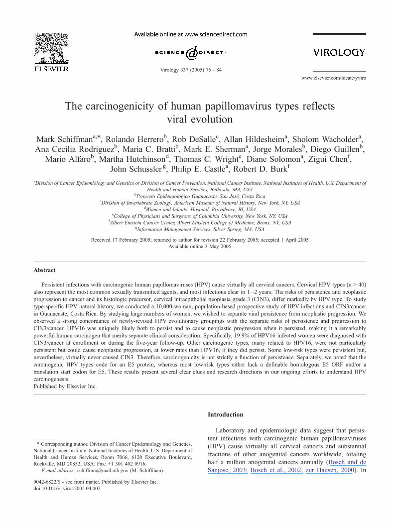
Fig. 1. Phylogenetic analysis of anogenital HPV types. Numbers on or near branches (nodes) indicate support indices from 100 bootstrap estimations using each of
themethods in the following order: Bayesian credibility value, parsimony bootstrap percentage based on nucleotide alignment, and parsimony bootstrap percentage
based on amino acid alignment. An asterisk indicates 100% agreement (all 100 estimations concordant for eachmethod for that branching). N reflects disagreement
between amethod and the reference Bayesian tree at a given node. ‘‘Risk’’ refers to the cancer risk categorization assigned by the IARC case-control data (Munoz, et
al., 2003); ‘‘?High’’ or ‘‘?Low’’ were assigned when the data were ambiguous. A homologous E5 gene is depicted as present (+) or absent (�).We indicated a value
of (+/�) when an E5 ORF was identified but did not have a translation start codon present. The number in brackets indicate the a species of the types.
M. Schiffman et al. / Virology 337 (2005) 76–8478
missed during the baseline screening), three showed
persistent HPV18 (one demonstrated by repeated testing
of the previously-negative enrollment specimen), one
persistent HPV45, one HPV18 only at diagnosis, and three
persistent HPV16. One tested HPV negative at both times.
In an ancillary analysis we divided the population into two
age groups, <30 and 30+, in order to see whether age
modified the trends we observed. HPV persistence rates
increased generally with age, as detailed in a more extensive
analysis published elsewhere (Castle et al., in press).
However, the ancillary analysis showed that relative, inter-
typic and inter-species differences in persistence and risk of
CIN3were not appreciably altered by broad age stratification.
Discussion
The combination of phylogenetic analysis and popula-
new insights. Phylogenetic grouping predicted the natural
history and carcinogenicity of individual HPV types, and
corroborated much of the recent cross-sectional data from
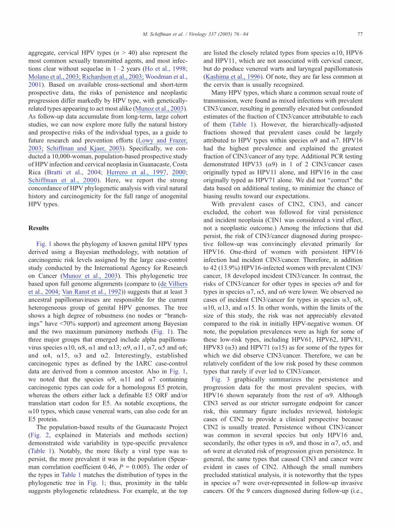
Fig. 2. Guanacaste study population. Of the 10,049 women enrolled in the Guanacaste cohort, we excluded 583 virgins, 300 women without enrollment HPV
results, and 630 women with hysterectomies. Among 8536 women screened at enrollment, there were 55 CIN2, 73 CIN3 and 12 invasive cancer cases. In the
prospective cohort, we excluded women without any follow-up PCR results (803 no follow-up, 165 inadequate or no PCR results for collected specimens). We
also excluded 290 women with possible CIN 2 or worse at enrollment (including the 140 with confirmed, prevalent �CIN2). There were 60 cases of incident
histologic CIN3 and 9 cases of cancer that occurred despite surveillance. For those censored during follow-up but prior to 5 years due to possible (even if not
confirmed) incident CIN2, CIN3, or cancer (n = 165) or for any other reason (e.g., death from other causes, relocation, n = 179), we analyzed the last specimen
prior to censoring and/or appropriate treatment.
M. Schiffman et al. / Virology 337 (2005) 76–84 79
case-control studies and case series of cervical cancer
(Munoz et al., 2003). As the most important novel
epidemiologic contributions, 1) we showed that population
prevalence of individual types is correlated with viral
persistence and 2) we distinguished viral persistence from
neoplastic progression given viral persistence. Persistence
and progression are highly associated (Ho et al., 1995;
Nobbenhuis et al., 1999; Schlecht et al., 2001) and large
numbers of subjects must be followed for several years to
disentangle the two. We observed that the types and species
with presumed low cancer risk defined a priori by their
absence (i.e., they are not found alone) in cancer case series
showed varying degrees of persistence, but did not progress
to CIN3/cancer. In contrast, the most carcinogenic HPV
types concentrated in species a9 and a7 were distinguished
by elevated risk of progression given persistence, rather than
persistence alone. We also showed clearly that the role of
E5, which is a transforming protein in some papillomavi-
ruses (DiMaio and Mattoon, 2001), deserves further study
regarding its activities in infected cervical cells.
HPV16 was uniquely carcinogenic by all important
standards of risk, i.e., attributable fraction of prevalent
CIN3/cancer, probability of persistence, and probability
of incident CIN3/cancer given persistence. It has been
reported previously based on cross-sectional evidence that
approximately 50% of cervical cancers, and an even higher
percentage of non-cervical HPV-induced cancers (e.g., HPV-
positive oropharyngeal cancers), are caused by HPV16
(Bosch and de Sanjose, 2003; Herrero et al., 2003). To this,
we add that persistent HPV16 infection represents a
carcinogen with a very high positive predictive value of
serious neoplasia, deserving clinical evaluation and follow-
up. Few other exposures cause precancer/cancer in approx-
imately 20% of those exposed. HPV16 is now the primary
target of HPV vaccine trials (Koutsky et al., 2002; Lowy and
Frazer, 2003), and our data indicate it should be distinguished
from other carcinogenic types in diagnostic kits, even if the
other types are not individually characterized. In previous
work, we and others have observed that most resolving
HPV16 infections cleared by 1–2 years after enrollment (Ho
et al., 1998; Richardson et al., 2003), suggesting a practical
endpoint to surveillance of persistently infected women
without evident lesions (yet). HPV18 also deserves special
attention because of its established association with glandular
lesions that are difficult to detect by cytology (Andersson et
al., 2001), shown perhaps by the presence of HPV18 (and
possibly HPV45, which is closely related) among the few
missed follow-up cancers in our study.
Despite the large size of this cohort, the stability of
estimates for the less common types was low and individual
confidence intervals were very broad. Also, it is possible that
some of the risk estimates for probability of persistence and
probability of CIN3/cancer could have been affected by
differential sensitivity of HPV testing method for different
HPV types (Gravitt et al., 2000). Specifically, the primer set
we used does not efficiently amplify some types, including
HPV68, which showed low rates of persistence. Furthermore,
our type-specific HPV-infected subcohorts were defined at
enrollment, without distinction between new (incident) and
already persistent infections. Perhaps, on average, some of
the types had already persisted longer than others and were,
therefore, at altered risk of subsequent extended persistence
and progression to CIN3/cancer. In ongoing work, we are
following newly-infected women, to gain a more complete
Population-based HPV natural history and CIN3/cancer risk data from Guanacaste
Species (alpha) Type # of infected
women
# Prevalent CIN3/cancer # and % of persistence among
women <CIN2 at enrollmentc# and % of CIN3/ICC among women
wit persistent type-specific HPVc,d
# AFa #adjb AFadj
b
10 6 50 1 0.6% 0 0.0% 4 10.0% 0 0.0%
10 11 24 2 2.1% 2 2.3% 0 0.0% N/A
10 74v 15 0 0.0% 0 0.0% 1 7.7% 0 0.0%
10 55 20 0 0.0% 0 0.0% 4 22.2% 0 0.0%
8 40 12 0 0.0% 0 0.0% 0 0.0% N/A
1 32 29 1 0.8% 0 0.0% 3 12.0% 0 0.0%
13 54 37 3 3.1% 0 0.0% 5 17.2% 0 0.0%
9 52 135 6 5.6% 0 0.0% 13 13.1% 4 30.8%
9 67 14 0 0.0% 0 0.0% 0 0.0% N/A
9 33 59 4 4.0% 1 1.2% 13 27.7% 3 23.1%
9 58 170 12 12.4% 10 11.1% 20 16.5% 1 5.0%
9 16 302 42 47.6% 42 47.6% 56 28.9% 18 32.1%
9 31 121 10 10.5% 9 10.2% 13 14.9% 2 15.4%
9 35 36 1 0.8% 0 0.0% 2 6.9% 1 50.0%
11 73 38 0 0.0% 0 0.0% 1 3.7% 0 0.0%
7 59 31 2 2.0% 0 0.0% 2 8.3% 1 50.0%
7 18 111 10 10.6% 5 5.7% 13 15.7% 4 30.8%
7 45 66 3 2.8% 0 0.0% 5 9.4% 1 20.0%
7 70 177 3 1.5% 0 0.0% 22 16.1% 1 4.6%
7 39 69 2 1.6% 0 0.0% 8 15.1% 0 0.0%
7 68 29 1 0.8% 1 1.2% 1 4.4% 0 0.0%
7 85 59 2 1.7% 0 0.0% 5 10.9% 0 0.0%
5 26 16 1 1.0% 0 0.0% 0 0.0% N/A
5 51 166 6 5.2% 3 3.4% 6 4.9% 2 33.3%
5 82v 31 1 0.8% 0 0.0% 3 12.5% 0 0.0%
6 53 200 3 1.2% 0 0.0% 10 6.6% 2 20.0%
6 56 75 3 2.7% 3 3.4% 9 14.8% 1 11.1%
6 66 68 1 0.4% 0 0.0% 5 9.4% 0 0.0%
15 71 204 1 0.0% 1 1.1% 29 16.9% 0 0.0%
3 61 208 2 0.0% 0 0.0% 25 13.9% 0 0.0%
3 72 25 0 0.0% 0 0.0% 5 21.7% 0 0.0%
3 62 150 2 0.6% 0 0.0% 7 5.6% 0 0.0%
3 81 103 2 1.2% 0 0.0% 10 11.6% 0 0.0%
3 83 100 2 1.2% 0 0.0% 11 13.9% 0 0.0%
3 89 21 0 0.0% 0 0.0% 0 0.0% N/A
3 84 58 1 0.5% 0 0.0% 4 8.2% 0 0.0%
? type 378 6 2.8% 6 6.0% N/A 3 0.9%e
Neg 6182 2 N/A 2 N/A N/A 21 0.4%e
a The fraction of prevalent cases of CIN3/cancer attributable to an HPV type was calculated as AF = % cases positive for that type � (1 – 1/OR) where OR
represents the odds ratio associating that type and prevalent risk of CIN3/cancer. OR for a type = (number of cases positive for that type divided by number of
cases negative for that type) divided by (number of controls positive for that type divided by number of controls negative for that type). Two types (71 and 61)
had slightly negative AF, which were set to zero.b In the hierarchical analysis of attributable fraction, women with types accounting for a higher fraction of CIN3/cancer were excluded. For example, women
with the type accounting for the highest fraction of CIN3/cancer (i.e., HPV16) were excluded to find the next most important type (i.e., HPV58), and women
with either type were excluded to find the third, etc.c Women with prevalent or incident CIN2 were excluded to provide diagnostic certainty, because they represented equivocal cases. As a result, the percentages
in the table can not be exactly calculated directly from the counts of numbers of infections and outcomes.d Four cases of CIN3 involved persistence of two types: 52/59, 16/33, 16/51, 51/70.e Some cases of incident CIN3 arose without observed viral persistence of a defined type (i.e., they were HPV negative or had an undefined type at
enrollment). In some cases, we possibly missed the period of persistence by infrequent (5–7 year interval) measurement.
M. Schiffman et al. / Virology 337 (2005) 76–8480
understanding of the comparative natural histories of incident
infections with different HPV types.
Despite these acknowledged limitations, the consistency
of the genealogical inference represented by Fig. 1 with the
patterns of natural history data was remarkable. The HPV
genome is small enough (8 Kb) to permit a comprehensive
analysis of all its components and functions on a population
level. Through the study of HPV types and variants, we
wish to define the critical, specific viral sequences and
polymorphisms associated with immune evasion, persis-
tence, transformation (Fehrmann and Laimins, 2003), and
possible interactions between HPV and humans (Wang and
Hildesheim, 2003). In particular, we want to identify and
understand the genetic determinants that set HPV16 apart
from other closely-related types in species a9 and from
long-persisting but benign types in other species groups
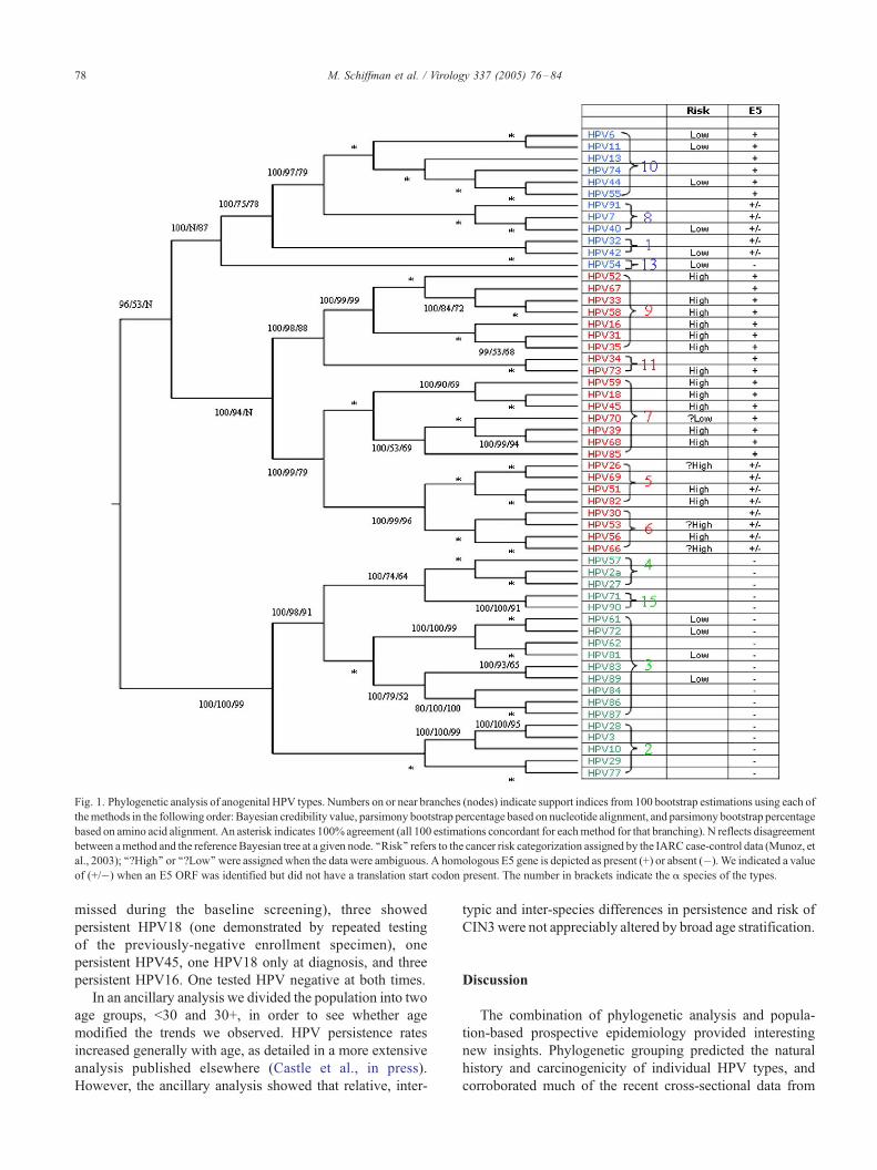
Fig. 3. Outcomes of infections in the 5–7 year study, by species. For the most prevalent species, we graphed the additive percentages of prevalent CIN3/cancer
(black), prevalent CIN2 (gray), incident CIN3/cancer given viral persistence (blue), incident CIN2 given viral persistence (red) and viral persistence without
progression to CIN2/CIN3/cancer (green). To highlight its uniqueness, we showed HPV16 separately from the rest of species a9. The addition of prevalent and
incident CIN3/cancer yields a rough estimate of the absolute risk of CIN3/cancer for each species. Prevalent cases were assigned hierarchically (see Materials
and methods and Table 1). The percentage of incident CIN2/CIN3/cancer was calculated among women with follow-up. We noted above each bar the number
of infections with types in that species.
M. Schiffman et al. / Virology 337 (2005) 76–84 81
(e.g., a3). HPV molecular epidemiology now presents an
unparalleled opportunity to study carcinogenesis from the
molecular to the population level.
Materials and methods
Phylogenetic analysis
Papillomavirus evolution is inferred from phylogenetic
analysis, in which the relatedness of viral DNA and
translated proteins from individual types is analyzed. Types
with smaller differences between them are presumed to be
more closely related, as displayed on the branchings of
evolutionary trees. We derived a phylogenetic tree using
standard approaches. Specifically, we used independent
Bayesian (Huelsenbeck, 2004) and maximum parsimony
methods (Swofford, 1998). The HPV genome is quite
small, permitting analysis based on most of its genes. All
component trees were based on the alignment of con-
catenated early and late open reading frames (E6, E7, E1,
E2, L2 and L1 ORF). The Bayesian tree was calculated
from a mixed data set containing alignments of both amino
acid and nucleotide sequences. Only informative sites were
kept for the two parsimony analyses, one based on
nucleotide sequences and the other based on amino acid
sequences. Bovine papillomavirus 1 was used as the
‘‘referent outgroup’’, meaning an independent point of
comparison to the human papillomaviruses under study. We
determined the presence of an E5 ORF since it is the only
ORF in the genome of genital HPVs showing significant
heterogeneity in its presence. (We consider the E5 ORF
identified in HPV16 as the bona fide E5, since the protein
has been identified in lesions (Kell et al., 1994)). HPV
genomes were examined for the presence of a homologous
E5 ORF based on the length and sequence similarity of
codon region between E2 and L2 ORFs by pairwise
alignment with the HPV16 E5 amino acid sequence. In
addition, we also checked the annotated file of each HPV
type in GenBank, NCBI.
Epidemiologic methods
The Guanacaste study population is outlined in Fig. 2.
The Guanacaste Project enrolled 10,049 (93.6% of eligible)
women by random sampling of the high-risk Costa Rican
province in 1993-1994, following approval by Costa Rican
and U.S. ethical boards and individual written informed
consent (Bratti et al., 2004; Herrero et al., 1997). A few,
intensively trained nurses screened adult women (age range
18–97) using cytology and cervicography (magnified
cervical images). The cytologic specimens were collected
with cervical brooms to create split-sample cytology
(conventional then liquid-based). Cells for HPV DNA
testing were collected using Dacron swabs placed into
Digene specimen transport medium, and kept at �70- for
long-term storage.
For both the cross-sectional (enrollment) and prospective
parts of this analysis, we excluded women with prior
hysterectomies (n = 630), who were virgins (n = 583),
who refused a pelvic exam (n = 291), or who were missing
enrollment HPV test results (n = 9). The remaining women
were categorized into several groups based on the results of
the screening exam; the groups were followed differently
according to perceived risk of developing CIN3/cancer. We
actively re-screened every 6–12 months those women
considered at elevated risk because of equivocal or mildly
abnormal cytology, HPV DNA positivity using the Hybrid
Capture Tube Test (Digene Corporation, Gaithersburg, MD),
a positive cervigram, or a lifetime report of >4 sexual
partners. A randomly-chosen, comparison group with
entirely normal findings at enrollment was also followed