This article appeared in a journal published by Elsevier. The attached copy is furnished to the author for internal non-commercial research and education use, including for instruction at the authors institution and sharing with colleagues. Other uses, including reproduction and distribution, or selling or licensing copies, or posting to personal, institutional or third party websites are prohibited. In most cases authors are permitted to post their version of the article (e.g. in Word or Tex form) to their personal website or institutional repository. Authors requiring further information regarding Elsevier’s archiving and manuscript policies are encouraged to visit: http://www.elsevier.com/copyright
Transcript
This article appeared in a journal published by Elsevier. The attachedcopy is furnished to the author for internal non-commercial researchand education use, including for instruction at the authors institution
and sharing with colleagues.
Other uses, including reproduction and distribution, or selling orlicensing copies, or posting to personal, institutional or third party
websites are prohibited.
In most cases authors are permitted to post their version of thearticle (e.g. in Word or Tex form) to their personal website orinstitutional repository. Authors requiring further information
regarding Elsevier’s archiving and manuscript policies areencouraged to visit:
aDipartimento di Geoscienze, Università di Padova, Via Gradenigo 6, I-35131 Padova, ItalybDepartment of Earth Sciences, University College London, Gower Street, London WC1E 6BT, UKcDipartimento di Scienze Chimiche, Università di Padova, Via Marzolo 1, I-35135 Padova, Italy
a r t i c l e i n f o
Article history:Received 13 August 2010Received in revised form23 May 2011Accepted 24 May 2011
We report here on a study of 57 fragments of wall painting excavated from the Temple of Venus(Pompeii). These samples were characterised by a wide range of analytical methods. Data showed thatthe palette is varied, although not so broad as that found in other buildings in Pompeii, and is consistentwith pigments used elsewhere in Pompeii and in the Roman Empire. It is composed of: natural earths,minerals and rare artificial pigments. Paintings are made up of thin paint layers (0.01e0.10 mm thick)strongly adhering to the underlying preparation layer. Nonetheless, in some cases thicker layers (up to0.40 mm) were recognised, often spread on other previous painting layers. Samples were also comparedwith the microstratigraphic criteria developed in Piovesan (2009) to distinguish wall painting tech-niques. This comparison demonstrated that both fresco and lime painting techniques were adopted.
� 2011 Elsevier Ltd. All rights reserved.
1. Introduction
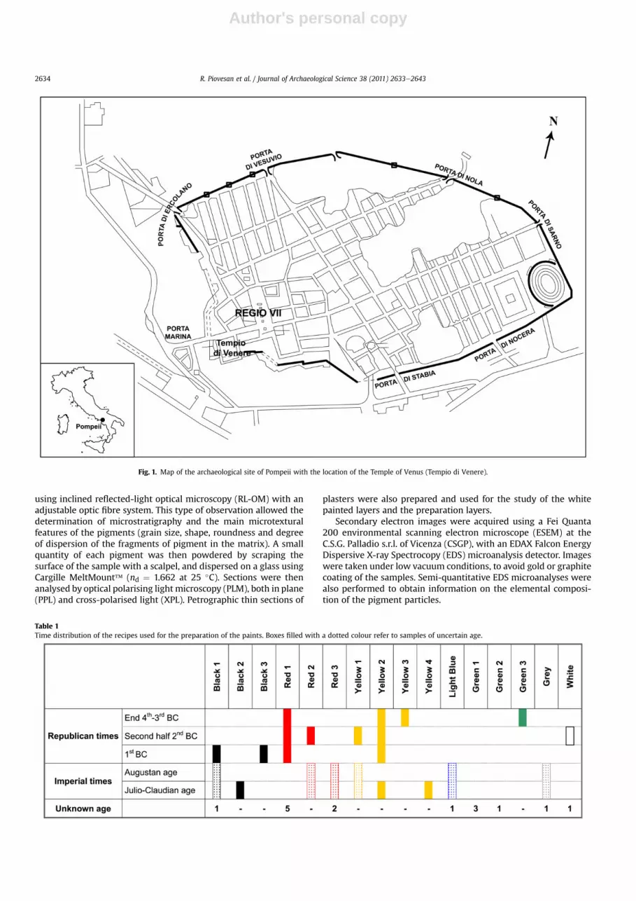
Roman pigments, particularly from Rome and Pompeii, havebeen studied using analytical techniques and a variety of pigmentsand mixtures have been recognised (Augusti, 1967; Fuchs andBéarat, 1997; Meggiolaro et al., 1997; Edwards et al., 2002; Walshet al., 2003). One of the most important questions in the study ofancient paintings is the identification of pigments and paint recipes,which give useful information about the material knowledge ofa culture and helps in identifying lines of trade. The site of Pompeii isthe most studied of the Roman Period, but many areas and buildingsremain unexplored; the Temple of Venus is one of those. Venus is thepatron divinity of Pompeii and her temple is one of the mostimportant buildings in the town, located in the south-west of the site(Fig. 1). The present building itself underwent numerous phases ofreconstruction and renovation. The area was probably a place ofworship since Archaic time and was occupied by the Sannites fromthe late 4the3rd centuries BC. Redesigned during definitive Roma-nisation in 130 BC and renovated during the Julio-Claudian Periods,the building was destroyed during the earthquake of AD 62. At the
time of the eruption of Vesuvius in AD 79, it was still under reno-vation (Curti, 2007, 2008).
The main aim of this work is the characterisation of the paint-ings from the Temple of Venus with a multi-analytical approachand to derive information about the artistic knowledge, technology,and probable trade routes of the Sannitic and the Roman in Pom-peii. Within the frame of this research, we also had the opportunityto test a new Mössbauer portable spectrometer.
2. Sample preparation and analytical approach
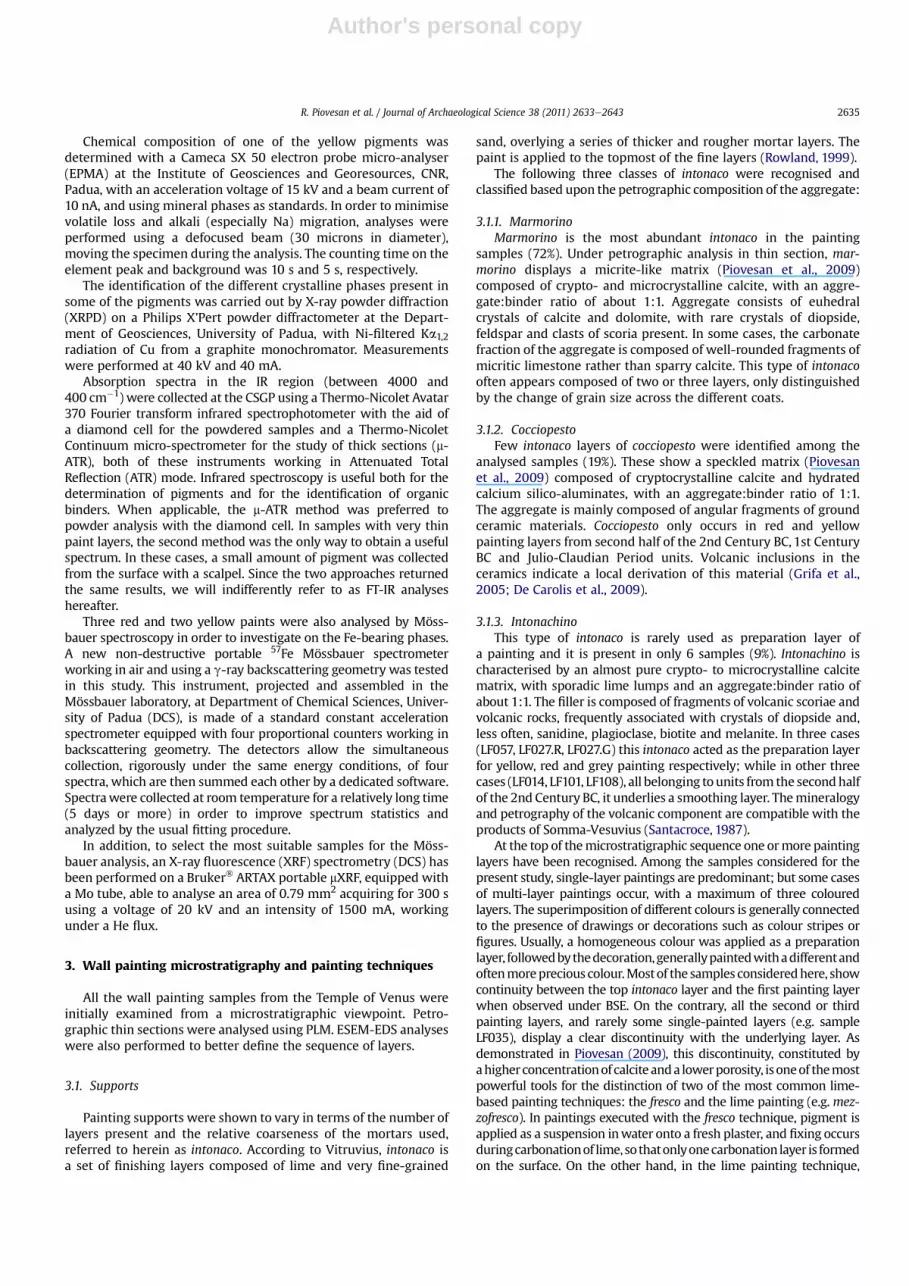
57 fragments of wall painting were collected predominantlyfrom debris and floor filling material representing five differentchronological units from Republican to Imperial times (Table 1). Intwo cases (samples LF057, LF117) samples were also collected fromtheir primary location, i.e. fromwalls of the 3rd Century BC and theend of the 2nd Century BC respectively. They represent the maincolour palette used for the wall paintings of the Temple of Venus.The dimensions of the fragments ranged from 1 to 10 cm2. Due tothe fragmentary nature and, in most of the cases, secondary loca-tion of the samples, it was impossible to make any technologicalcomments about the wall paintings as a whole; nevertheless theanalysis of these samples is a useful method for the identification ofpigments and recipes used in the Temple.
The samples were observed under an optical microscope inthick and thin section, and also as pigment dispersions. Thicksections were polished and studied under the optical microscope
using inclined reflected-light optical microscopy (RL-OM) with anadjustable optic fibre system. This type of observation allowed thedetermination of microstratigraphy and the main microtexturalfeatures of the pigments (grain size, shape, roundness and degreeof dispersion of the fragments of pigment in the matrix). A smallquantity of each pigment was then powdered by scraping thesurface of the sample with a scalpel, and dispersed on a glass usingCargille MeltMount� (nd ¼ 1.662 at 25 �C). Sections were thenanalysed by optical polarising light microscopy (PLM), both in plane(PPL) and cross-polarised light (XPL). Petrographic thin sections of
plasters were also prepared and used for the study of the whitepainted layers and the preparation layers.
Secondary electron images were acquired using a Fei Quanta200 environmental scanning electron microscope (ESEM) at theC.S.G. Palladio s.r.l. of Vicenza (CSGP), with an EDAX Falcon EnergyDispersive X-ray Spectrocopy (EDS) microanalysis detector. Imageswere taken under low vacuum conditions, to avoid gold or graphitecoating of the samples. Semi-quantitative EDS microanalyses werealso performed to obtain information on the elemental composi-tion of the pigment particles.
Fig. 1. Map of the archaeological site of Pompeii with the location of the Temple of Venus (Tempio di Venere).
Table 1Time distribution of the recipes used for the preparation of the paints. Boxes filled with a dotted colour refer to samples of uncertain age.
R. Piovesan et al. / Journal of Archaeological Science 38 (2011) 2633e26432634
Author's personal copy
Chemical composition of one of the yellow pigments wasdetermined with a Cameca SX 50 electron probe micro-analyser(EPMA) at the Institute of Geosciences and Georesources, CNR,Padua, with an acceleration voltage of 15 kV and a beam current of10 nA, and using mineral phases as standards. In order to minimisevolatile loss and alkali (especially Na) migration, analyses wereperformed using a defocused beam (30 microns in diameter),moving the specimen during the analysis. The counting time on theelement peak and background was 10 s and 5 s, respectively.
The identification of the different crystalline phases present insome of the pigments was carried out by X-ray powder diffraction(XRPD) on a Philips X’Pert powder diffractometer at the Depart-ment of Geosciences, University of Padua, with Ni-filtered Ka1,2radiation of Cu from a graphite monochromator. Measurementswere performed at 40 kV and 40 mA.
Absorption spectra in the IR region (between 4000 and400 cm�1) were collected at the CSGP using a Thermo-Nicolet Avatar370 Fourier transform infrared spectrophotometer with the aid ofa diamond cell for the powdered samples and a Thermo-NicoletContinuum micro-spectrometer for the study of thick sections (m-ATR), both of these instruments working in Attenuated TotalReflection (ATR) mode. Infrared spectroscopy is useful both for thedetermination of pigments and for the identification of organicbinders. When applicable, the m-ATR method was preferred topowder analysis with the diamond cell. In samples with very thinpaint layers, the second method was the only way to obtain a usefulspectrum. In these cases, a small amount of pigment was collectedfrom the surface with a scalpel. Since the two approaches returnedthe same results, we will indifferently refer to as FT-IR analyseshereafter.
Three red and two yellow paints were also analysed by Möss-bauer spectroscopy in order to investigate on the Fe-bearing phases.A new non-destructive portable 57Fe Mössbauer spectrometerworking in air and using a g-ray backscattering geometry was testedin this study. This instrument, projected and assembled in theMössbauer laboratory, at Department of Chemical Sciences, Univer-sity of Padua (DCS), is made of a standard constant accelerationspectrometer equipped with four proportional counters working inbackscattering geometry. The detectors allow the simultaneouscollection, rigorously under the same energy conditions, of fourspectra, which are then summed each other by a dedicated software.Spectrawere collected at room temperature for a relatively long time(5 days or more) in order to improve spectrum statistics andanalyzed by the usual fitting procedure.
In addition, to select the most suitable samples for the Möss-bauer analysis, an X-ray fluorescence (XRF) spectrometry (DCS) hasbeen performed on a Bruker� ARTAX portable mXRF, equipped witha Mo tube, able to analyse an area of 0.79 mm2 acquiring for 300 susing a voltage of 20 kV and an intensity of 1500 mA, workingunder a He flux.
3. Wall painting microstratigraphy and painting techniques
All the wall painting samples from the Temple of Venus wereinitially examined from a microstratigraphic viewpoint. Petro-graphic thin sections were analysed using PLM. ESEM-EDS analyseswere also performed to better define the sequence of layers.
3.1. Supports
Painting supports were shown to vary in terms of the number oflayers present and the relative coarseness of the mortars used,referred to herein as intonaco. According to Vitruvius, intonaco isa set of finishing layers composed of lime and very fine-grained
sand, overlying a series of thicker and rougher mortar layers. Thepaint is applied to the topmost of the fine layers (Rowland, 1999).
The following three classes of intonaco were recognised andclassified based upon the petrographic composition of the aggregate:
3.1.1. MarmorinoMarmorino is the most abundant intonaco in the painting
samples (72%). Under petrographic analysis in thin section, mar-morino displays a micrite-like matrix (Piovesan et al., 2009)composed of crypto- and microcrystalline calcite, with an aggre-gate:binder ratio of about 1:1. Aggregate consists of euhedralcrystals of calcite and dolomite, with rare crystals of diopside,feldspar and clasts of scoria present. In some cases, the carbonatefraction of the aggregate is composed of well-rounded fragments ofmicritic limestone rather than sparry calcite. This type of intonacooften appears composed of two or three layers, only distinguishedby the change of grain size across the different coats.
3.1.2. CocciopestoFew intonaco layers of cocciopesto were identified among the
analysed samples (19%). These show a speckled matrix (Piovesanet al., 2009) composed of cryptocrystalline calcite and hydratedcalcium silico-aluminates, with an aggregate:binder ratio of 1:1.The aggregate is mainly composed of angular fragments of groundceramic materials. Cocciopesto only occurs in red and yellowpainting layers from second half of the 2nd Century BC, 1st CenturyBC and Julio-Claudian Period units. Volcanic inclusions in theceramics indicate a local derivation of this material (Grifa et al.,2005; De Carolis et al., 2009).
3.1.3. IntonachinoThis type of intonaco is rarely used as preparation layer of
a painting and it is present in only 6 samples (9%). Intonachino ischaracterised by an almost pure crypto- to microcrystalline calcitematrix, with sporadic lime lumps and an aggregate:binder ratio ofabout 1:1. The filler is composed of fragments of volcanic scoriae andvolcanic rocks, frequently associated with crystals of diopside and,less often, sanidine, plagioclase, biotite and melanite. In three cases(LF057, LF027.R, LF027.G) this intonaco acted as the preparation layerfor yellow, red and grey painting respectively; while in other threecases (LF014, LF101, LF108), all belonging tounits from the secondhalfof the 2nd Century BC, it underlies a smoothing layer. Themineralogyand petrography of the volcanic component are compatible with theproducts of Somma-Vesuvius (Santacroce, 1987).
At the top of themicrostratigraphic sequence one ormore paintinglayers have been recognised. Among the samples considered for thepresent study, single-layer paintings are predominant; but some casesof multi-layer paintings occur, with a maximum of three colouredlayers. The superimposition of different colours is generally connectedto the presence of drawings or decorations such as colour stripes orfigures. Usually, a homogeneous colour was applied as a preparationlayer, followedbythedecoration,generallypaintedwithadifferentandoftenmoreprecious colour.Mostof the samples consideredhere, showcontinuity between the top intonaco layer and the first painting layerwhen observed under BSE. On the contrary, all the second or thirdpainting layers, and rarely some single-painted layers (e.g. sampleLF035), display a clear discontinuity with the underlying layer. Asdemonstrated in Piovesan (2009), this discontinuity, constituted byahigherconcentrationof calcite anda lowerporosity, isoneof themostpowerful tools for the distinction of two of the most common lime-based painting techniques: the fresco and the lime painting (e.g.mez-zofresco). In paintings executed with the fresco technique, pigment isapplied as a suspension inwater onto a fresh plaster, and fixing occursduringcarbonationof lime, sothatonlyonecarbonation layer is formedon the surface. On the other hand, in the lime painting technique,
R. Piovesan et al. / Journal of Archaeological Science 38 (2011) 2633e2643 2635
pigment is mixedwith a bindingmaterial such as limewash (lime andwater), and then applied on a dry lime plaster. In this case, a carbon-ation layer isobservedonthesurfaceof theplaster, andasecondoneonthe top of the painted layer, derived from the limewash.
3.2. Painting techniques
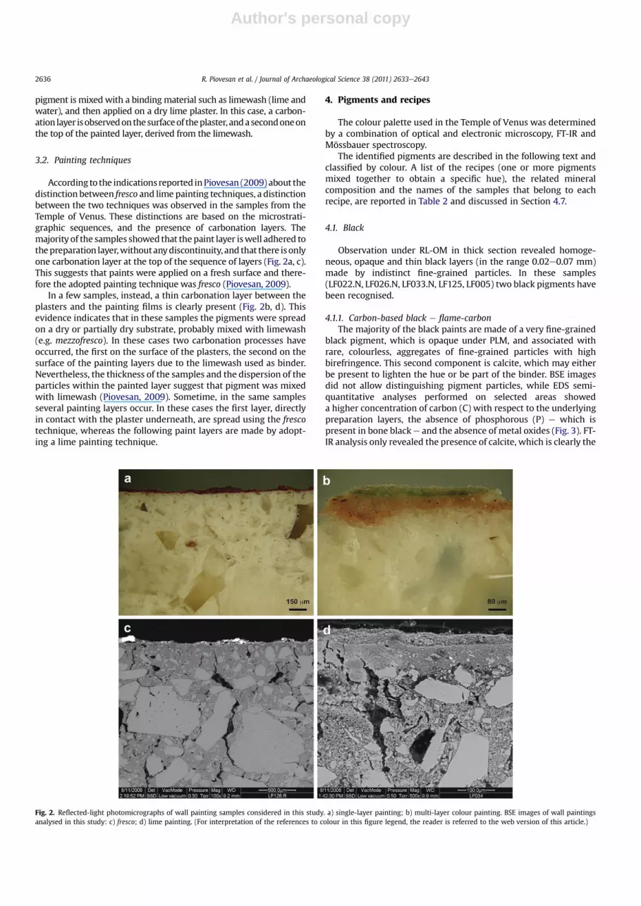
According to the indications reported inPiovesan (2009) about thedistinction between fresco and lime painting techniques, a distinctionbetween the two techniques was observed in the samples from theTemple of Venus. These distinctions are based on the microstrati-graphic sequences, and the presence of carbonation layers. Themajority of the samples showed that thepaint layer iswell adhered tothepreparation layer,withoutanydiscontinuity, and that there isonlyone carbonation layer at the top of the sequence of layers (Fig. 2a, c).This suggests that paints were applied on a fresh surface and there-fore the adopted painting technique was fresco (Piovesan, 2009).
In a few samples, instead, a thin carbonation layer between theplasters and the painting films is clearly present (Fig. 2b, d). Thisevidence indicates that in these samples the pigments were spreadon a dry or partially dry substrate, probably mixed with limewash(e.g. mezzofresco). In these cases two carbonation processes haveoccurred, the first on the surface of the plasters, the second on thesurface of the painting layers due to the limewash used as binder.Nevertheless, the thickness of the samples and the dispersion of theparticles within the painted layer suggest that pigment was mixedwith limewash (Piovesan, 2009). Sometime, in the same samplesseveral painting layers occur. In these cases the first layer, directlyin contact with the plaster underneath, are spread using the frescotechnique, whereas the following paint layers are made by adopt-ing a lime painting technique.
4. Pigments and recipes
The colour palette used in the Temple of Venus was determinedby a combination of optical and electronic microscopy, FT-IR andMössbauer spectroscopy.
The identified pigments are described in the following text andclassified by colour. A list of the recipes (one or more pigmentsmixed together to obtain a specific hue), the related mineralcomposition and the names of the samples that belong to eachrecipe, are reported in Table 2 and discussed in Section 4.7.
4.1. Black
Observation under RL-OM in thick section revealed homoge-neous, opaque and thin black layers (in the range 0.02e0.07 mm)made by indistinct fine-grained particles. In these samples(LF022.N, LF026.N, LF033.N, LF125, LF005) two black pigments havebeen recognised.
4.1.1. Carbon-based black e flame-carbonThe majority of the black paints are made of a very fine-grained
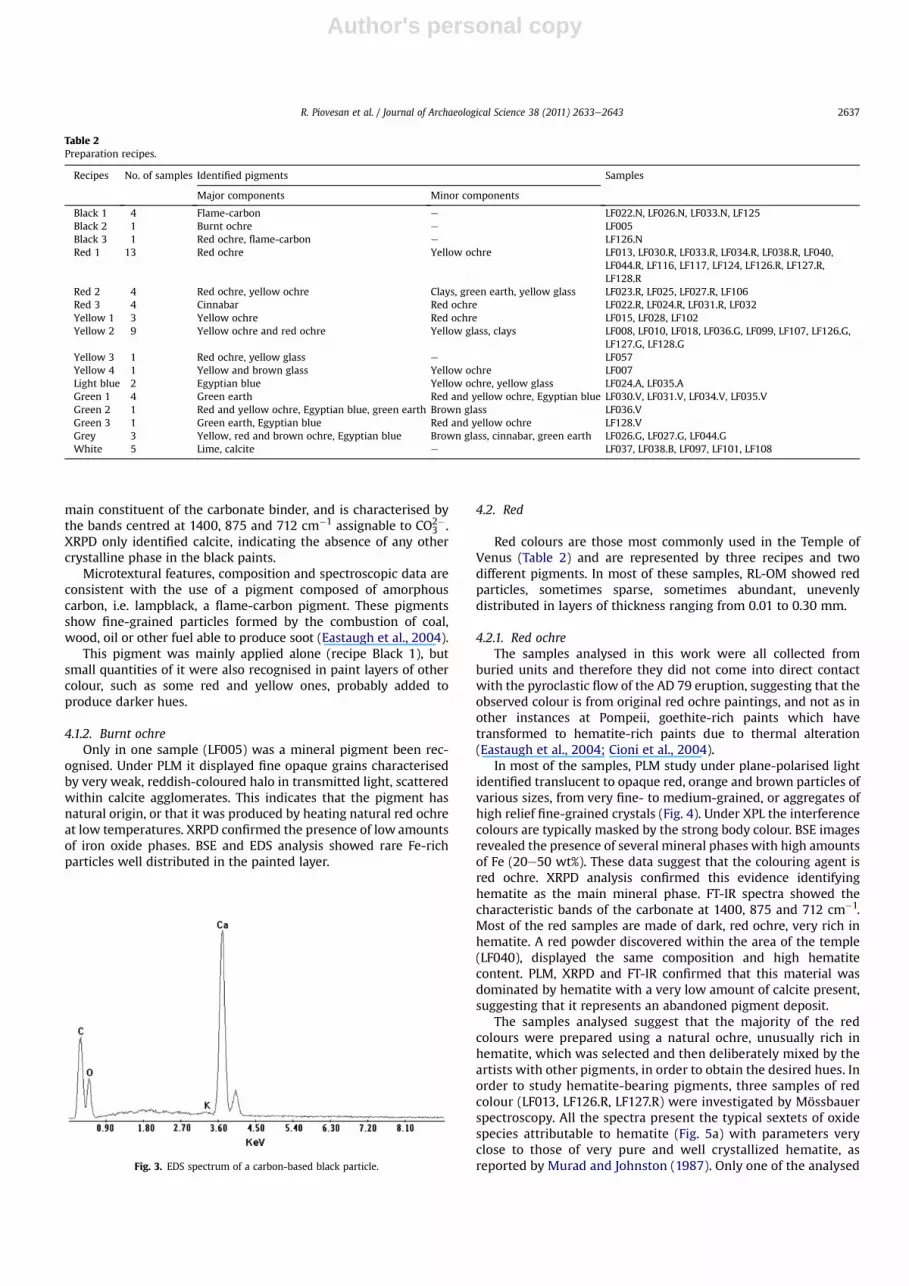
black pigment, which is opaque under PLM, and associated withrare, colourless, aggregates of fine-grained particles with highbirefringence. This second component is calcite, which may eitherbe present to lighten the hue or be part of the binder. BSE imagesdid not allow distinguishing pigment particles, while EDS semi-quantitative analyses performed on selected areas showeda higher concentration of carbon (C) with respect to the underlyingpreparation layers, the absence of phosphorous (P) e which ispresent in bone blacke and the absence of metal oxides (Fig. 3). FT-IR analysis only revealed the presence of calcite, which is clearly the
Fig. 2. Reflected-light photomicrographs of wall painting samples considered in this study. a) single-layer painting; b) multi-layer colour painting. BSE images of wall paintingsanalysed in this study: c) fresco; d) lime painting. (For interpretation of the references to colour in this figure legend, the reader is referred to the web version of this article.)
R. Piovesan et al. / Journal of Archaeological Science 38 (2011) 2633e26432636
main constituent of the carbonate binder, and is characterised bythe bands centred at 1400, 875 and 712 cm�1 assignable to CO2�
3 .XRPD only identified calcite, indicating the absence of any othercrystalline phase in the black paints.
Microtextural features, composition and spectroscopic data areconsistent with the use of a pigment composed of amorphouscarbon, i.e. lampblack, a flame-carbon pigment. These pigmentsshow fine-grained particles formed by the combustion of coal,wood, oil or other fuel able to produce soot (Eastaugh et al., 2004).
This pigment was mainly applied alone (recipe Black 1), butsmall quantities of it were also recognised in paint layers of othercolour, such as some red and yellow ones, probably added toproduce darker hues.
4.1.2. Burnt ochreOnly in one sample (LF005) was a mineral pigment been rec-
ognised. Under PLM it displayed fine opaque grains characterisedby very weak, reddish-coloured halo in transmitted light, scatteredwithin calcite agglomerates. This indicates that the pigment hasnatural origin, or that it was produced by heating natural red ochreat low temperatures. XRPD confirmed the presence of low amountsof iron oxide phases. BSE and EDS analysis showed rare Fe-richparticles well distributed in the painted layer.
4.2. Red
Red colours are those most commonly used in the Temple ofVenus (Table 2) and are represented by three recipes and twodifferent pigments. In most of these samples, RL-OM showed redparticles, sometimes sparse, sometimes abundant, unevenlydistributed in layers of thickness ranging from 0.01 to 0.30 mm.
4.2.1. Red ochreThe samples analysed in this work were all collected from
buried units and therefore they did not come into direct contactwith the pyroclastic flow of the AD 79 eruption, suggesting that theobserved colour is from original red ochre paintings, and not as inother instances at Pompeii, goethite-rich paints which havetransformed to hematite-rich paints due to thermal alteration(Eastaugh et al., 2004; Cioni et al., 2004).
In most of the samples, PLM study under plane-polarised lightidentified translucent to opaque red, orange and brown particles ofvarious sizes, from very fine- to medium-grained, or aggregates ofhigh relief fine-grained crystals (Fig. 4). Under XPL the interferencecolours are typically masked by the strong body colour. BSE imagesrevealed the presence of several mineral phases with high amountsof Fe (20e50 wt%). These data suggest that the colouring agent isred ochre. XRPD analysis confirmed this evidence identifyinghematite as the main mineral phase. FT-IR spectra showed thecharacteristic bands of the carbonate at 1400, 875 and 712 cm�1.Most of the red samples are made of dark, red ochre, very rich inhematite. A red powder discovered within the area of the temple(LF040), displayed the same composition and high hematitecontent. PLM, XRPD and FT-IR confirmed that this material wasdominated by hematite with a very low amount of calcite present,suggesting that it represents an abandoned pigment deposit.
The samples analysed suggest that the majority of the redcolours were prepared using a natural ochre, unusually rich inhematite, which was selected and then deliberately mixed by theartists with other pigments, in order to obtain the desired hues. Inorder to study hematite-bearing pigments, three samples of redcolour (LF013, LF126.R, LF127.R) were investigated by Mössbauerspectroscopy. All the spectra present the typical sextets of oxidespecies attributable to hematite (Fig. 5a) with parameters veryclose to those of very pure and well crystallized hematite, asreported by Murad and Johnston (1987). Only one of the analysed
Table 2Preparation recipes.
Recipes No. of samples Identified pigments Samples
Major components Minor components
Black 1 4 Flame-carbon e LF022.N, LF026.N, LF033.N, LF125Black 2 1 Burnt ochre e LF005Black 3 1 Red ochre, flame-carbon e LF126.NRed 1 13 Red ochre Yellow ochre LF013, LF030.R, LF033.R, LF034.R, LF038.R, LF040,
Red 2 4 Red ochre, yellow ochre Clays, green earth, yellow glass LF023.R, LF025, LF027.R, LF106Red 3 4 Cinnabar Red ochre LF022.R, LF024.R, LF031.R, LF032Yellow 1 3 Yellow ochre Red ochre LF015, LF028, LF102Yellow 2 9 Yellow ochre and red ochre Yellow glass, clays LF008, LF010, LF018, LF036.G, LF099, LF107, LF126.G,
LF127.G, LF128.GYellow 3 1 Red ochre, yellow glass e LF057Yellow 4 1 Yellow and brown glass Yellow ochre LF007Light blue 2 Egyptian blue Yellow ochre, yellow glass LF024.A, LF035.AGreen 1 4 Green earth Red and yellow ochre, Egyptian blue LF030.V, LF031.V, LF034.V, LF035.VGreen 2 1 Red and yellow ochre, Egyptian blue, green earth Brown glass LF036.VGreen 3 1 Green earth, Egyptian blue Red and yellow ochre LF128.VGrey 3 Yellow, red and brown ochre, Egyptian blue Brown glass, cinnabar, green earth LF026.G, LF027.G, LF044.GWhite 5 Lime, calcite e LF037, LF038.B, LF097, LF101, LF108
Fig. 3. EDS spectrum of a carbon-based black particle.
R. Piovesan et al. / Journal of Archaeological Science 38 (2011) 2633e2643 2637
samples (LF013) showed also a paramagnetic doublet attributableto silicate and/or nanosized oxide particles. Considering that g-raybackscattering Mössbauer information derives from a layer as thinas the painting film and Si content measured on the surface bynon-destructive XRF is negligible, doublet is most probably due tonanosized oxide particles.
4.2.2. CinnabarFour of the red samples (recipe Red 3 in Table 2) are mercury
sulphide, HgS (the mineral cinnabar). Under RL-OM thesespigments appear as very thin, brilliant-red painted layers,0.02e0.04mm thick, with very fine-grained particles (Fig. 6). UnderPLM in PPL, particles exhibited strong red-orange body colour,weakly pleochroic from pale to deep red-orange and withextremely high relief. Particles sometimes display rectangular orcolumnar habits but predominantly the shape is irregular withconchoidal fractures. Under XPL particles glow a deep red due tohigh interference colours masked by the body colour and the stronginternal reflections. XRPD and FT-IR identified traces of cinnabar;only small amounts of this pigment are necessary to obtain a brightred painting. ESEM-EDS analysis showed several particles, whichwere rich in Hg and S, confirming the presence of cinnabar, and thepresence of a carbonate binder.
In two samples (LF022.R, LF031.R) cinnabar occurred alone. Inthe other two cases (samples LF024.R, LF032), cinnabar was used inconjunctionwith hematite presumably to both extend this valuablepigment and brighten hematite red.
4.3. Yellow
The majority of the yellow paintings appear under RL-OM asa thin pale yellow layer from 0.02 to 0.05 mm thick, although insome cases (samples LF107, LF0126.G) the painted layer is irregular,with thickness up to 0.3 mm. Four recipes (Table 2) and two yellowpigments constitute yellow colours.
4.3.1. Yellow ochrePLM analysis under PPL showed a pigment made from trans-
lucent yellow particles with fine to medium grain size andmoderate relief, associated with few scattered medium-grainedorange particles. Under XPL, pigment grains display high birefrin-gence, and interference colours are strongly masked by the bodycolour. XRPD analysis identified calcite, goethite and other ironhydroxides. FT-IR spectra showed the characteristic bands of
Fig. 4. Polarising light photomicrograph of red ochre agglomerate (PPL). (For inter-pretation of the references to colour in this figure legend, the reader is referred to theweb version of this article.)
Fig. 5. Backscattering Mössbauer spectra. a) sample LF127.R; dots: experimental datapoints; solid line: hematite. b) sample LF015; dots: experimental data points; solidline: microcrystalline goethite; dotted line: nanosized oxyhydroxide particles. Fig. 6. Polarising light photomicrograph of a crystal of cinnabar (PPL).
R. Piovesan et al. / Journal of Archaeological Science 38 (2011) 2633e26432638
Author's personal copy
carbonate (1400, 875, 712 cm�1). EDS revealed high concentrationof Fe (between 15 and 35 wt%), confirming that pigment is mainlyderived from iron oxide hydroxides.
Two samples (LF015, LF128.G) containing yellow ochre werealso studied by Mössbauer spectroscopy. Sample LF128.G presentsonly a low field (37.7 T) sextet typical of oxyhydroxide species. Theparameters well fit those of goethite, as described by Murad andJohnston (1987). In addition to the goethite sextet, sample LF015shows a doublet (Fig. 5b) attributed to nanosized oxyhydroxideparticles because silicates were not detected on the paintingsurface by XRPD, FT-IR, EDS and non-destructive XRF analyses.
4.3.2. Yellowebrown glassIn a few samples, glassy yellowebrown fragments, displaying
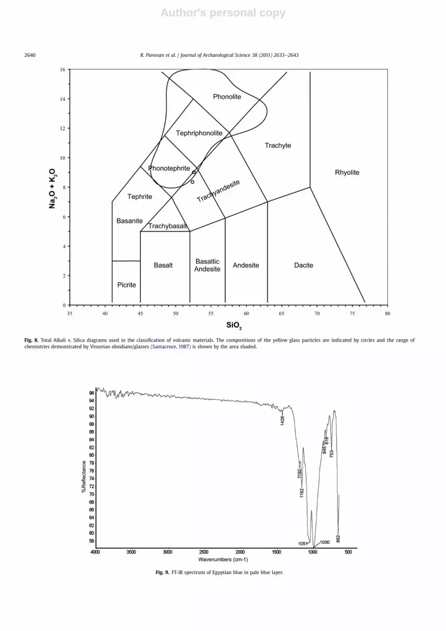
irregular shape and conchoidal fractures (Fig. 7), have been rec-ognised both under PLM and RL-OM. Under XPL, these particleswere isotropic. XRPD and FT-IR did not recognised any crystallinephase (except for the calcite of the binder), while EDSmicroanalysisshowed that pigment particles contained Si, Al, Fe, Mg, Ca, Na, Kand traces of other elements. EMPA microanalyses were performedin situ on some glassy yellow grains and their compositions werecompatible with those of trachyandesitic-phonotephritic glassesfrom Somma-Vesuvius (Fig. 8). This indicated that these glassesused as pigments probably derived from a natural volcanic glass,which was deliberately ground by the artist to produce a yellowpigment. This pigment was not been previously recorded.
4.4. Blue
The paints bearing this pigment are pale blue, green and grey.The former are characterised under RL-OM by the presence of fine-to medium-grained blue particles (up to 0.07 mm) homogeneouslydistributed in a thin layer with a thickness of 0.05e0.15 mm. Thelast two by smaller blue particles (up to 0.02 mm in diameter)mixed with medium-grained red, yellow, green, and brown parti-cles in thick layers of 0.1e0.2 mm with a bluish-greenish shade.
4.4.1. Egyptian bluePLM observations in PPL revealed the presence of blue particles,
pleochroic from blue to pale blue with high relief, occasionallydisplaying reddish patches. In the majority of the samples, theseparticles have anhedral habits and are angular; more rarely theyshowed euhedral shape and visible cleavage. In XPL, the rare well-crystallised blue fragments show high birefringence partially
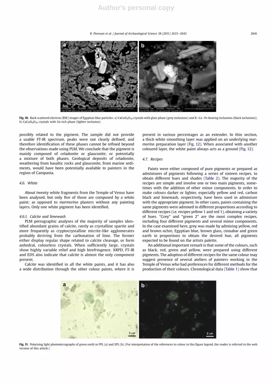
masked by the body colour. When observed through a Chelseafilter, all blue particles transmit a dull red colour. FT-IR spectrashowed, besides the typical carbonate bands, two medium-intensebands at 1000 and 1056 cm�1 and other two bands at 1162 and1192 cm�1 with low intensity, which are typical of the Egyptianblue (Fig. 9).
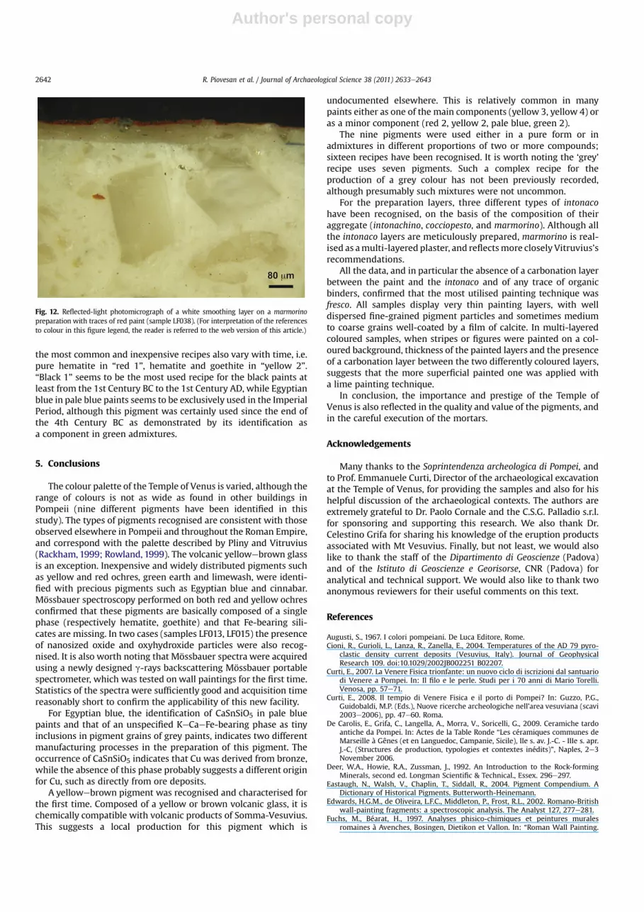
Blue particles in BSE images were also distinguishable on thebasis of their atomic number contrast. Phases with high atomicnumber, which appear bright in BSE images, were composed, inorder of decreasing abundance, of C, O, Si, Cu and Ca, which ischaracteristic of the calcium copper tetrasilicate CaCuSi4O10 (anal-ogous to mineral cuprorivaite), the main component of the Egyp-tian blue (Fig.10) mixedwith the calcium carbonate (CaCO3) binder.Slightly darker inclusions in BSE images display a similar compo-sition but with higher silica content, suggesting the presence ofa glass (Fig. 10a). More rarely, and only in the pale blue samples,a phase containing tin (Sn) has been identified in small inclusionswithin the CaCuSi4O10 crystals, which was CaSnSiO5 (analogous tomineral malayaite) (Fig. 10b). The identification of this componentin the Egyptian blue suggests that the source of the copper wasfrom bronze (Hatton et al., 2008). Egyptian blue particles in greysamples showed tiny KeCaeFe-bearing inclusions (Fig.10a) and theabsence of CaSnSiO5; these evidences suggest that the source ofCopper for the pigment preparation probably derived directly fromcopper ore or from copper metal (Hatton et al., 2008), indicatingdifferent manufacturing techniques for the production of Egyptianblue. By the 1st Century AD, manufactories of Egyptian blue weredeveloped throughout the Roman Empire, however there is insuf-ficient archaeological evidencewhichmay be reliably used to locatemanufacturing technologies and materials to industrial centres(Tite et al., 1984, 1987; Eastaugh et al., 2004). Because of theextremely fine grain size of the Egyptian blue particles in greensamples, more precise determination of their composition was notpossible.
4.5. Green
Only one green pigment was recognised, which is the maincolouring agent for all the green colour recipes (Table 2). Themajority of the green samples appear under RL-OM as a thin palegreen paint layer with few well-distinguishable particles and rareyellow and red fine particles. The thickness of the painted layer isconstant; 0.03 mm. In one sample where the green colour isapplied on a previous red painting layer, thickness reaches about0.05 mm.
4.5.1. Green earthPLM study revealed abundant rounded and sub-rounded parti-
cles with a colour varying from bright emerald green to pale green(Fig. 11a). Green particles display a low relief and are made ofa polycrystalline aggregate as also shown by mottled extinction(Fig. 11b). In XPL they show moderate birefringence and second tothird order interference colours, sometimes masked by the highbody colours. All these characteristics indicate that the greenpigment is a green earth. XRPD identified calcite, aragonite, quartz,and celadonite or glauconite the primary components of the greenearth. Celadonite and glauconite are green ferromagnesian silicatemineral belonging to the mica group. Although associated withvery different geological environments, these phases are indistin-guishable using PLM (Deer et al., 1992). The relative proportions ofthe different chemical components in the EDS spectrum confirmedthe presence of at least one of these minerals. FT-IR spectra showedthe presence of carbonates, due to the CaCO3 of the binder (bandscentred at 1400, 875 and 712 cm�1 assignable to CO3
�) and silicates(bands centred at 1000 cm�1 assignable to the SieOeSi stretching),
Fig. 7. Polarising light photomicrograph of two particles of yellow glass (PPL). Theseparticles are isotropic in XPL. (For interpretation of the references to colour in thisfigure legend, the reader is referred to the web version of this article.)
R. Piovesan et al. / Journal of Archaeological Science 38 (2011) 2633e2643 2639
Fig. 8. Total Alkali v. Silica diagrams used in the classification of volcanic materials. The compositions of the yellow glass particles are indicated by circles and the range ofchemistries demonstrated by Vesuvian obsidians/glasses (Santacroce, 1987) is shown by the area shaded.
Fig. 9. FT-IR spectrum of Egyptian blue in pale blue layer.
R. Piovesan et al. / Journal of Archaeological Science 38 (2011) 2633e26432640
Author's personal copy
possibly related to the pigment. The sample did not providea usable FT-IR spectrum, peaks were not clearly defined, andtherefore identification of these phases cannot be refined beyondthe observations made using PLM. We conclude that the pigment ismainly composed of celadonite or glauconite, or potentiallya mixture of both phases. Geological deposits of celadonite,weathering from basaltic rocks and glauconite, from marine sedi-ments, would have been potentially available to painters in theregion of Campania.
4.6. White
About twenty white fragments from the Temple of Venus havebeen analysed, but only five of those are composed by a whitepaint; as opposed to marmorino plasters without any paintinglayers. Only one white pigment has been identified.
4.6.1. Calcite and limewashPLM petrographic analyses of the majority of samples iden-
tified abundant grains of calcite, rarely as crystalline sparite andmore frequently as cryptocrystalline micrite-like agglomeratesprobably deriving from the carbonation of lime. The formereither display regular shape related to calcite cleavage, or formanhedral, colourless crystals. When sufficiently large, crystalsshow highly variable relief and high birefringence. XRPD, FT-IRand EDS also indicate that calcite is almost the only componentpresent.
Calcite was identified in all the white paints, and it has alsoa wide distribution through the other colour paints, where it is
present in various percentages as an extender. In thin section,a thick white smoothing layer was applied on an underlying mar-morino preparation layer (Fig. 12). When associated with anothercoloured layer, the white paint always acts as a ground (Fig. 12).
4.7. Recipes
Paints were either composed of pure pigments or prepared asadmixtures of pigments following a series of sixteen recipes, toobtain different hues and shades (Table 2). The majority of therecipes are simple and involve one or two main pigments, some-times with the addition of other minor components. In order tomake colours darker or lighter, especially yellow and red, carbonblack and limewash, respectively, have been used in admixturewith the appropriate pigment. In other cases, paints containing thesame pigments were admixed in different proportions according todifferent recipes (i.e. recipes yellow 1 and red 1), obtaining a varietyof hues. “Grey” and “green 2” are the most complex recipes,including four different pigments and several minor components.In the case examined here, grey was made by admixing yellow, redand brown ochre, Egyptian blue, brown glass, cinnabar and greenearth in proportions to obtain the desired hue, all pigmentsexpected to be found on the artists palette.
An additional important remark is that some of the colours, suchas black, red, green and yellow, were prepared using differentpigments. The adoption of different recipes for the same colourmaysuggest presence of several ateliers of painters working in theTemple of Venus who had preferences for different methods for theproduction of their colours. Chronological data (Table 1) show that
Fig. 10. Back-scattered electron (BSE) images of Egyptian blue particles: a) CaCuSi4O10 crystals with glass phase (grey inclusions) and KeCaeFe-bearing inclusions (black inclusions);b) CaCuSi4O10 crystals with Sn-rich phase (lighter inclusion).
Fig. 11. Polarising light photomicrographs of green earth in PPL (a) and XPL (b). (For interpretation of the references to colour in this figure legend, the reader is referred to the webversion of this article.)
R. Piovesan et al. / Journal of Archaeological Science 38 (2011) 2633e2643 2641
Author's personal copy
the most common and inexpensive recipes also vary with time, i.e.pure hematite in “red 1”, hematite and goethite in “yellow 2”.“Black 1” seems to be the most used recipe for the black paints atleast from the 1st Century BC to the 1st Century AD, while Egyptianblue in pale blue paints seems to be exclusively used in the ImperialPeriod, although this pigment was certainly used since the end ofthe 4th Century BC as demonstrated by its identification asa component in green admixtures.
5. Conclusions
The colour palette of the Temple of Venus is varied, although therange of colours is not as wide as found in other buildings inPompeii (nine different pigments have been identified in thisstudy). The types of pigments recognised are consistent with thoseobserved elsewhere in Pompeii and throughout the Roman Empire,and correspond with the palette described by Pliny and Vitruvius(Rackham, 1999; Rowland, 1999). The volcanic yellowebrown glassis an exception. Inexpensive and widely distributed pigments suchas yellow and red ochres, green earth and limewash, were identi-fied with precious pigments such as Egyptian blue and cinnabar.Mössbauer spectroscopy performed on both red and yellow ochresconfirmed that these pigments are basically composed of a singlephase (respectively hematite, goethite) and that Fe-bearing sili-cates are missing. In two cases (samples LF013, LF015) the presenceof nanosized oxide and oxyhydroxide particles were also recog-nised. It is also worth noting that Mössbauer spectra were acquiredusing a newly designed g-rays backscattering Mössbauer portablespectrometer, which was tested on wall paintings for the first time.Statistics of the spectra were sufficiently good and acquisition timereasonably short to confirm the applicability of this new facility.
For Egyptian blue, the identification of CaSnSiO5 in pale bluepaints and that of an unspecified KeCaeFe-bearing phase as tinyinclusions in pigment grains of grey paints, indicates two differentmanufacturing processes in the preparation of this pigment. Theoccurrence of CaSnSiO5 indicates that Cu was derived from bronze,while the absence of this phase probably suggests a different originfor Cu, such as directly from ore deposits.
A yellowebrown pigment was recognised and characterised forthe first time. Composed of a yellow or brown volcanic glass, it ischemically compatible with volcanic products of Somma-Vesuvius.This suggests a local production for this pigment which is
undocumented elsewhere. This is relatively common in manypaints either as one of the main components (yellow 3, yellow 4) oras a minor component (red 2, yellow 2, pale blue, green 2).
The nine pigments were used either in a pure form or inadmixtures in different proportions of two or more compounds;sixteen recipes have been recognised. It is worth noting the ‘grey’recipe uses seven pigments. Such a complex recipe for theproduction of a grey colour has not been previously recorded,although presumably such mixtures were not uncommon.
For the preparation layers, three different types of intonacohave been recognised, on the basis of the composition of theiraggregate (intonachino, cocciopesto, and marmorino). Although allthe intonaco layers are meticulously prepared, marmorino is real-ised as amulti-layered plaster, and reflectsmore closely Vitruvius’srecommendations.
All the data, and in particular the absence of a carbonation layerbetween the paint and the intonaco and of any trace of organicbinders, confirmed that the most utilised painting technique wasfresco. All samples display very thin painting layers, with welldispersed fine-grained pigment particles and sometimes mediumto coarse grains well-coated by a film of calcite. In multi-layeredcoloured samples, when stripes or figures were painted on a col-oured background, thickness of the painted layers and the presenceof a carbonation layer between the two differently coloured layers,suggests that the more superficial painted one was applied witha lime painting technique.
In conclusion, the importance and prestige of the Temple ofVenus is also reflected in the quality and value of the pigments, andin the careful execution of the mortars.
Acknowledgements
Many thanks to the Soprintendenza archeologica di Pompei, andto Prof. Emmanuele Curti, Director of the archaeological excavationat the Temple of Venus, for providing the samples and also for hishelpful discussion of the archaeological contexts. The authors areextremely grateful to Dr. Paolo Cornale and the C.S.G. Palladio s.r.l.for sponsoring and supporting this research. We also thank Dr.Celestino Grifa for sharing his knowledge of the eruption productsassociated with Mt Vesuvius. Finally, but not least, we would alsolike to thank the staff of the Dipartimento di Geoscienze (Padova)and of the Istituto di Geoscienze e Georisorse, CNR (Padova) foranalytical and technical support. We would also like to thank twoanonymous reviewers for their useful comments on this text.
References
Augusti, S., 1967. I colori pompeiani. De Luca Editore, Rome.Cioni, R., Gurioli, L., Lanza, R., Zanella, E., 2004. Temperatures of the AD 79 pyro-
clastic density current deposits (Vesuvius, Italy). Journal of GeophysicalResearch 109. doi:10.1029/2002JB002251 B02207.
Curti, E., 2007. La Venere Fisica trionfante: un nuovo ciclo di iscrizioni dal santuariodi Venere a Pompei. In: Il filo e le perle. Studi per i 70 anni di Mario Torelli.Venosa, pp. 57e71.
Curti, E., 2008. Il tempio di Venere Fisica e il porto di Pompei? In: Guzzo, P.G.,Guidobaldi, M.P. (Eds.), Nuove ricerche archeologiche nell’area vesuviana (scavi2003e2006), pp. 47e60. Roma.
De Carolis, E., Grifa, C., Langella, A., Morra, V., Soricelli, G., 2009. Ceramiche tardoantiche da Pompei. In: Actes de la Table Ronde “Les céramiques communes deMarseille à Gênes (et en Languedoc, Campanie, Sicile), IIe s. av. J.-C. - IIIe s. apr.J.-C, (Structures de production, typologies et contextes inédits)”, Naples, 2e3November 2006.
Deer, W.A., Howie, R.A., Zussman, J., 1992. An Introduction to the Rock-formingMinerals, second ed. Longman Scientific & Technical., Essex. 296e297.
Eastaugh, N., Walsh, V., Chaplin, T., Siddall, R., 2004. Pigment Compendium. ADictionary of Historical Pigments. Butterworth-Heinemann.
Edwards, H.G.M., de Oliveira, L.F.C., Middleton, P., Frost, R.L., 2002. Romano-Britishwall-painting fragments: a spectroscopic analysis. The Analyst 127, 277e281.
Fuchs, M., Béarat, H., 1997. Analyses phisico-chimiques et peintures muralesromaines à Avenches, Bosingen, Dietikon et Vallon. In: “Roman Wall Painting.
Fig. 12. Reflected-light photomicrograph of a white smoothing layer on a marmorinopreparation with traces of red paint (sample LF038). (For interpretation of the referencesto colour in this figure legend, the reader is referred to the web version of this article.)
R. Piovesan et al. / Journal of Archaeological Science 38 (2011) 2633e26432642
Materials, Techniques, Analysis and Conservation” Proceedings of the Interna-tional Workshop on Roman Wall Painting, Fribourg, 7e9 March 1996. Instituteof Mineralogy and Petrography Fribourg University, pp. 181e191.
Grifa, C., Langella, A., Morra, V., Soricelli, G., 2005. Pantellerian Ware from Miseno(Campi Flegrei, Napoli). Periodico di Mineralogia 74 (1), 69e86.
Hatton, G.D., Shortland, A.J., Tite, M.S., 2008. The production technology of Egyptianblue and green frits from second millennium BC Egypt and Mesopotamia.Journal of Archaeological Science 35, 1591e1604.
Meggiolaro, V., Molin, G., Pappalardo, U., Vergerio, P., 1997. Contribution to studieson Roman wall painting materials and technique in Greece: Corinth, thesoutheast building. In: “Roman Wall Painting. Materials, Techniques, Analysisand Conservation” Proceedings of the International Workshop on Roman WallPainting, Fribourg, 7e9 March 1996. Institute of Mineralogy and PetrographyFribourg University, pp. 105e117.
Murad, E., Johnston, J.H., 1987. Iron oxides and hydroxides. In: Long, G.J. (Ed.),Mössbauer Spectroscopy Applied to Inorganic Chemistry, Vol. 2. Plenum Press,New York.
Piovesan, R., 2009. Archaeometrical Investigations on Mortars and Paintings atPompeii and Experiments for the Determination of the Painting Technique.Ph.D. Thesis, University of Padua, Italy.
Piovesan, R., Curti, E., Grifa, C., Maritan, L., Mazzoli, C., 2009. Ancient plaster tech-nology: petrographic and microstratigraphic analysis of plaster-based building
materials from the Temple of Venus, Pompeii. In: Quinn, P.S. (Ed.), InterpretingSilent Artefacts: Petrographic Approaches to Archaeological Ceramics.Archaeopress, Oxford, pp. 65e79.
Rackham, H., 1999. Loeb Classical Library (trans.). Harvard University Press, Cam-bridge, Massachusetts. Pliny (1st cent AD/Rackham 1952). Pliny Natural History.
Rowland, I.D., 1999. Vitruvius the Ten Books on Architecture (trans.). CambridgeUniversity Press. Vitruvius (1st cent BC/Rowland 1999).
Santacroce, R. (Ed.), 1987. Somma-Vesuvius, Quaderni de “La Ricerca Scientifica”Progetto finanziato “Geodinamica”. Monografie finali, 8. CNR Roma.
Tite, M.S., Bimson, M., Cowell, M.R., 1984. Technological examination of Egyptianblue. In: Archaeological Chemistry III Advances in Chemistry Series, Vol. 205.American Chemical Society, Washington DC., pp. 215e242.
Tite, M.S., Bimson, M., Cowell, M.R., 1987. The technology of Egyptian blue. In:Bimson, M., Freestone, I.C. (Eds.), Early Vitreous Materials, British MuseumOccassional Paper, 56. British Museum, London, pp. 39e46.
Walsh, V., Siddall, R., Eastaugh, N., Chaplin, T., 2003. Pigmenti di Pompeii: verso ladefinizione di uno standard di riferimento per la ricerca sui pigmenti romani.In: “Scienze e archeologia” Proceedings of Giornate di Studio: “Le scienze el’ambiente” e “Le scienze chimico fisiche”, Pompeii, 23 October and 19November 2003. Università del Sannio-dipartimento di studi geologici eambientali and Università di Modena e Reggio Emilia-dipartimento di chimica,pp. 181e196.
R. Piovesan et al. / Journal of Archaeological Science 38 (2011) 2633e2643 2643