SAND20XX-XXXXR LDRD PROJECT NUMBER: 178849 LDRD PROJECT TITLE: A Novel Application of Synthetic Biology and Directed Evolution to Engineer Phage-based Antibiotics PROJECT TEAM MEMBERS: Meiye Wu (PI), Nancy Villa ABSTRACT: The emergence of multiple drug resistant bacteria poses threats to human health, agriculture and food safety. Annually over 100,000 deaths and up to $20 billion loss to the U.S. economy are attributed to multiple drug resistant bacteria. With only four new chemical antibiotics in the drug development pipeline, we are in dire need of new solutions to address the emerging threat of multiple drug resistance. We propose a paradigm-changing approach to address the multi-drug resistant bacteria problem by utilizing Synthetic Biology (SynBio) methodologies to create and evolve “designer” bacteriophages or phages – viruses that specifically infect bacteria – to infect and kill newly emerging pathogenic bacterial strains WITHOUT the need for chemical antibiotics. A major advantage of using phage to combat pathogenic bacteria is that phages can co-evolve with their bacterial host, and Sandia can be the first in the world to establish an industrial scale Synthetic Biology pipeline for phage directed evolution for safe, targeted, customizable solution to bacterial drug resistance. Since there is no existing phage directed evolution effort within or outside of Sandia, this proposal is suitable as a high-risk LDRD effort to create the first pipeline for such an endeavor. The high potential reward nature of this proposal will be the immediate impact in decontamination and restoration of surfaces and infrastructure, with longer term impact in human or animal therapeutics. The synthetic biology and screening approaches will lead to fundamental knowledge of phage/bacteria co-evolution, making Sandia a world leader in directed evolution of bacteriophages. INTRODUCTION: Synthetic Biology is an emerging engineering discipline at the intersection of Biology, Chemical Engineering, Chemistry, Electrical engineering, or Computer Science 1 . In layman’s terms, SynBio is “genetic engineering on steroids” - where traditional genetic engineering examines one mutant at a time, SynBio screens billions of mutants in an iterative fashion to select for desired traits. SynBio is made possible by recent advancements in ultra-high throughput technologies, some of which are developed and used at Sandia, including enzyme directed evolution and ultra-high throughput enzyme screening platform 2-4 . Sandia’s existing SynBio efforts have been focused on enzyme engineering and metabolic engineering for bioenergy applications. Using Sandia’s existing expertise in SynBio as a launching pad, this LDRD project will be the first in the SynBio field to establishing a phage directed evolution pipeline (figure 1). SAND2014-17407R
Transcript
SAND20XX-XXXXR LDRD PROJECT NUMBER: 178849 LDRD PROJECT TITLE: A Novel Application of Synthetic Biology and Directed Evolution to Engineer Phage-based Antibiotics PROJECT TEAM MEMBERS: Meiye Wu (PI), Nancy Villa ABSTRACT: The emergence of multiple drug resistant bacteria poses threats to human health, agriculture and
food safety. Annually over 100,000 deaths and up to $20 billion loss to the U.S. economy are
attributed to multiple drug resistant bacteria. With only four new chemical antibiotics in the drug
development pipeline, we are in dire need of new solutions to address the emerging threat of
multiple drug resistance. We propose a paradigm-changing approach to address the multi-drug
resistant bacteria problem by utilizing Synthetic Biology (SynBio) methodologies to create and
evolve “designer” bacteriophages or phages – viruses that specifically infect bacteria – to infect
and kill newly emerging pathogenic bacterial strains WITHOUT the need for chemical
antibiotics. A major advantage of using phage to combat pathogenic bacteria is that phages can
co-evolve with their bacterial host, and Sandia can be the first in the world to establish an
industrial scale Synthetic Biology pipeline for phage directed evolution for safe, targeted,
customizable solution to bacterial drug resistance. Since there is no existing phage directed
evolution effort within or outside of Sandia, this proposal is suitable as a high-risk LDRD effort
to create the first pipeline for such an endeavor. The high potential reward nature of this proposal
will be the immediate impact in decontamination and restoration of surfaces and infrastructure,
with longer term impact in human or animal therapeutics. The synthetic biology and screening
approaches will lead to fundamental knowledge of phage/bacteria co-evolution, making Sandia a
world leader in directed evolution of bacteriophages.
INTRODUCTION:
Synthetic Biology is an emerging engineering discipline at the intersection of Biology, Chemical
Engineering, Chemistry, Electrical engineering, or Computer Science1. In layman’s terms,
SynBio is “genetic engineering on steroids” - where traditional genetic engineering examines
one mutant at a time, SynBio screens billions of
mutants in an iterative fashion to select for desired traits. SynBio is made possible by recent
advancements in ultra-high throughput technologies, some of which are developed and used at
Sandia, including enzyme directed evolution and ultra-high throughput enzyme screening
platform2-4
. Sandia’s existing SynBio efforts have been focused on enzyme engineering and
metabolic engineering for bioenergy applications. Using Sandia’s existing expertise in SynBio
as a launching pad, this LDRD project will be the first in the SynBio field to establishing a phage
directed evolution pipeline (figure 1).
SAND2014-17407R
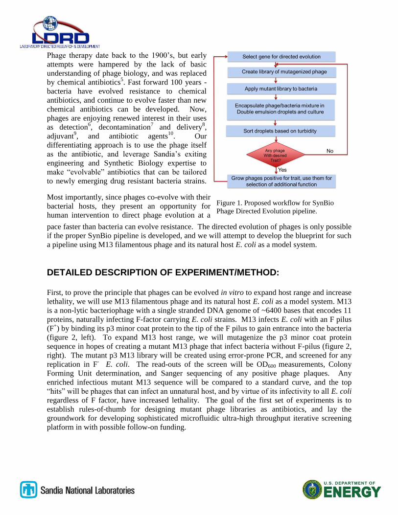
Phage therapy date back to the 1900’s, but early
attempts were hampered by the lack of basic
understanding of phage biology, and was replaced
by chemical antibiotics5. Fast forward 100 years -
bacteria have evolved resistance to chemical
antibiotics, and continue to evolve faster than new
chemical antibiotics can be developed. Now,
phages are enjoying renewed interest in their uses
as detection6, decontamination
7 and delivery
8,
adjuvant9, and antibiotic agents
10. Our
differentiating approach is to use the phage itself
as the antibiotic, and leverage Sandia’s exiting
engineering and Synthetic Biology expertise to
make “evolvable” antibiotics that can be tailored
to newly emerging drug resistant bacteria strains.
Most importantly, since phages co-evolve with their
bacterial hosts, they present an opportunity for
human intervention to direct phage evolution at a
pace faster than bacteria can evolve resistance. The directed evolution of phages is only possible
if the proper SynBio pipeline is developed, and we will attempt to develop the blueprint for such
a pipeline using M13 filamentous phage and its natural host E. coli as a model system.
DETAILED DESCRIPTION OF EXPERIMENT/METHOD: First, to prove the principle that phages can be evolved in vitro to expand host range and increase
lethality, we will use M13 filamentous phage and its natural host E. coli as a model system. M13
is a non-lytic bacteriophage with a single stranded DNA genome of ~6400 bases that encodes 11
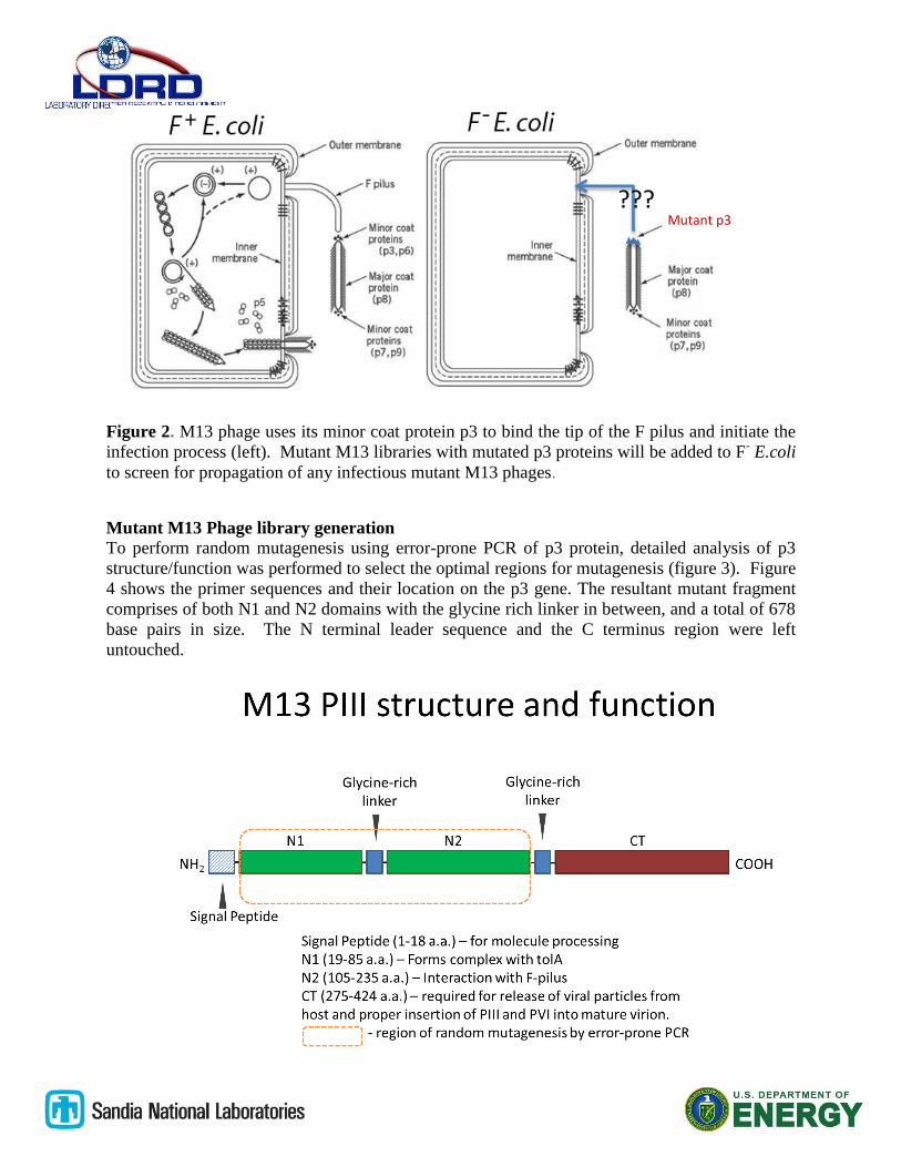
proteins, naturally infecting F-factor carrying E. coli strains. M13 infects E. coli with an F pilus
(F+) by binding its p3 minor coat protein to the tip of the F pilus to gain entrance into the bacteria
(figure 2, left). To expand M13 host range, we will mutagenize the p3 minor coat protein
sequence in hopes of creating a mutant M13 phage that infect bacteria without F-pilus (figure 2,
right). The mutant p3 M13 library will be created using error-prone PCR, and screened for any
replication in F- E. coli. The read-outs of the screen will be OD600 measurements, Colony
Forming Unit determination, and Sanger sequencing of any positive phage plaques. Any
enriched infectious mutant M13 sequence will be compared to a standard curve, and the top
“hits” will be phages that can infect an unnatural host, and by virtue of its infectivity to all E. coli
regardless of F factor, have increased lethality. The goal of the first set of experiments is to
establish rules-of-thumb for designing mutant phage libraries as antibiotics, and lay the
groundwork for developing sophisticated microfluidic ultra-high throughput iterative screening
platform in with possible follow-on funding.
Figure 1. Proposed workflow for SynBio
Phage Directed Evolution pipeline.
Figure 2. M13 phage uses its minor coat protein p3 to bind the tip of the F pilus and initiate the
infection process (left). Mutant M13 libraries with mutated p3 proteins will be added to F- E.coli
to screen for propagation of any infectious mutant M13 phages.
Mutant M13 Phage library generation
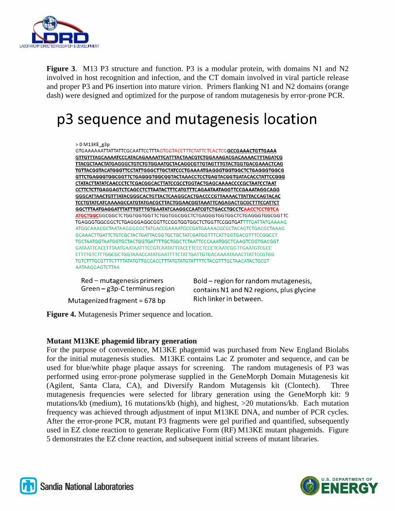
To perform random mutagenesis using error-prone PCR of p3 protein, detailed analysis of p3
structure/function was performed to select the optimal regions for mutagenesis (figure 3). Figure
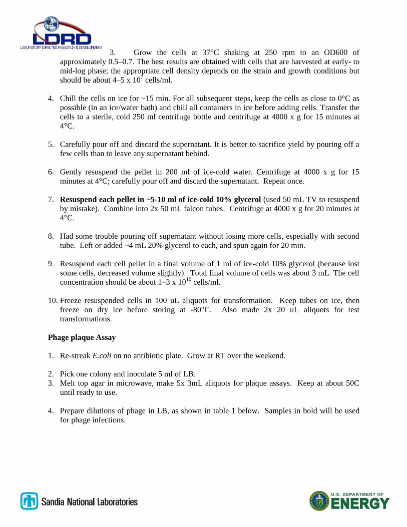
4 shows the primer sequences and their location on the p3 gene. The resultant mutant fragment
comprises of both N1 and N2 domains with the glycine rich linker in between, and a total of 678
base pairs in size. The N terminal leader sequence and the C terminus region were left
untouched.
Mutant p3
???
Figure 3. M13 P3 structure and function. P3 is a modular protein, with domains N1 and N2
involved in host recognition and infection, and the CT domain involved in viral particle release
and proper P3 and P6 insertion into mature virion. Primers flanking N1 and N2 domains (orange
dash) were designed and optimized for the purpose of random mutagenesis by error-prone PCR.
Figure 4. Mutagenesis Primer sequence and location.
Mutant M13KE phagemid library generation
For the purpose of convenience, M13KE phagemid was purchased from New England Biolabs
for the initial mutagenesis studies. M13KE contains Lac Z promoter and sequence, and can be
used for blue/white phage plaque assays for screening. The random mutagenesis of P3 was
performed using error-prone polymerase supplied in the GeneMorph Domain Mutagenesis kit
(Agilent, Santa Clara, CA), and Diversify Random Mutagensis kit (Clontech). Three
mutagenesis frequencies were selected for library generation using the GeneMorph kit: 9
mutations/kb (medium), 16 mutations/kb (high), and highest, >20 mutations/kb. Each mutation
frequency was achieved through adjustment of input M13KE DNA, and number of PCR cycles.
After the error-prone PCR, mutant P3 fragments were gel purified and quantified, subsequently
used in EZ clone reaction to generate Replicative Form (RF) M13KE mutant phagemids. Figure
5 demonstrates the EZ clone reaction, and subsequent initial screens of mutant libraries.
Figure 5. The EZ clone reaction is illustrated on the left (Adapted from Agilent product insert).
The mutant PCR products generated by error-prone PCR is used as a megaprimer and annealed
to donor M13KE phagemid, and extended in the EZClone reaction. Following amplification,
Dpn I, a restriction enzyme that digests methylated DNA is added to destroy the non-mutated
donor phagemid, leaving only synthetic double stranded M13KE phagemids containing the
mutated P3 sequences. The mutant libraries were then electroporated into F- E. coli cells and
their growth is monitored by OD600 readings and blue/white plaque assays.
Electrocompetent F- bacteria generation
Make electrocompetent E. coli TB-1 cells. These are F-, and have Lac Z promoter. Will be
used for screening of mutant libraries.
Procedure:
1. Inoculate 2 ml of a fresh overnight E. coli culture into 4x 200 ml of LB in 500 mL flask.
2. Cool rotor, centrifuge bottles, water, and 10% glycerol to 4°C or place on ice.
3. Grow the cells at 37°C shaking at 250 rpm to an OD600 of
approximately 0.5–0.7. The best results are obtained with cells that are harvested at early- to
mid-log phase; the appropriate cell density depends on the strain and growth conditions but
should be about 4–5 x 10⁷ cells/ml.
4. Chill the cells on ice for ~15 min. For all subsequent steps, keep the cells as close to 0°C as
possible (in an ice/water bath) and chill all containers in ice before adding cells. Transfer the
cells to a sterile, cold 250 ml centrifuge bottle and centrifuge at 4000 x g for 15 minutes at
4°C.
5. Carefully pour off and discard the supernatant. It is better to sacrifice yield by pouring off a
few cells than to leave any supernatant behind.
6. Gently resuspend the pellet in 200 ml of ice-cold water. Centrifuge at 4000 x g for 15
minutes at 4°C; carefully pour off and discard the supernatant. Repeat once.
7. Resuspend each pellet in ~5-10 ml of ice-cold 10% glycerol (used 50 mL TV to resuspend
by mistake). Combine into 2x 50 mL falcon tubes. Centrifuge at 4000 x g for 20 minutes at
4°C.
8. Had some trouble pouring off supernatant without losing more cells, especially with second
tube. Left or added ~4 mL 20% glycerol to each, and spun again for 20 min.
9. Resuspend each cell pellet in a final volume of 1 ml of ice-cold 10% glycerol (because lost
some cells, decreased volume slightly). Total final volume of cells was about 3 mL. The cell
concentration should be about 1–3 x 10¹⁰
cells/ml.
10. Freeze resuspended cells in 100 uL aliquots for transformation. Keep tubes on ice, then
freeze on dry ice before storing at -80°C. Also made 2x 20 uL aliquots for test
transformations.
Phage plaque Assay
1. Re-streak E.coli on no antibiotic plate. Grow at RT over the weekend.
2. Pick one colony and inoculate 5 ml of LB.
3. Melt top agar in microwave, make 5x 3mL aliquots for plaque assays. Keep at about 50C
until ready to use.
4. Prepare dilutions of phage in LB, as shown in table 1 below. Samples in bold will be used
for phage infections.
Table 1. Dilutions of phage for plaque assay.
5. When the culture in Step 1 reaches mid-log phase, dispense 200 μl into microfuge tubes,
one for each phage dilution.
6. To carry out infection, add 10 μl of each phage dilution to each tube, mix briefly, and
incubate at room temperature for 5 minutes.
7. Transfer the infected cells one infection at a time to culture tubes containing warm Top Agar.
Vortex briefly and IMMEDIATELY pour culture onto a LB/IPTG/X-gal agar plate. Gently
tilt and rotate plate to spread top agar evenly.
8. Allow the plates to cool for 30 minutes, invert, and incubate overnight at 37°C.
9. Count plaques. Calculate titer.
RESULTS:
Mutagenesis Primer Optimization
5 sets of mutagenesis primers (table 2) were designed and tested to find the optimal sequences
and annealing temperatures for error-prone PCR. The primers were purchased from Integrated
DNA Technologies (Coralville, IA).
Table 2. M13 P3 mutagenesis primer sets.
g3p_Nterm_1F TCGCAATTCCTTTAGTGGTACCTT
g3p_Nterm_1R CAAAATCACCGGAACCAGAGC
g3p_Nterm_2F GCAATTCCTTTAGTGGTACCTTTCT
g3p_Nterm_2R AATCACCGGAACCAGAGCCA
g3p_Nterm_3F TTCGCAATTCCTTTAGTGGTACCTT
g3p_Nterm_3R AAAATCACCGGAACCAGAGC
g3p_Nterm_4F GCAATTCCTTTAGTGGTACCTTTC
g3p_Nterm_4R AAAATCACCGGAACCAGAGCC
g3p_Nterm_5F GTGGTACCTTTCTATTCTCACTCG
Sample Cells DilutionSerial
DilutionLB (uL) Phage (uL)
Total
Volume (uL)
Stock - 0 0 - - -
A ER2537 (F-) 1x10² 100 49.5 0.5 50
B ER2537 (F-) 1x10⁴ 100 198 2 200
C ER2537 (F-) 1x10⁶ 100 198 2 200
D ER2537 (F-) 1x10⁸ 100 198 2 200
E ER2537 (F-) 1x10⁹ 10 90 10 100
F ER2537 (F-) 5x10¹⁰ 2 50 50 100
Negative (no phage) ER2537 (F-) 0 0 0 0 50
g3p_Nterm_5R GCCAGCATTGACAGGAGGTT
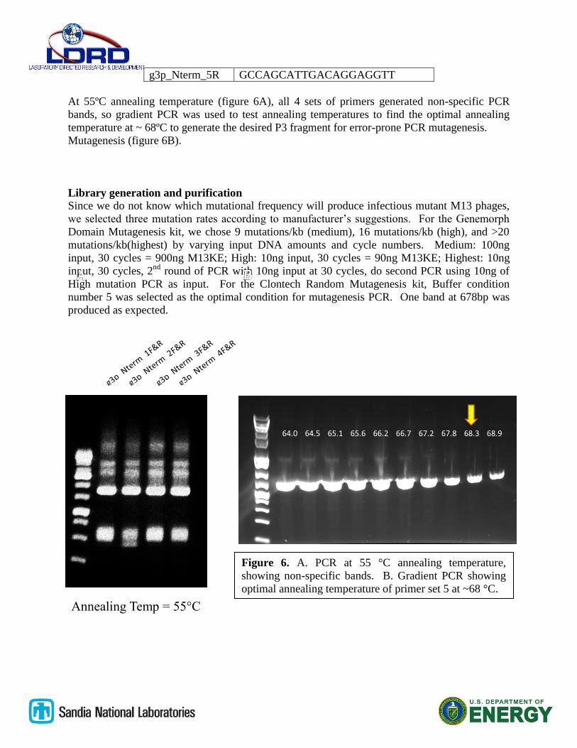
At 55ºC annealing temperature (figure 6A), all 4 sets of primers generated non-specific PCR
bands, so gradient PCR was used to test annealing temperatures to find the optimal annealing
temperature at ~ 68ºC to generate the desired P3 fragment for error-prone PCR mutagenesis.
Mutagenesis (figure 6B).
Library generation and purification
Since we do not know which mutational frequency will produce infectious mutant M13 phages,
we selected three mutation rates according to manufacturer’s suggestions. For the Genemorph
Domain Mutagenesis kit, we chose 9 mutations/kb (medium), 16 mutations/kb (high), and >20
mutations/kb(highest) by varying input DNA amounts and cycle numbers. Medium: 100ng
round of PCR with 10ng input at 30 cycles, do second PCR using 10ng of
High mutation PCR as input. For the Clontech Random Mutagenesis kit, Buffer condition
number 5 was selected as the optimal condition for mutagenesis PCR. One band at 678bp was
produced as expected.
Figure 6. A. PCR at 55 °C annealing temperature,
showing non-specific bands. B. Gradient PCR showing
optimal annealing temperature of primer set 5 at ~68 °C.
Annealing Temp = 55°C
A. B.
EZ Clone Phagemid generation
EZ Clone reaction from the Genemorph kit was used to generate double stranded replicative
form (RF) of M13KE phagemid for all libraries. The megaprimer reaction was digested with
Dpn1 to destroy all methylated double stranded M13KE phagemid, leaving only synthetic RF
M13KE with mutant megaprimer incorporated. Figure 8 below shows all 4 mutant libraries
before and after digestion with Dpn1 restriction enzyme.
Figure 7. A. lane 1. Medium mutation
rate fragment. Lane 2. High mutation
rate fragment. B. Highest mutation rate
fragment. Fragments shown in A and B
are generated using Genemorph kit
from Agilent Technologies. C.
Fragment generated using Clontech
Diversify mutagenesis kit, buffer
condition number 5.
678 bp
C.
678 bp
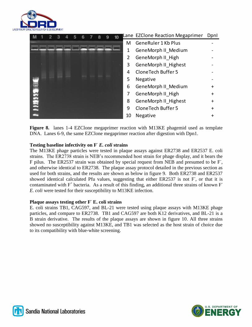
Figure 8. lanes 1-4 EZClone megaprimer reaction with M13KE phagemid used as template
DNA. Lanes 6-9, the same EZClone megaprimer reaction after digestion with Dpn1.
Testing baseline infectivity on F- E. coli strains
The M13KE phage particles were tested in plaque assays against ER2738 and ER2537 E. coli
strains. The ER2738 strain is NEB’s recommended host strain for phage display, and it bears the
F pilus. The ER2537 strain was obtained by special request from NEB and presumed to be F-,
and otherwise identical to ER2738. The plaque assay protocol detailed in the previous section as
used for both strains, and the results are shown as below in figure 9. Both ER2738 and ER2537
showed identical calculated Pfu values, suggesting that either ER2537 is not F-, or that it is
contaminated with F+ bacteria. As a result of this finding, an additional three strains of known F
-
E. coli were tested for their susceptibility to M13KE infection.
Plaque assays testing other F- E. coli strains
E. coli strains TB1, CAG597, and BL-21 were tested using plaque assays with M13KE phage
particles, and compare to ER2738. TB1 and CAG597 are both K12 derivatives, and BL-21 is a
B strain derivative. The results of the plaque assays are shown in figure 10. All three strains
showed no susceptibility against M13KE, and TB1 was selected as the host strain of choice due
to its compatibility with blue-white screening.
Lane EZClone Reaction Megaprimer DpnI
M GeneRuler 1 Kb Plus -
1 GeneMorph II_Medium -
2 GeneMorph II_High -
3 GeneMorph II_Highest -
4 CloneTech Buffer 5 -
5 Negative -
6 GeneMorph II_Medium +
7 GeneMorph II_High +
8 GeneMorph II_Highest +
9 CloneTech Buffer 5 +
10 Negative +
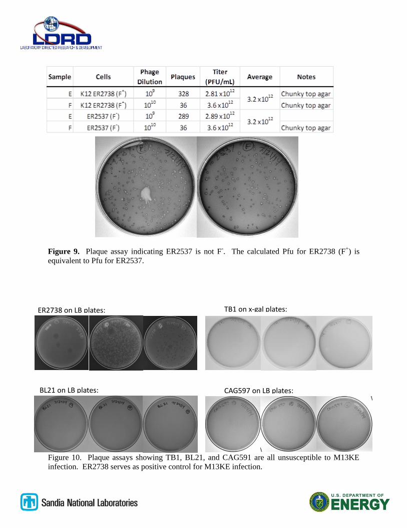
Figure 9. Plaque assay indicating ER2537 is not F-. The calculated Pfu for ER2738 (F
+) is
equivalent to Pfu for ER2537.
Figure 10. Plaque assays showing TB1, BL21, and CAG591 are all unsusceptible to M13KE
infection. ER2738 serves as positive control for M13KE infection.
ER2738 on LB plates:
BL21 on LB plates: CAG597 on LB plates:
TB1 on x-gal plates:
K12 derivative TB-1 as F- strain host of choice for screening. Electrocompetent TB-1 cells were
generated and tested for transformation efficiency (TE).
TE = (# colonies) / (ug DNA) / (fraction of total recovery vol plated = colony forming units/ ug
DNA
= 78/0.0005 ug DNA/ (1/1020) = 1.6 x 10⁹ cfu/ug
= 133/0.0005 ug DNA/ (2/1020) = 1.36 x 10⁹ cfu/ug
= 311/0.0005 ug DNA/ (5/1020) = 1.27 x 10⁹ cfu/ug
average TE ~1.41 x 10⁹ cfu/ug
Figure 11. Electrocompetent TB-1 cells were tested for their transformation efficiency by
electroporation with pET SUMO/CAT on Kanamycin resistant plates. The average TE of the
TB-1 electrocompetent cells was calculated to be ~1.41 x 10⁹ cfu/ug.
M13KE mutant library screening
A novel method was developed for screening of mutant phages. The mutant phagemid library
was transformed by electroporation into host TB1 cells and one round of phage particle
production was expected. The supernatant from the TB1 cells was recovered and tested using
Plaque assay against ER2738 bacteria to check for presence of phage particles. As indicated in
row A of figure 12, there was live, infectious phages present in the supernatant. For screening
the entire phage particle library, the supernatant was concentrated using Amicon columns and
applied to top agar containing TB1 cells to screen for presence of phage plaques formed by
mutant phage that can infect F- TB1 E. coli. So far, no positive mutant has been found .
311 colonies 133 colonies 78 colonies
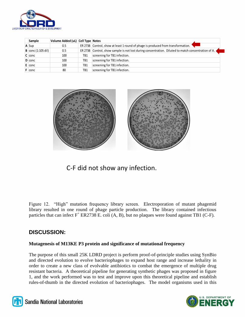
Figure 12. “High” mutation frequency library screen. Electroporation of mutant phagemid
library resulted in one round of phage particle production. The library contained infectious
particles that can infect F+ ER2738 E. coli (A, B), but no plaques were found against TB1 (C-F).
DISCUSSION:
Mutagenesis of M13KE P3 protein and significance of mutational frequency
The purpose of this small 25K LDRD project is perform proof-of-principle studies using SynBio
and directed evolution to evolve bacteriophages to expand host range and increase lethality in
order to create a new class of evolvable antibiotics to combat the emergence of multiple drug
resistant bacteria. A theoretical pipeline for generating synthetic phages was proposed in figure
1, and the work performed was to test and improve upon this theoretical pipeline and establish
rules-of-thumb in the directed evolution of bacteriophages. The model organisms used in this
Sample Volume Added (uL) Cell Type Notes
A Sup 0.5 ER 2738 Control, show at least 1 round of phage is produced from transformation.
B conc (1:105 dil) 0.5 ER 2738 Control, show sample is not lost during concentration. Diluted to match concentration of A.
C conc 100 TB1 screening for TB1 infection.
D conc 100 TB1 screening for TB1 infection.
E conc 100 TB1 screening for TB1 infection.
F conc 80 TB1 screening for TB1 infection.
C-F did not show any infection.
LDRD effort was M13KE, a modified version of M13 coliphage that
naturally infects F+ E. coli bacteria. M13 naturally infects host by binding its P3 coat protein to
the conjugative or F pilus, and as the pilus retracts into the cell body, the M13 is brought along
with it and domains of the P3 protein makes contact with TolA protein in the bacterial membrane
and initiates the translocation process by which the M13 phage enters the host and begins its
replication process. In the beginning of the project, existing structure/function information
regarding the P3 protein was used to select for the most relevant region in the P3 for randomized
mutagenesis to produce a mutant phage that can bind and infect a host without the presence of
the F pilus (Figure 2). The P3 protein itself is a modular protein as indicated in figure 3, with a
leader sequence that is required for expression, 2 N-terminal domains and a C terminal domain,
all separated by glycine-rich linkers. The two N-terminal domains are known to be involved in
host recognition and infections, whereas the C-terminal domain is known to be essential for
proper mature P3 protein insertion into progeny phages at the end of the phage reproductive
cycle. Since no previous attempts at random mutagenesis and directed evolution of M13 phages
have been attempted, the logical starting point for random mutagenesis was the two N-terminal
domains. Mutagenesis primers were designed to flank the two N-terminal domains and the
glycine-rich linker in between, with total length of 678 bp (figure 4), and mutant M13KE
phagemid libraries containing mutated P3 gene was successfully done using the EZClone
reaction supplied by the GeneMorph kit (Figure 5). The mutagenesis primers were optimized to
produce one visible product band (figure 6), and error-prone PCR reactions were used to
generate randomly mutated libraries of P3 mutant N-terminal fragments (figure 7). What was
not obvious at the beginning of the project was that multiple mutational frequencies would need
to be made in order to screen for the desired mutant. According to Agilent, selection of the
appropriate mutation frequency is very important to the success of a particular application. For
analyzing protein structure-function relationships, the desired mutation frequency is very low, at
1-2 nuleotides/gene. In directed evolution studies, 2-7 nucleotide/gene is commonly used, and
sometimes, highly mutagenized libraries with >20 mutations/gene have yielded improved protein
function. Hence, for P3 directed evolution studies in this effort, mutational frequencies of 9/kb
(medium), 16/kb(high), and >20/kb (highest) were chosen to optimize the chance of creating the
desired mutant infectious M13KE phage. Clontech’s suggested mutational frequency for
directed evolution echoed that of Agilent. Clontech mutagenesis rate for directed evolution was
suggested to be 2-6 mutations/gene, and a mutant library with 4.6 mutations/kb was generated
with the Clontech diversify random mutagenesis kit. All four mutant phagemid libraries were
incorporated into M13KE using the EZClone reaction, and digested using DpnI to eliminate any
WT M13KE in the library (figure 8) prior to electroporation into E. coli. Since four libraries
needs to be screened to ascertain the optimal mutagenesis frequency for M13 directed evolution
for future mutagenesis efforts, the screening process became much more labor-intensive.
Choice of F- E. coli host
The choice of an F- host E. coli strain that is not naturally infected by M13 was made with
consultation with New England Biolabs (NEB). The strain ER2537 is reportedly a parental F-
strain to the F+ ER2738 which NEB recommends as the host strain of choice for M13KE phage
display studies. However, upon initial experiments using phage plaque assays to establish
baseline infectivity using both ER2537 and ER2738 (Table 1, figure 9). Since ER2738 and
ER2537 showed the same Pfu values, the only conclusion is that either ER2537 is F+, or it is
contaminated with F+ bacteria, rendering it useless in this study. A significant effort was put
forth to test other F- E. coli strains to find a suitable host strain that is not
naturally infected by M13, and use it as target host for directed evolution of mutant M13KE.
This unexpected interlude cost the project valuable time and resource, and the lesson here is not
to rely on commercial vendors to provide reliable reagents when no contract or payment has
been put in place to ensure accountability on the part of the vendor. Three additional F- strains:
TB-1, BL-21, and CAT597 were tested and compared to ER2738 for their susceptibility to
M13KE infection using plaque assays (figure 10). All three strains tested showed no plaques
even at the highest phage concentration, and the K12 derivative TB-1 was selected as F- strain of
choice due to its suitability for blue/white screening. TB-1 cells were grown overnight, and
prepared as electrocompetent stocks, and their transformation efficiency was tested by
electroporation of pET SUMO/CAT (Invitrogen) plasmid bearing Kanamycin resistance (Figure
11). The average TE of plasmid was calculated to be ~1.41 x 10⁹ cfu/µg.
Screening of M13KE mutant library
When the phagemid library was electroporated into TB-1 E. coli host, the RF mutant phagemids
produced one cycle of phages to yield phage particles that can be measured using phage plaque
assay. The resultant phage particles were concentrated using Amicon spin column concentrators,
and applied to bacterial lawns for plaque assays. The mutant library contained infectious phage
plaques when applied to the F+ ER2738 strain (A, B figure 12), but no infectivity was found
against the TB-1 strain (C-F Figure 12). Since the LDRD project budget was only a modest
25K, we only had 10 weeks to perform all the work described in this report. We did not have
time to complete screening for all four libraries, and the diversity of each mutant library far
exceeds the volume of transformation we were able to manually perform. In addition, for the
sake of convenience, we chose to use M13KE phages for the initial pilot experiments, and since
M13KE is a modified phage used for phage display and sequencing applications, it is not a
wildtype M13 coliphage, and therefore has lower infectivity than the natural M13 bacteriophage.
In the future, only natural phages should be used for mutagenesis and directed evolution to
ensure that the resultant mutants have the highest chance of acquiring desired traits.
ANTICIPATED IMPACT:
Scientific lessons learned
Mutagenesis and mutant library preparation is very easily accomplished using the newest
Directed Evolution reagents.
The anticipated impact of this 25K LDRD includes technical lessons learned regarding the
suitability of bacteriophages as subjects of directed evolution. At least with the case of the DNA
bacteriophage M13, bacteriophages can be seamlessly incorporated into the newest SynBio
methodologies available from commercial sources. The existing reagents and methodologies
employed to enhance enzyme catalytic activity can be directly used, without modification, to
randomly mutagenize any selected region of phage genome without the need to isolate and purify
phage genes and proteins. This alone is a significant finding that opens the door to large scale,
industrial level SynBio efforts for phage directed evolution. Hands-on manipulation time for the
mutagenesis and library preparation can be accomplished easily within 1-2 weeks, and existing
mutazymes and cloning kits easily produces replicative phage genomes that
can be translated into functional phage particles within a day.
Ultra high-throughput technology for mutant phage
library screening
Another significant technical finding is that the sheer
volume of phage screening required for even a small
evolutionary change in phage infection phenotype is
overwhelming when conducted by traditional plaque
assay methods and manual manipulations. An ultra
high-throughput technology for the library screens will
be absolutely necessary. A candidate technology that
was proposed for a larger LDRD budget, but could not
be accommodated by the smaller approved budget of
25K was the use of microfluidic double emulsion
droplets. The key technological advance will be the
development and implementation of ultra-high
throughput double-emulsion microfluidic droplet
platform, which will generate water-in-oil-in-water
(W/O/W) droplets that can encapsulate phage and
bacteria for culture and automated screening and
sorting in a commercial flow cytometer (figure 13).
The use of double emulsion droplets as microreactors
of cell studies have been reported in the literature 11,12
,
but none have been reported for use in phage directed
evolution. The double emulsion generator will be
fabricated using fused silica, and the droplet generator
chip will be compatible with Sandia’s MICA platform13,14
, capitalizing on the precise fluidic
controls and automated valves attributed to MICA. The droplet-based microfluidic platform will
allow us to rapidly screen billions of candidate phages and isolate lytic phage, bypassing
laborious low-throughput manual plaque assays. After several rounds of automated
mutagenesis/culture/ screening, candidate phages will be sequenced, and BLASTed to identify
reoccurring genetic motifs and uncover patterns of co-evolution that will provide insights for
rationally designing phages that will specifically target a range of pathogenic bacteria strains
without causing harm to the beneficial microbiome. In addition, incremental directed evolution
of phages that can survive exposure to various body fluids can also be performed using the
microfluidic droplet platform to select for designer phages that can be safely and effectively
delivered to multiple organs.
After development of the double emulsion droplet screening platform, directed evolution of the
“designer” phages enabled by microfluidic droplet platform can be used to test “designer”
phage’s ability to infect and lyse clinical isolates of pathogenic E. coli from UC Davis’s
infectious disease clinic. Ultimately, our goal is to expand designer phage development for
combating different species of multiple drug resistant select agent bacteria.
Potential External Sponsors
Figure 13. Double emulsion droplet
generator chip. A. A glass chip with 2 T-
junctions for forming double emulsions.
B. First T-junction to form water-in-oil
droplets containig bacteria/phage
mixture. C. Second junction to form
W/O/W droplets.
As it stands, Defense Threat Reduction Agency (DTRA) is the most likely external sponsor to be
interested in funding the development of synthetic phages for biodefense applications. The
preliminary data generated with the 25K project will be combined with the design of the double-
emulsion droplet microfluidic platform to generate quad chart and white paper to be used for
solicitation of sponsorship from DTRA. I will be working closely with IAT lead Blake Simmons
and Senior Scientist Paula Imbro to craft strategies for finding external sponsors at DTRA.
CONCLUSION:
In conclusion, the key accomplishments of this LDRD are as follows:
1. The establishment of the SynBio pipeline for the directed evolution of bacteriophages
with increased host range and infectivity for applications as novel evolvable antibiotics
2. The establishment of rules-of-thumb when designing phage mutagenesis experiments for
directed evolution.
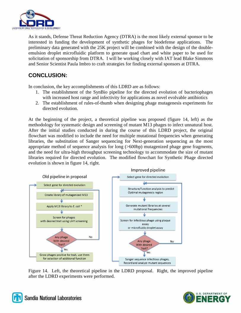
At the beginning of the project, a theoretical pipeline was proposed (figure 14, left) as the
methodology for systematic design and screening of mutant M13 phages to infect unnatural host.
After the initial studies conducted in during the course of this LDRD project, the original
flowchart was modified to include the need for multiple mutational frequencies when generating
libraries, the substitution of Sanger sequencing for Next-generation sequencing as the most
appropriate method of sequence analysis for long (>600bp) mutagenized phage gene fragments,
and the need for ultra-high throughput screening technology to accommodate the size of mutant
libraries required for directed evolution. The modified flowchart for Synthetic Phage directed
evolution is shown in figure 14, right.
Figure 14. Left, the theoretical pipeline in the LDRD proposal. Right, the improved pipeline
after the LDRD experiments were performed.
The rules-of-thumb for designing synthetic phage directed evolution studies learned from these
initial pilot experiments include:
1). Use natural phage backbone instead of modified phagemids in order to preserve maximal
infectivity of phages.
2). The selection of mutagenesis target in the phage genome requires substantial knowledge of
the structure/function of that segment.
3). Multiple mutational frequencies need to be used in order to maximize the probability of
finding the mutant with desirable characteristics.
4). The screening efforts to provide adequate coverage of the mutation library diversity is
beyond that of traditional manual plaque assay. Ultra high-throughput methodologies MUST be
developed and employed. A continuous mutagenesis and screening platform that minimizes
manual manipulations will be key to establishing a successful implementation of Synthetic Phage
genesis.
The initial feasibility studies into Synthetic Phage directed evolution produces some valuable
results that promises a true solution to the emerging threat of bacterial drug resistance. The fact
that phages genes can be easily mutated, inserted, and translated into infectious phage particles
using common commercial mutagenesis reagents is very promising in terms of establishing a
new area in SynBio to include whole organism directed evolution in a potentially rapid and
automatable fashion. The emergence of multiple drug resistance in bacteria due to the overuse
and misuse of chemical antibiotics is exacerbated by the lateral gene transfer mechanisms that
confer new antibiotic resistance across multiple species of pathogenic bacteria, and what was
once opportunistic infections in hospitals are now becoming community acquired infections in
healthy people. The underlying obstacle in developing new effective countermeasure against
bacterial pathogens is the bacteria’s ability to evolve and respond to evolutionary pressure
exerted by the chemical antibiotics we have used heavily for the past 8 decades. The speed at
which bacteria can evolve resistance far exceeds our ability to decipher the genetic and
biochemical mechanisms behind their resistance and generate new chemical antibiotics as
alternative treatments. Since evolution and the ability to adapt is key to the bacteria’s survival,
an obvious conclusion is that in order to win the war of evolution, we must employ an arsenal
capable of can co-evolve and out-evolve the bacterial enemy. Bacteriophages naturally infect
and either kill or retard bacteria growth, and specifically only infect bacteria, they make the
perfect candidates for human intervention and usage as the evolvable antibiotics we will need to
permanently address the pathogenic bacteria problem. The results from this LDRD indicate that
bacteriophages can be easily manipulated and subjected to SynBio efforts and be subject of
automated directed evolution process to become the custom tailored “live” antibiotics that are
safe the human and animal patients.
Sandia National Laboratories is a multi-program laboratory managed and operated by Sandia Corporation, a wholly owned subsidiary of Lockheed Martin Corporation, for the U.S. Department of Energy’s National Nuclear
Security Administration under Contract DE-AC04-94AL85000.
REFERENCES 1. Peccoud, J. & Isalan, M. The PLOS ONE synthetic biology collection: six years and