British Journal of Industrial Medicine 1986;43:18-28 Asbestos content of lung tissue in asbestos associated diseases: a study of 110 cases VL ROGGLI,1 PC PRATT,' AND AR BRODY2 From the Department of Pathology,' Duke University and Durham Veterans Administration Medical Centers, Durham, North Carolina 27710, and Laboratory of Pulmonary Pathobiology,2 National Institute of Environmental Health Sciences, Research Triangle Park, NC 27709, USA ABSTRACT Diseases associated with asbestos exposure include asbestosis, malignant mesothelioma, carcinoma of the lung, and parietal pleural plaques. In this study the asbestos content of lung tissue was examined in groups of cases representing each of these diseases and in several cases with non-occupational idiopathic pulmonary fibrosis. Asbestos bodies (AB), which are the hallmark of asbestos exposure, were present in the lungs of virtually everyone in the general population and present at increased levels in individuals with asbestos associated diseases. The highest numbers of AB occurred in individuals with asbestosis, all of whom had levels > 2000 ABs/g wet lung tissue. Every case with a content of 100 000 ABs/g or higher had asbestosis. Intermediate levels occurred in individuals with malignant mesothelioma and the lowest levels in patients with parietal pleural plaques. There was no overlap between the asbestos content of lung tissue from patients with asbestosis and those with idiopathic pulmonary fibrosis. Lung cancer was present in half the patients with asbestosis, and the distribution of histological patterns did not differ from that in patients with lung cancer without asbestosis. The asbestos body content in patients with lung cancer was highly variable. Control cases had values within our previously established normal range (0-20 ABs/g). There was a significant correlation (p < 0-001) between AB counted by light microscopy and AB and uncoated fibres counted by scanning electron microscopy. The previous observation that the vast majority of asbestos bodies isolated from human tissues have an amphibole core was confirmed. Asbestos exposure has been associated with several diseases, including asbestosis, mesothelioma of the pleura and peritoneum, lung carcinoma, and parietal pleural plaques.' -I Asbestos bodies, the hallmark of exposure to asbestos, are formed by the coating of partially phagocytosed asbestos fibres with an iron protein mucopolysaccharide complex.4 When sufficiently sensitive digestion techniques are used, these structures may be extracted from the lung tissue of virtually every adult in industrialised nations, indi- cating low level contamination of the environ- ment.5 - 10 Only a portion of the asbestos fibres within the lung are coated, however, so that studies of the correlation between the asbestos content of lung tissue and various asbestos associated diseases require deter- mination of both the coated and uncoated fibre con- tent of the lung using quantitative techniques. In the present study the asbestos concentration of lung tissue from 110 cases of asbestos associated dis- eases was examined to attempt to correlate lung asbes- Accepted 2 April 1985 tos burdens with specific pathological changes. Furthermore, the asbestos concentrations within the lung were compared with the occupational exposure history so that, in cases where exposure was unknown or unavailable, an assessment could be made regard- ing an approximate level of exposure-for example, environmental v low level occupational v long term occupational. In addition, the relation between the asbestos body concentration estimated by light microscopy (LM) and the type and numbers of coated and uncoated fibres observed by scanning electron microscopy (SEM) was studied. Such a comparison should provide information on the comparability of asbestos body counts using different analytical tech- niques, and the relation between asbestos bodies and total fibre or uncoated fibre counts as well as the types of fibres present. Materials and methods PATIENTS The study group included all cases of asbestosis, mesothelioma, parietal pleural plaques, and lung can- 18 group.bmj.com on June 15, 2017 - Published by http://oem.bmj.com/ Downloaded from
Transcript
British Journal of Industrial Medicine 1986;43:18-28
Asbestos content of lung tissue in asbestos associateddiseases: a study of 110 casesVL ROGGLI,1 PC PRATT,' AND AR BRODY2From the Department of Pathology,' Duke University and Durham Veterans Administration Medical Centers,Durham, North Carolina 27710, and Laboratory of Pulmonary Pathobiology,2 National Institute ofEnvironmental Health Sciences, Research Triangle Park, NC 27709, USA
ABSTRACT Diseases associated with asbestos exposure include asbestosis, malignant mesothelioma,carcinoma of the lung, and parietal pleural plaques. In this study the asbestos content of lung tissuewas examined in groups of cases representing each of these diseases and in several cases withnon-occupational idiopathic pulmonary fibrosis. Asbestos bodies (AB), which are the hallmark ofasbestos exposure, were present in the lungs of virtually everyone in the general population andpresent at increased levels in individuals with asbestos associated diseases. The highest numbers ofAB occurred in individuals with asbestosis, all of whom had levels > 2000 ABs/g wet lung tissue.Every case with a content of 100 000 ABs/g or higher had asbestosis. Intermediate levels occurredin individuals with malignant mesothelioma and the lowest levels in patients with parietal pleuralplaques. There was no overlap between the asbestos content of lung tissue from patients withasbestosis and those with idiopathic pulmonary fibrosis. Lung cancer was present in half the patientswith asbestosis, and the distribution of histological patterns did not differ from that in patients withlung cancer without asbestosis. The asbestos body content in patients with lung cancer was highlyvariable. Control cases had values within our previously established normal range (0-20 ABs/g).There was a significant correlation (p < 0-001) between AB counted by light microscopy and ABand uncoated fibres counted by scanning electron microscopy. The previous observation that thevast majority of asbestos bodies isolated from human tissues have an amphibole core was confirmed.
Asbestos exposure has been associated with severaldiseases, including asbestosis, mesothelioma of thepleura and peritoneum, lung carcinoma, and parietalpleural plaques.' -I Asbestos bodies, the hallmark ofexposure to asbestos, are formed by the coating ofpartially phagocytosed asbestos fibres with an ironprotein mucopolysaccharide complex.4 Whensufficiently sensitive digestion techniques are used,these structures may be extracted from the lung tissueof virtually every adult in industrialised nations, indi-cating low level contamination of the environ-ment.5 - 10 Only a portion of the asbestos fibres withinthe lung are coated, however, so that studies of thecorrelation between the asbestos content oflung tissueand various asbestos associated diseases require deter-mination of both the coated and uncoated fibre con-tent of the lung using quantitative techniques.
In the present study the asbestos concentration oflung tissue from 110 cases of asbestos associated dis-eases was examined to attempt to correlate lung asbes-
Accepted 2 April 1985
tos burdens with specific pathological changes.Furthermore, the asbestos concentrations within thelung were compared with the occupational exposurehistory so that, in cases where exposure was unknownor unavailable, an assessment could be made regard-ing an approximate level of exposure-for example,environmental v low level occupational v long termoccupational. In addition, the relation between theasbestos body concentration estimated by lightmicroscopy (LM) and the type and numbers of coatedand uncoated fibres observed by scanning electronmicroscopy (SEM) was studied. Such a comparisonshould provide information on the comparability ofasbestos body counts using different analytical tech-niques, and the relation between asbestos bodies andtotal fibre or uncoated fibre counts as well as the typesof fibres present.
Materials and methods
PATIENTSThe study group included all cases of asbestosis,mesothelioma, parietal pleural plaques, and lung can-
18
group.bmj.com on June 15, 2017 - Published by http://oem.bmj.com/Downloaded from
Asbestos content of lung tissue in asbestos associated diseases: a study of 110 cases
cer with a suspected asbestos aetiology seen at DukeUniversity Medical Center or Durham VeteransAdministration Medical Center (57 cases) or referredin consultation to one of the authors (VLR, 53 cases)from July 1980 to April 1984. To be included in thestudy, tissue had to be available for determination ofasbestos content. Thirty cases of asbestosis wereincluded in the study, defined histologically as thepresence in tissue sections of both asbestos bodies andperibronchiolar fibrosis, with or without fibrosis ofthe alveolar septa and with or without honey-combing.11 The severity of asbestosis was judged his-tologically using a previously reported gradingscheme 1' that takes into account both the proportionof bronchioles affected and the severity of the disease.Nineteen cases of diffuse (malignant) mesotheliomawere studied, the diagnosis being based on the grossdistribution of tumour, typical histological pattern,and the absence of any other primary site."12 Eigh-teen of these cases were confirmed at necropsy. Fortyeight cases of parietal pleural plaques without asbes-tosis were examined, plaques being defined as ivorycoloured, circumscribed foci of pleural thickening,-with or without calcification, most often affecting theposterolateral chest wall and domes of the diaphragm,and exhibiting microscopic features of layers ofalmost acellular hyalinised collagen."'314 Finally,there were 17 cases of primary lung carcinomas withneither plaques nor asbestosis. These were classifiedhistologically according to the criteria proposed bythe World Health Organisation.'5A "control" group included 10 cases with idio-
pathic pulmonary fibrosis (cryptogenic fibrosing alve-olitis) and 10 cases with normal lungs. Idiopathicpulmonary fibrosis (IPF) was defined as diffuse bilat-eral interstitial fibrosis with varying degrees ofinflammation for which there was no apparentaetiology. These cases were diagnosed by open lungbiopsy (5 cases) or necropsy (5 cases). Asbestos bodieswere not seen in tissue sections, and there was noevidence of pleural plaques. In the 10 cases with nor-mal lungs no fibrosis, emphysema, or consolidation,and minimal pigmentation, was evident on grossinspection at necropsy.
Occupational information and smoking historywere obtained by a review of the medical recordswithout prior knowledge of the asbestos content ofthe lung tissue. The age and sex of each patient werealso recorded.
rISSUE DIGESTION TECHNIQUEAsbestos was recovered from the lung by digesting thetissue in 5 25% sodium hypochlorite solution as pre-viously described.'6 A sample weighing 4 5-5 5 g wasselected (one to four samples a case, depending ontissue availability), blotted briefly on a paper towel,
and minced with a clean scalpel blade. After digestionwas complete and the contents allowed to settle for atleast 72 hours, the supernatant was carefully pipettedand the sediment suspended in 40ml of a 1:1 (v/v)mixture of chloroform and 50% ethanol. The sus-pension was centrifuged at 10 000 rpm for 30 minutes,the supernatant discarded, and the sediment sus-pended in 95% ethanol. The sediment was then col-lected on a Nuclepore filter (pore size 0 4 pm) that wasmounted on a glass slide for asbestos bodyquantification by LM.
This method works well for asbestos bodies andlarger uncoated amphibole fibres but studies in ourlaboratory, using a rat model of chrysotile inhalationexposure, indicated that a variable and sometimessubstantial proportion of small chrysotile fibres arelost during the centrifugation step at the chloroform-ethanol interface (unpublished observations). Fur-thermore, the use of large sample sizes in patients withheavy asbestos exposure results in filters that areunusable because of large accumulations of fibres.Therefore, we devised a hypochlorite digestion tech-nique (modified after Williams etal'7) that does notrequire centrifugation, permits quantitative recoveryofchrysotile asbestos fibres, and is suitable for smallersample sizes (0-1-0-4 g wet weight).'8 Organic resi-dues are minimised with this technique by successiverinsing of the filter with oxidising agents (8-0% oxalicacid, 5 25% sodium hypochlorite). In most cases,before the samples were digested, tissue sections werescreened for asbestos body content. In cases whereasbestos bodies were absent or infrequent, the tech-nique using centrifugation and a large tissue sample(4.5-5.5 g) was used to determine the asbestos bodycontent. In cases where asbestos bodies were numer-ous or the tissue sample was limited (<1 g), the tech-nique not requiring centrifugation"8 was used; it wasalways used for SEM studies. Both techniques givecomparable results for quantification of asbestos bod-ies by LM. In 10 cases for which both techniques wereused the mean ratio of asbestos body counts by thecentrifugation technique to that by the non-centrifugation technique was 1-10 (range, 0-31-3-53).
In 21 cases wet fixed tissue was not available and itwas necessary to digest tissue recovered from aparaffin block. The blocks were deparaffinised inxylene and then rehydrated to 95% ethanol, fromwhich a wet weight was obtained. Since a portion oftissue that has been dehydrated through a series oflipid solvents will weigh less than its formalin fixed wetweight, it was necessary to determine a conversionfactor so that the asbestos counts on tissue obtainedfrom paraffin blocks would be comparable to thoseobtained from wet fixed tissue. We determined that,on average, a deparaffinised lung section rehydratedto 95% ethanol weighs 70% as much as the same
19
group.bmj.com on June 15, 2017 - Published by http://oem.bmj.com/Downloaded from
formalin fixed section before paraffin embedding.Therefore, all asbestos body and fibre counts fromtissues recovered from paraffin blocks were multipliedby a factor of 0 70.
ASBESTOS QUANTIFICATIONAsbestos bodies were counted on Nuclepore filters byLM at a magnification of x 200, and the resultsexpressed as asbestos bodies per gram of wet lungtissue. Only bodies with typical dumbbell, javelin, orsegmented morphologies and thin transparent coreswere included in the counts.4 Non-asbestos fer-ruginous bodies (pseudoasbestos bodies)'9 withbroad yellow cores or dark brown to black cores werefrequently encountered but were not included in thecalculations. In most cases they were far less numer-ous than the true asbestos bodies. The analytical sen-sitivity of the technique is one asbestos body per filter,with a detection limit of 0-2 asbestos bodies per gramof wet lung tissue.
Analytical SEM with asbestos fibre identificationand enumeration was performed in 59 cases. TheNuclepore filter was mounted on a carbon disc withcolloidal graphite, sputter coated with gold, andexamined in a SEM (JEOL type JSM35) equippedwith a Kevex energy dispersive spectrometer at amagnification of x 1000. This magnification wasselected because it is low enough to detect the entirerange of asbestos body sizes, yet high enough to iden-tify the vast majority of fibres 5pm or greater inlength. Coated and uncoated fibres were counted sep-arately. All the fibres whose centres fell withinsequential fields were counted until a total of 200fibres or 100 fields (whichever came first) were encoun-tered. The total number of coated and uncoated fibreson the filter could then be calculated, and the resultsexpressed per gram of lung tissue. The analytical sen-sitivity is 125 fibres a filter, with a theoretical detectionlimit of400 fibres a gram for a 0 3 gram tissue sample.Samples were examined at 00 tilt, with a constantworking distance of 15 mm between the specimen andthe objective lens.
In each case examined by SEM 10-20 fibres wereanalysed by energy dispersive x ray analysis to deter-mine the types of fibres present. Consecutive fibresand asbestos bodies with sufficiently exposed cores topermit analysis were identified at x 1500magnification and analysed using the spot mode at20kV accelerating voltage and acquisition time of10-100 sec (average 60 sec). Chrysotile was recognisedby its often curly morphology, small diameter, andelemental content of Mg and Si only. The amphiboleswere straight fibres, sometimes with longitudinalgrooves, diameters somewhat greater than chrysotile,and distinctive chemical compositions (fig 1). Thechemical compositions of unknown fibres were com-
Roggli, Pratt, and Brody
pared with samples prepared from the UICC asbestosstandards (kindly provided by Dr V Timbrell, MRCPneumoconiosis Unit, Penarth, Cardiff, United King-dom).
STATISTICAL METHODSThe relation between histological grade of asbestosisand the asbestos concentration in lung tissue, smokinghistory, age, duration of asbestos exposure, anduncoated to coated fibre ratio was examined by linearregression analysis and determination of the cor-relation coefficient r. This method was also used toexamine the relation between dimensions of pleuralplaques and asbestos body content, asbestos bodycounts by LM as compared with SEM, and coated vuncoated fibre counts by SEM. Non-parametricanalysis (Wilcoxon signed rank test) was used to com-pare the asbestos content of the lung in patients withasbestosis with and without lung cancer. Results wereaccepted as statistically significant when p < 0 05.
Results
NORMAL LUNGSOccupational information for the 10 patients withnormal lungs at necropsy is given in table 1 and theasbestos body concentrations for these cases sum-marised in table 2. These values compare well with ourpreviously established normal range of 0-20ABs/gm.'216
ASBESTOSISAll 30 patients with asbestosis were men, with a meanage of 60-6 + 9-1 years. Occupational informationwas available for 29 (table 1) and all had workeddirectly with asbestos or asbestos containing productsfor periods ranging from five to 44 years (mean 27 5years). Smoking history was available for 26: all weresmokers or ex-smokers (one smoked cigars only).Four had malignant mesothelioma (3 pleural, 1 peri-toneal) and 15 had carcinoma of the lung (see below).
Table 2 shows the asbestos content of the lung tis-sue in these 30 cases. All patients had at least 2000asbestos bodies per gram of wet lung (ABs/g), with amedian concentration exceeding 100000 ABs/g. Inevery patient with 100 000 or more ABs/g checked byLM, asbestosis was confirmed histologically. Simi-larly, every patient with 500000 or more uncoatedfibres greater than or equal to 5 gm in length hadasbestosis. There was no overlap in the asbestos bodyor uncoated fibre concentrations between asbestosisand either idiopathic pulmonary fibrosis cases or nor-mal lungs (table 2).
The relation between the histological grade ofasbestosis and the asbestos body count (LM andSEM), uncoated fibre count (SEM), and total fibre
group.bmj.com on June 15, 2017 - Published by http://oem.bmj.com/Downloaded from
Asbestos content of lung tissue in asbestos associated diseases: a study of 110 cases
KEVEX e700mi r~~il
MI cR o -
M. R A riiveeEX7000 MI CRO - X
Fig 1 Energy dispersive x ray spectra offour different amphibole asbestos fibres. (a) Amosite has peaks for Si, Fe, Mg,and sometimes Mn. (b) Crocidolite has peaks for Si, Fe, Na, and Mg. (c) Anthophyllite has peaks for Si, Mg, and Fe. (d)Tremolite has peaks for Si, Mg, and Ca. Peak in each spectrwn inmediately to right of Si is due to Au used to coatspecimen.
count (SEM) was examined. When only cases withthree or more histological sections of lung were con-sidered, there was a significant (p < 0.05) correlationbetween the grade of asbestosis and each of the fourasbestos content parameters. The best correlationswere obtained for histological grade of asbestosis vtotal fibre count by SEM (r = 0 57) and v uncoatedfibre count by SEM (r = 0-56, fig 2). There was nosignificant correlation between histological grade ofasbestosis and uncoated to coated fibre ratio (r =0 08), age (r = 0-15), or duration ofexposure to asbes-tos (r = 0-23). Interestingly, there was a correlationbetween histological grade of asbestosis and smokinghistory by pack-years (n = 15, r = 0-53, p < 0 05).
MESOTHELIOMANineteen patients (18 men, I woman) had meso-thelioma, four of whom also had asbestosis asdescribed above. The mean age was 57-8 + 11-5 years.Occupational information was available for all 19
(table 1). Fifteen (including the four with asbestosis)had been exposed to asbestos or asbestos containingproducts for periods ranging from one to 40 years(mean 21-0 years). The remaining four were manuallabourers (maintenance, heavy machinery operator,construction) and could conceivably have beenexposed to asbestos containing materials. Smokinghistory was available for 14; 10 were smokers or ex-smokers. There were 16 pleural and three peritonealtumours. Among the 16 cases for whom histologicalsections were available for review, there were threeepithelial, six sarcomatous, and seven biphasic (mixedepithelial and sarcomatous) tumours.
Table 2 shows the asbestos content of the lung tis-sue of the 15 with mesothelioma without asbestosis.The asbestos body counts exceeded our previouslyestablished normal range of 0-20 ABs/g'216 in 10 ofthese cases, nine ofwhom had a definite occupationalexposure to asbestos. In five patients the asbestosbody count was within our normal range, although
21
group.bmj.com on June 15, 2017 - Published by http://oem.bmj.com/Downloaded from
'Patients with mesothelioma without asbestosis."Patients with pleural plaque without asbestosis or mesothelioma.'Patients with lung cancer without asbestosis or pleural plaques.dNumber of cases that are smokers/number of cases for which smoking history available.'Magnification 1000 x -includes mainly fibres >55pm in length.*Values reported as median, with range indicated in parentheses underneath.tMedian value below range of detection.ND = Data unavailable.
one of these was probably exposed to asbestos (brakerepairman, > 40 years). The highest counts were seenin the four patients who also had asbestosis (mediancount 380 000 ABs/g, range 28000-684000 ABs/g).SEM was performed in 10 of the 15 patients withoutasbestosis (table 2). These patients had on averageabout 10% as many uncoated fibres per gram as thepatients with asbestosis.
PARIETAL PLEURAL PLAQUESThe 48 patients with parietal pleural plaques had nei-ther asbestosis on histological examination nor meso-thelioma. Forty six were men with a mean age of 62-4+ 9 4 years. Occupational information was obtainedfor 44 (table 1). Eleven were exposed to asbestosoccupationally, 15 were manual labourers with possi-ble exposure, and 18 had no known exposure to asbes-tos. Smoking history was available for 38 and 32 weresmokers or ex-smokers (including one pipe smoker
and one cigar smoker). Plaques were bilateral in 33patients, unilateral in 12, and ofunknown distributionin three. Six had carcinoma of the lung (see below).Twenty five of the 48 cases of plaques included in thepresent study have been reported previously.'3The asbestos body content of the lung tissue of all
48 patients with pleural plaques is summarised in table2. The asbestos body content exceeded our normalrange of 0-20 ABs/g in a greater proportion of the 33patients with bilateral plaques (26/33, or 79%) thanunilateral plaques (6/12, or 50%), although thisdifference is not significant. The median count forpatients with bilateral plaques was 170 ABs/g (range1 2-27 500) as compared with 46 ABs/g (range06-1420) in patients with unilateral plaques. Therewas no significant correlation between the asbestosbody content of lung parenchyma and the maximumdimension (n = 19, r = 0-14) or the total area (n =14, r = 0-03) of plaque cases for whom this data was
22 Roggli, Pratt, and Brody
group.bmj.com on June 15, 2017 - Published by http://oem.bmj.com/Downloaded from
Asbestos content of lung tissue in asbestos associated diseases: a study of 110 cases
i7 k
105
* Asbestosis only (nu7)o Asbestosis luno cancer (n- 8 )tog ya 108x .5-2ArO-956 (p'c005)
0
0
0
oy o~~0
0
/:o 0
0
1 2 3 I. 5 6 7 8 9 10 11 12Gmde of asbestouis
Fig 2 Correlation between uncoated fibre count byscanning electron microscopy and histological assessment ofseverity of asbestosis using grading scheme of CAP andNIOSH11 for 15 cases with asbestosis (r = 0-56,p < 0 05).
available. SEM was performed in five instances (table2) and these patients had on average about 3% asmany uncoated fibres per gram as the patients withmesothelioma.
LUNG CANCERThere were 38 patients with carcinoma of the lung,including 15 with asbestosis, six with pleural plaques,and 17 with neither plaques nor asbestosis. Most ofthe latter cases were examined for asbestos content oflung tissue because of clinical suspicion of asbestosexposure. There were 37 men, and the mean age was60-8 + 9-6 years. All 15 patients with asbestosisworked directly with asbestos. Of the six patients withplaques (but no asbestosis) and lung cancer, one wasan asbestos insulator, two were manual labourers, andthree had no known exposure to asbestos. Among theremaining 17, nine were exposed occupationally toasbestos or asbestos containing products, three weremanual labourers, two had no history of exposure
to asbestos, and occupational information wasunavailable in the remaining three. Smoking historywas available in 34 cases; all were smokers or ex-
smokers (including one pipe smoker and one cigarsmoker).
Table 2 shows the asbestos content of the lung tis-sue for the 17 patients with neither plaques nor asbes-tosis. The LM asbestos body concentrations weresimilar for patients with lung cancer and those withparietal pleural plaques. Asbestos body counts wereincreased in 12 of the 17 (71%). Nevertheless, SEMstudies (performed in 10 cases) yielded median coatedand uncoated fibre counts about 10 times higher thanin plaque cases, although the range of values is similar(table 2). Table 3 shows the distribution of histologi-cal patterns of lung cancer of cases with asbestosis,without asbestosis (but with increased lung asbestosbody content), and with normal asbestos body con-tent. There is no apparent trend in the distribution ofhistological types among these three catagories.Among patients with asbestosis, there was nosignificant difference in the asbestos body content oflung tissue for those with lung cancer as comparedwith those without lung cancer (p = 0 74 by Wilcoxonsigned rank test, median values of 118 000 and 90 000ABs/g, respectively).
OTHER NEOPLASIASeveral tumours other than lung carcinoma wereencountered in this study. There were 15 cases ofmalignancy in this group with other neoplasia, all butone of which had parietal pleural plaques (see above).None had asbestosis histologically. There were fourcases with laryngeal carcinoma, five with gastro-intestinal carcinoma, and four with haematopoieticmalignancies. The gastrointestinal carcinomasincluded two squamous cell carcinomas of theoesophagus, two adenocarcinomas of the colon, andone rectal adenocarcinoma. One patient with colonicadenocarcinoma had neither plaques nor asbestosisand does not appear in tables I or 2. This 55 year oldman had been a shipfitter for 30 years and had 22 000ABs/g of lung tissue. The haematopoietic malig-nancies included one patient with primary pulmonarylymphoma,20 one with chronic granulocytic leu-kaemia, one with nodular poorly differentiated lym-phocytic lymphoma, and one with acutemyelomonocytic leukaemia. The remaining twopatients included one case of hepatoma and one withthree malignancies: carcinoma of the lung, prostate,and kidney. The median asbestos body concentrationfor this group was 380 ABs/g (range 10-20000ABs/g), which is greater than the median value forparietal pleural plaque cases as a group (table 2).Among the 14 cases of other neoplasia with plaques,12 were bilateral and two unilateral.
ASBESTOS BODY CONTENT OF LUNGV OCCUPATIONAL CATEGORYThe highest levels of asbestos body concentrationwere found in patients whose occupation entailed
23
group.bmj.com on June 15, 2017 - Published by http://oem.bmj.com/Downloaded from
Multiple tumours in one individual (one case from each column): adenosquamous + small cell carcinoma, squamous + adenocarcinoma, andsquamous + small cell carcinoma.ABs = Asbestos bodies per gram of wet lung.
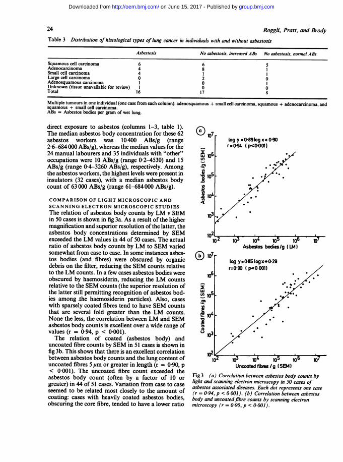
direct exposure to asbestos (columns 1-3, table 1). 00The median asbestos body concentration for these 62 107asbestos workers was 10400 ABs/g (range log y 0-89log x 0902-6-684 000 ABs/g), whereas the median values for the _ r zo94 (p<0001)24 manual labourers and 35 individuals with "other" 2 106occupations were 10 ABs/g (range 0.2-4530) and 15 #.)ABs/g (range 04-3260 ABs/g), respectively. Among JIMthe asbestos workers, the highest levels were present in 105 *insulators (32 cases), with a median asbestos body ..count of 63 000 ABs/g (range 61-684 000 ABs/g).
COMPARISON OF LIGHT MICROSCOPIC ANDSCANNING ELECTRON MICROSCOPIC STUDIESThe relation of asbestos body counts by LM v SEM 103.in 50 cases is shown in fig 3a. As a result of the highermagnification and superior resolution of the latter, theasbestos body concentrations determined by SEM 102exceeded the LM values in 44 of 50 cases. The actual 102 i03 o 0 iratio of asbestos body counts by LM to SEM varied Asbestos bodies/g (LM)somewhat from case to case. In some instances asbes- 107tos bodies (and fibres) were obscured by organic ' log ya085 logx.029debris on the filter, reducing the SEM counts relative r:0o90 (pO0001)to the LM counts. In a few cases asbestos bodies were 106obscured by haemosiderin, reducing the LM counts .relative to the SEM counts (the superior resolution of z .0the latter still permitting recognition of asbestos bod- 5ies among the haemosiderin particles). Also, cases ,with sparsely coated fibres tend to have SEM counts *that are several fold greater than the LM counts. . 410None the less, the correlation between LM and SEM 10asbestos body counts is excellent over a wide range of a
values (r = 0-94, p < 0-001). 3The relation of coated (asbestos body) and 10
uncoated fibre counts by SEM in 51 cases is shown infig 3b. This shows that there is an excellent correlation ____/___, ________between asbestos body counts and the lung content of 102 ;r- io0 105 106 1o7uncoated fibres 5pm or greater in length (r = 090, p Uncooted fibas/g (SEM)< 0-001). The uncoated fibre count exceeded theasbestos body count (often by a factor of 10 or Fig3 (a) Correlation between asbestos body counts bygreater) in 44 of 51 cases. Variation from case to case light and scanning electron microscopy in 50 cases ofgreater) in4of 51elasedost closely to the amountofasbestos associated diseases. Each dot represents one caseseemed to be related most closely to the amountof (r = 0 94, p < 0 001). (b) Correlation between asbestoscoating: cases with heavily coated asbestos bodies, body and uncoatedfibre counts by scanning electronobscuring the core fibre, tended to have a lower ratio microscopy (r = 0 90, p < 0-001).
24 Roggli, Pratt, and Brody
-r l r - s9J
group.bmj.com on June 15, 2017 - Published by http://oem.bmj.com/Downloaded from
Asbestos content of lung tissue in asbestos associated diseases: a study of 110 cases
of uncoated to coated fibres, whereas cases withsparsely coated bodies tended to have a higher ratio.
CHEMICAL COMPOSITION OF FIBRESThe results of energy dispersive x ray analysis of 809fibres from 57 cases are summarised in table 4. Analy-sis of 407 asbestos body cores shows that 98 5% arein fact nucleated on asbestos, and non-asbestos coreswere rare, being found in only one case. In thisinstance six fibres with a chemical composition ofSi-Al-K-Ca-Fe-Mg were identified as constituting thecores of thin, high aspect ratio coated fibres from anasbestos cement worker. The vast majority (93-9%) ofasbestos bodies were nucleated on commericalamphibole (amosite or crocidolite) cores, whereas2 5% and 2-2% had cores of non-commercial amphi-boles (anthophyllite, tremolite, or actinolite) andchrysotile, respectively. Analysis of 404 uncoatedfibres 5pm or greater in length shows that most ofthese (88 1 %) are also asbestos, with 78 1% commer-cial amphiboles, 4 5% non-commercial amphiboles,and 5-5% chrysotile. In cases with high content ofamphibole fibres (100 000 or more per gram of wetlung) chrysotile fibres are difficult to identify by SEM.In cases with low amphibole content-for example,the four cases with idiopathic pulmonary fibrosis-afew fibres identified were more often chrysotile ornon-asbestos fibres. The latter include fibreglass, talc,silica, rutile, kaolinite,. mica, and assorted silicates notfurther classified (table 4).
Discussion
In the present study the asbestos content of lung tissuein patients with asbestosis, mesothelioma, and pleuralplaques was found to correlate well with present con-cepts of the epidemiology of these diseases. Patientswith asbestosis have the highest levels of exposure toasbestos, whereas mesothelioma (in the absence of
asbestosis) can occur in individuals with much lessexposure." The relatively greater asbestos content ofthe lung in asbestosis as compared with cases ofmeso-thelioma is consistent with this observation. Similarly,parietal pleural plaques are the most common lesionsobserved in populations exposed to asbestos,'32' andin patients with plaques in the absence of asbestosisthe asbestos content of lung is relatively low in thisstudy and previous ones.2224 Unilateral parietalpleural plaques may be related to asbestos exposure,but these lesions can also be related to infection ortrauma.24 The asbestos body content of lung tissuetends to be much higher in patients who work directlywith asbestos compared with manual labourers and"other" occupational groups, although there is con-siderable overlap among occupational categories.Individuals in the other occupational category withasbestos body content exceeding 100 ABs/g probablyhave remote, undetected prior exposure to asbes-tos.413
Previous studies have noted a correlation betweenthe degree of interstitial fibrosis and the asbestos fibrecount by phase contrast microscopy.2526 Morerecently Warnock et al examined this relation usingtransmission electron microscopy.27 Their data (table2)27 show a fairly good correlation between the esti-mated degree of fibrosis and asbestos body and com-mercial amphibole content of lung tissue, but not fortotal fibre counts, non-commercial amphiboles, orchrysotile content. The results of our study, usingscanning electron microsocopy and the asbestosisgrading scheme of the Pneumoconiosis Committee ofthe College of American Pathologists and theNational Institute for Occupational Safety andHealth show a correlation between the severity ofasbestosis and the total (coated and uncoated) fibrecount (r = 0 57, p < 0-05) and the uncoated fibrecount (r = 0 56, p < 0 05) for fibres 5pm or greaterin length." Several studies have indicated that longer
Table 4 Energy dispersive x ray analysis data on 809 fibres from 57 cases
No Commercial Non-commercial Chrysotile Other* Totalamphiboles amphiboles
fibres are more fibrogenic than shorter ones,28 - 30 andit is these longer fibres that are measured under thecurrent regulatory standards.31 Although the degreeof correlation in our study is less than impressive, itwould probably improve with more extensive histo-logical and mineralogical sampling of the lungs andthe expression of the data as total lung burden ratherthan concentration. Accumulation of collagen andother cellular components as a result of the scarringprocess increases the weight of the lungs and hencedilutes the concentration of fibres in the parenchyma,a point often overlooked in dust analysis studies.32An additional finding in our study was a correlation
between the grade of asbestosis and smoking historyin pack-years (r = 0 53, p < 0-05). This observationhas been noted previously in radiological studies,33and it has been suggested that this is due to inter-ference with dust clearance mechanisms by cigarettesmoke. Our data, however, did not show a correlationbetween pack-years of smoking and uncoated fibrecontent of lung tissue (n = 19, r = 0-28, p > 005).The mechanism of interaction between asbestos andcigarette smoke in increasing interstitial fibrosisdeserves further study.Lung cancer occurred in 15 of the patients with
asbestosis in our study. Among the patients withasbestosis, those with lung cancer were older (medianage of 63 v 57) and had a higher average cigaretteconsumption (mean of 48-8 v 29 pack-years) thanthose without cancer. The latter observation was alsonoted in the study by Warnock etaL.27 The histologi-cal patterns of lung cancer did not differ amongpatients with asbestosis, with increased asbestos con-tent without asbestosis, or with normal asbestos con-tent (table 4). This finding is in keeping with theobservation of Ives etal that no specific histologicalpattern of lung cancer is associated with asbestosexposure.34 Although our study does not permit acalculation of the incidence of lung cancer in patientswith asbestosis due to biases in referral of cases, itshould be noted that other authors have reported thatmore than half the patients with asbestosis willdevelop lung cancer.35 In our experience this is muchgreater than the incidence of lung cancer in patientswith idiopathic pulmonary fibrosis, and indeed onlyone case in ten with idiopathic pulmonary fibrosis inour study had lung cancer. Thus mechanisms otherthan the scarring process per se are probably oper-ative in the pathogenesis of lung cancer in patientsexposed to asbestos.36
Relatively few reports have dealt with the lung con-tent of asbestos in patients with mesothelioma. Whit-well et al in a series of 100 patients with mesotheliomareported that 95% of those with asbestos inducedmesotheliomas had over 50 000 fibres/g of dried lungby phase contrast microscopy compared with only
Roggli, Pratt, and Brody
15% of the control series.26 In a study of 99 meso-thelial tumours in North America McDonald etalnoted equal numbers of chrysotile fibres in cases vcontrols, whereas there were increased numbers ofamphibole fibres by transmission electron microscopyin a greater percentage of cases compared with con-trols.37 More recently, Churg and Wiggs reported onnumbers and sizes of fibres from the lungs of 10patients who had an amphibole induced malignantpleural mesothelioma,38 and found an approximately250-fold increase in commercial amphiboles by anal-ytical transmission electron microscopy in the patientswith mesothelioma compared with the general popu-lation. Two studies have reported data concerningasbestos fibre counts by SEM in patients with meso-thelioma.22 39 Gylseth et al found two million or morefibres per gram of dried lung in all 15 patients withmesothelioma studied.22 Friedrichs and Otto studied34 cases of occupationally associated mesotheliomas,and found more than three times as many fibres inthose with asbestosis than in those without.39The present study shows that our patients with
mesothelioma fall into three broad categories. Thosewho also have asbestosis have among the highest val-ues of asbestos body and uncoated fibre counts wehave observed. Those who do not have asbestosis butdo have an occupational exposure history almostalways have raised asbestos body counts and about10% as many uncoated fibres greater than 5.pm inlength compared with cases of asbestosis. Those whohave normal asbestos body counts do not have asbes-tosis and usually do not give a history of exposure toasbestos. Others have reported such cases and haveattributed them as being "spontaneous" meso-theliomas.263940 These cases with a lung asbestoscontent within the normal range and with no demon-strable occupational exposure to asbestos are proba-bly non-asbestos related mesotheliomas and accountfor 20-30% of all cases.41 Alternatively, these casesmay represent mesotheliomas in a susceptible hostdue to environmental rather than occupational asbes-tos exposure.
Several epidemiological studies have shown anassociation between exposure to asbestos and gastro-intestinal carcinoma,24243 laryngeal carcinoma,2144and haematopoietic malignances,20 although theseassociations have not remained unchallenged.3145Analysis of the asbestos content of lung tissue in suchcases can document exposure but does not prove cau-sation. None the less, it is of interest to examine lungtissue from individuals with such diseases and histor-ies of asbestos exposure to try to estimate degrees ofexposure. None of our cases with histologicallyproved asbestosis had any of these neoplasms. Amongour cases of pleural plaques, however, were 12 withone of these three categories of malignancy. The 12
group.bmj.com on June 15, 2017 - Published by http://oem.bmj.com/Downloaded from
Asbestos content of lung tissue in asbestos associated diseases: a study of 110 cases
had a higher median asbestos body count than theremaining 21 with bilateral pleural plaques. Thesedata suggest the possibility that these diseases mayoccur in individuals with moderate exposures toasbestos, and further studies are needed to examinethis matter more fully.The present study has dealt with the asbestos con-
tent of lung tissue in a series of patients with diseasesthat have been associated with exposure to asbestos.It is important to emphasise the value and the limi-tations of asbestos body quantification in these dis-eases. As has been noted by Churg, determination ofasbestos body content is a relatively quick and easyprocedure.4 Bodies with the typical beadedconfiguration and a thin transparent central core arevirtually always nucleated on asbestos fibres as shownby energy dispersive x ray analysis and selected areaelectron diffraction.419 The vast majority are com-mercial amphiboles (amosite or crocidolite), bothamong individuals with asbestos associated diseases(table 4) and members of the general population, withthe exception that non-commercial amphibole cores(tremolite or anthophyllite) are fairly common inwomen from the general population.46 Furthermore,it is primarily fibres 20pm or more in length thatbecome coated.47 Although there are virtually alwaysmore uncoated than coated fibres by electron micros-copy, the correlation between asbestos body countsand uncoated fibres 5 gm or greater in length is excel-lent in the population we studied (fig 3). Thesefindings are essentially indentical to those reported byMorgan and Holmes, who used phase contrastmicroscopy to count coated and uncoated fibres.48Thus asbestos body content is a reasonably reliablemarker for levels of long amphibole fibres. On theother hand, the correlation between asbestos bodycounts and concentration of chrysotile or non-commercial amphibole fibres is poor,49 the vastmajority of these fibres being 5pm or less in length.Although occasional asbestos bodies with chrysotilefibres may be encountered (table 4), asbestos bodiesgive little or no indication of the chrysotile content ofthe lung. In the present study we did not evaluate theshort fibres (<5 pm) and thus cannot comment ontheir possible association with these diseases. Thepathogenicity of such short fibres has been ques-tioned,50 and their role (if any) in asbestos associateddiseases has yet to be defined.3"
We gratefully acknowledge the following physicianswho referred case material for study: Doctors JAdams, Chattanooga, TN; F B Askin, Chapel Hill,NC; A Churg, Vancouver, BC; D Dail, Seattle, WA;J R Edgar, Savannah, GA; J C Franco, Fayetteville,NC; B Gylseth, Oslo, Norway; S Harris, Greensboro,NC; W B Helwig, J C Maddox, J Legier, J C Davis,
Jr, and F Q Wingfield, Newport News, VA; R AHeyer, Charlotte, NC; R V Joel, Jacksonville, FL; EKagan, Washington, DC; D Kaminsky, RanchoMirage, CA; Marie-Claire Marroum, Charlotte, NC;C T O'Connell, Hampton, VA; J H Riddick, Jr,Chesapeake, VA; W Stopford, Durham, NC; PWarga, Salisbury, NC; B Woodard, Anderson, SC;and Elsa Yap, Concord, NC. Dr R T Vollmer helpedwith the statistical analyses and Diane Evans providedexpert help in preparing the manuscript for publica-tion.
Requests for reprints to: Victor L Roggli, MD,Department of Pathology, Post Office Box 3712,Duke University Medical Center, Durham, NC27710, USA.
References
Becklake MR. Asbestos-related diseases of the lung and otherorgans: their epidemiology and implications for clinical practice.Am Rev Respir Dis 1976;114:187-227.
2Selikoff IJ, Lee DHK. Asbestos and disease. New York: AcademicPress, 1978.
3Wagner JC, Sleggs CA, Marchand P. Diffuse pleural mesotheliomaand asbestos exposure in the North Western Cape province. BrJ Ind Med 1960;17:260-71.
'Churg AM, Warnock ML. Asbestos and other ferruginous bodies:their formation and clinical significance. Am J Pathol1981 ;102:447-56.
'Smith MJ, Naylor B. A method of extracting ferruginous bodiesfrom sputum and pulmonary tissue. Am J Clin Pathol1972;58:250-4.
6Roggli VL, Greenberg SD, Seitzman LH, et al. Pulmonary fibrosis,carcinoma, and ferruginous body counts in amosite asbestosworkers: a study of six cases. Am J Clin Pathol 1980;73:496-503.
7Churg A, Warnock ML. Correlation of quantitative asbestos bodycounts and occupation in urban patients. Arch Pathol Lab Med1977;101:629-34.
8Bignon J, Goni J, Bonnaud G, Jaurand MC, Dufour G, PinchonMC. Incidence of pulmonary ferruginous bodies in France. Envi-ron Res 1970;3:430-42.
9 Bhagavan BS, Koss LG. Secular trends in prevalence and concen-tration of pulmonary asbestos bodies-1940 to 1972: a necropsystudy. Arch Pathol Lab Med 1976;100:539-41.
Rosen P, Melamed M, Savino A. The "ferruginous body" contentof lung tissue: a quantitative study of eighty-six patients. ActaCytol 1972;16:207-1 1.
Craighead JE, Abraham JL, Churg A, et al. The pathology ofasbestos-associated diseases of the lungs and pleural cavities:diagnostic criteria and proposed grading schema. (Report of thePneumoconiosis Committee of the College of American Pathol-ogists and the National Institute for Occupational Safety andHealth.) Arch Pathol Lab Med 1982;106:544-96.
12Roggli VL, McGavran MH, Subach J, Sybers HD, Greenberg SD.Pulmonary asbestos body content and electron probe analysis ofasbestos body cores in patients with mesothelioma: a study of 25cases. Cancer 1982;50:2423-32.
13Wain SL, Roggli VL, Foster WL. Parietal pleural plaques, asbestosbodies, and neoplasia: a clinical, pathological, and radiographiccorrelation of 25 consecutive cases. Chest 1984;86:707-13.
14Meurman L. Asbestos bodies and pleural plaques in a Finnishseries of autopsy cases. Acta Pathol Microbiol Immunol Scand[Suppi] 1966;181:1-107.
s World Health Organisation. The World Health Organisation his-
27
group.bmj.com on June 15, 2017 - Published by http://oem.bmj.com/Downloaded from
tologic typing of lung tumours, 2nd ed. Am J Clin Pathol1982;77:123-36.
16Roggli VL, Shelburne JD. New concepts in the diagnosis of min-eral pneumoconioses. Seminars in Respiratory Medicine1982;4:138-48.
7Williams MG, Dodson RF, Corn C, Hurst GA. A procedure forthe isolation of amosite asbestos and ferruginous bodies fromlung tissue and sputum. J Toxicol Environ Health1982;10:627-38.
Roggli VL, Brody AR. Changes in numbers and dimensions ofchrysotile asbestos fibers in lungs of rats following short-termexposure. Exp Lung Res 1984;7:133-47.
'9Churg A, Warnock ML, Green N. Analysis of the cores of fer-ruginous (asbestos) bodies from the general population. II. Trueasbestos bodies and pseudoasbestos bodies. Lab Invest1979;40:31-8.
20Kagan E, Jacobson RJ. Lymphoid and plasma cell malignances:asbestos-related disorders of long latency. Am J Clin Pathol1983;80:14-20.
21 Hillerdal G. Pleural plaques: occurrence, exposure to asbestos, andclinical importance. Uppsala: Offsetcenter ab, 1980.
22Gylseth B, Mowe G, Skaug V, Wannag A. Inorganic fibers in lungtissue from patients with pleural plaques or malignant meso-thelioma. Scand J Work Environ Health 1981;7:109-13.
23 Warnock ML, Prescott BT, Kuwahara TJ. Numbers and types ofasbestos fibers in subjects with pleural plaques. Am J Pathol1982;109:37-46.
24Churg A. Asbestos fibers and pleural plaques in a general autopsypopulation. Am J Pathol 1982;109:88-96.
2Ashcroft T, Heppleston AG. The optical and electron microscopicdetermination of pulmonary asbestos fibre concentration and itsrelation to the human pathological reaction. J Clin Pathol1973;26:224-34.
26 Whitwell F, Scott J, Grimshaw M. Relationship betweenoccupations and asbestos-fibre content of the lungs in patientswith pleural mesothelioma, lung cancer, and other diseases. Tho-rax 1977;32:377-86.
27 Warnock ML, Kuwahara TJ, Wolery G. The relation of asbestosburden to asbestosis and lung cancer. Pathol Annu1983;18:109-45, part 2.
28Davis JMG, Beckett ST, Bolton RE, Collings P, Middleton AP.Mass and number of fibres in the pathogenesis of asbestos-related lung disease in rats. Br J Cancer 1978;37:673-88.
29 Vorwald AJ, Durkan TM, Pratt PC. Experimental studies ofasbes-tosis. Arch Ind Hyg Occup Med 1951;3:1-43.
30 Wright GW, Kuschner M. The influence of varying lengths ofglassand asbestos fibres on tissue response in guinea pigs. In: WaltonWH, ed. Inhaled particles IV. Oxford: Pergammon Press,1977:455-74.
31 Craighead JE, Mossman BT. The pathogenesis of asbestos-associated disease. N Engl J Med 1982;306:1446-55.
32pratt PC. Role of silica in progressive massive fibrosis in coal
Roggli, Pratt, and Brodyworkers' pneumoconiosis. Arch Environ Health 1968;16:734-7.
3McMillan GHG, Pethybridge RJ, Sheers G. Effect of smoking onattack rates of pulmonary and pleural lesions related to exposureto asbestos dust. Br J Ind Med 1980;37:268-72.
34Ives JC, Buffler PA, Greenberg SD. Environmental associationsand histologic patterns of carcinoma of the lung: the challengeand dilemma in epidemiologic studies. Am Rev Respir Dis1983;128:195-209.
3S Buchanan WD. Asbestosis and primary intrathoracic neoplasms.Ann NY Acad Sci 1965;132:507-18.
36Mossman BT, Craighead JE. Mechanisms of asbestos carcino-genesis. Environ Res 1981;25:269-80.
37 McDonald AD, McDonald JC, Pooley FD. Mineral fibre contentof lung in mesothelial tumours in North America. Ann OccupHyg 1982;26:417-22.
38Churg A, Wiggs B. Fiber size and number in amphibole asbestos-induced mesothelioma. Am J Pathol 1984;115:437-42.
39Friedrichs KH, Otto H. Fibers in human lung dust samples: ascanning electron microscope study. Am Ind Hyg Assoc J1981;42:150-6.
"Peterson JT, Greenberg SD, Buffler PA. Non-asbestos-relatedmalignant mesothelioma: a review. Cancer 1984;54:951-60.
41Chahinian AP, Pajak TF, Holland JF, Norton L, Ambinder RM,Mandel EM. Diffuse malignant mesothelioma: prospective eval-uation of 69 patients. Ann Intern Med 1982;96:746-55.
42Selikoff IJ, Hammond EC, Seidman H. Mortality experience ofinsulation workers in the United States and Canada, 1943-1976.In: Selikoff IJ, Hammond EC, eds. Health hazards and asbestosexposure. Ann NY Acad Sci 1979;330:91-1 16.
43Finkelstein MM. Mortality among employees of an Ontarioasbestos-cement factory. Am Rev Respir Dis 1984;129:754-61.
"Stell PM, McGill T. Asbestos and laryngeal carcinoma. Lancet1973;ii:416-7.
'5McCullagh SF, Aresini G, Browne K, et al. Criteria for the diagno-sis of asbestosis and considerations in the attribution of lungcancer and mesothelioma to asbestos exposure. Int Arch OccupEnviron Health 1982;49:357-61.
"Churg A, Warnock ML. Analysis of the cores of ferruginous(asbestos) bodies from the general population. III. Patients withenvironmental exposure. Lab Invest 1979;40:622-6.
Morgan A, Holmes A. Concentrations and dimensions of coatedand uncoated asbestos fibres in the human lung. Br J Ind Med1980;37:25-32.
Morgan A, Holmes A. Distribution and characteristics of amphi-bole asbestos fibres, measured with the light microscope, in theleft lung of an insulation worker. Br J Ind Med 1983;40:45-50.
49Warnock ML, Prescott BT, Kuwahara TJ. Correlation of asbestosbodies and fibers in lungs ofsubjects with and without asbestosis.In: Johari 0, Becker RP, eds. Scanning electron microscopy II.AMF O'Hare, III: SEM, Inc, 1982:845-57.
30 Gross P. Is short-fibered asbestos dust a biological hazard? ArchEnviron Health 1974;29:1 15-7.
group.bmj.com on June 15, 2017 - Published by http://oem.bmj.com/Downloaded from