13
Color plates

Color plates

Plate 1. Intraoperative picture of cardioplication with proximal suture visible and the
plication apparent (See also Fig. 3 in article by Rosen and Ponsky.)
Plate 2. A retroflexed endoscopic view of an endoscopic intraluminal valvuloplasty in a
baboon at 6 months postprocedure (See also Fig. 10 in article by DeMeester.)

Plate 1
Plate 2

Plate 3. Porcine LES and gastric cardia demonstrating circular muscle lesion as pale
staining region on H and E stain (See also Fig. 5A in article by Utley.)
Plate 4. Absent staining region on diaphorase staining with intact mucosa (See also Fig. 5B
in article by Utley.)
Plate 5. Acute thermal lesion within human gastric cardia. Patient treated under human
subjects protocol several hours before esophagogastrectomy for midesophageal adeno-
carcinoma. Thermal lesion within circular muscle, with overlying intact mucosa (See also
Fig. 6 in article by Utley.)

Plate 5
Plate 3 Plate 4

Plate 6. Endoscopic appearance of the gastroesophageal junction (GEJ) before and
immediately after Stretta1. (Left) Retroflexed view of GEJ. (Right) Mucosal burn marks
straddling the GEJ. These marks disappear within 10 days after Stretta1 (See also Fig. 2 in
article by Triadafilopoulos.)
Plate 7. Injection of PMMA microspheres to the submucosa of the lower esophagus (See
also Fig. 1 in article by Feretis et al.)

Plate 7
Plate 6
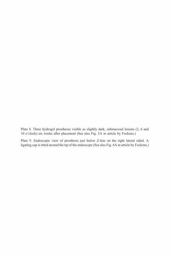
Plate 8. Three hydrogel prostheses visible as slightly dark, submucosal lesions (2, 6 and
10 o’clock) six weeks after placement (See also Fig. 3A in article by Fockens.)
Plate 9. Endoscopic view of prosthesis just below Z-line on the right lateral sided. A
ligating cap is titted around the tip of the endoscope (See also Fig. 6A in article by Fockens.)

Plate 8
Plate 9

Plate 10. Incision of overlying mucosa with needle knife (See also Fig. 6C in article
by Fockens.)
Plate 11. Aspirated hydrogel prosthesis (black) in ligating cap of endoscope (See also
Fig. 6D in article by Fockens.)

Plate 10
Plate 11

Plate 12. Squamocolumnar junction with prominent distal esophageal mucosal cushions
estimating the esophageal endoluminal area of the lower esophageal sphincter (LES)
(See also Fig. 1A in article by Ramage et al.) (Copyright 1995, Ciba-Geigy Corporation,
with permission.)
Plate 13. Esophagogastric junctional area visually noted as the border between the linear
mucosal distal esophageal vessels and the upper ends of the rugal folds (See also Fig. 2 in
article by Ramage et al.)
Plate 14. Retroflexed view of the cardia demonstrating the collar of tissue that comprises
the angle of His and the open area within the lesser curvature (See also Fig. 3 in article by
Ramage et al.)
Plate 15. Patulous cardia of a patient with gastroesophageal reflux disease (See also Fig. 4
in article by Ramage et al.)

Plate 12 Plate 13
Plate 14 Plate 15


