1 Dosimetry (Dose Estimation) of Internal Emitters. Lawrence E. Williams, PhD City of Hope National Medical Center Duarte CA 91010 [email protected]Outline 1. Dose Estimation Formula D = S*à 2. Determination of A(t) a. six methods b. errors in A 3. Integration of A to form à a. Open Model b. Closed Model 4. Calculation of Dose 5. Errors in Dose due to A, Ã, and S errors. Estimation of Dose and not Dosimetry • Dosimetry is the measurement of absorbed dose in erg/g or Joules/kg. This isn’t easily or ethically done in living tissues. Thus, use of the term is not appropriate in the context of radiation therapy. • We can only estimate the internal emitter dose. Our limitation is similar to that found in external beam work. “They don’t do dosimetry either”. For Radiation Effects, is Dose the only Answer? • Because of biological effectiveness, a QF (quality factor) may be multiplied by dose (Gray) values to yield a result in Sieverts. Alpha ray examples. • If this is done, however, the reader must be shown both values – not just the equivalent dose (Sv). • Effective dose is not appropriate for specific patient risk calculations and is intended as a comparison parameter to use for stochastic calculations.
• Dosimetry is themeasurement of absorbeddosein erg/gor Joules/kg.This isn’ t easilyor ethicallydonein living tissues.Thus,useof theterm is notappropriate in the context of radiation therapy.
• We canonly estimate theinternalemitter dose.Our limitation is similar to thatfoundin externalbeamwork. “Theydon’ t do dosimetry either”.
For Radiation Effects,is Dosetheonly Answer?
• Becauseof biologicaleffectiveness,a QF(quality factor)maybe multipliedby dose (Gray)values to yield a resultin Sieverts. Alpharay examples.
• If this is done,however,thereadermustbeshown bothvalues– not just theequivalentdose(Sv).
• Effectivedoseis not appropriatefor specific patient riskcalculationsand is intendedasa comparisonparametertousefor stochasticcalculations.
2
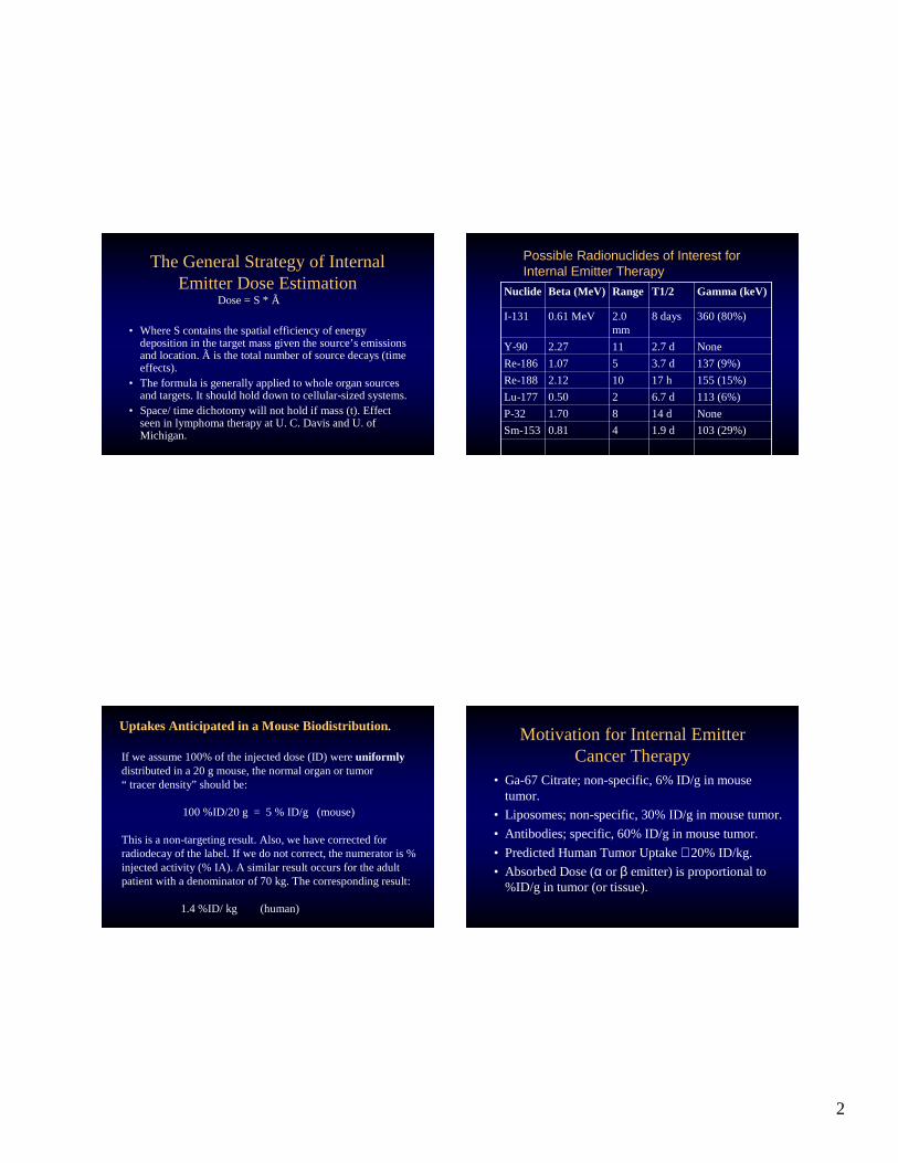
TheGeneral Strategy of InternalEmitter DoseEstimation
Dose= S * Ã
• Where S contains thespatialefficiency of energydepositionin thetargetmass giventhesource’s emissionsand location. Ã is thetotal number of sourcedecays (timeeffects).
• Theformulais generally appliedto wholeorgansourcesand targets.It should hold downto cellular-sizedsystems.
• Space/timedichotomywill not hold if mass (t). Effectseen in lymphoma therapyat U. C. Davisand U. ofMichigan. 103 (29%)1.9d40.81Sm-153
None14 d81.70P-32
113 (6%)6.7d20.50Lu-177
155 (15%)17 h102.12Re-188
137 (9%)3.7d51.07Re-186
None2.7d112.27Y-90
360 (80%)8 days2.0mm
0.61MeVI-131
Gamma (keV)T1/2RangeBeta (MeV)Nuclide
Possible Radionuclides of Interest forInternal Emitter Therapy
UptakesAnti cipated in a MouseBiodistri bution.
If we assume100%of theinjecteddose(ID) wereuniformlydistributedin a 20 g mouse,thenormalorganor tumor“ tracerdensity” shouldbe:
100%ID/20 g = 5 % ID/g (mouse)
This is a non-targeting result.Also, we havecorrectedforradiodecayof thelabel.If we do not correct,thenumeratoris %injectedactivity (% IA). A similar resultoccurs for the adultpatient with a denominatorof 70 kg. Thecorresponding result:
1.4%ID/ kg (human)
Motivationfor InternalEmitterCancerTherapy
• Ga-67 Citrate; non-specific, 6%ID/g in mousetumor.
• Liposomes;non-specific,30%ID/g in mousetumor.
• Antibodies;specific, 60%ID/g in mouse tumor.
• PredictedHuman TumorUptake≅ 20%ID/kg.
• AbsorbedDose(α or β emitter)is proportionalto%ID/g in tumor (or tissue).
3
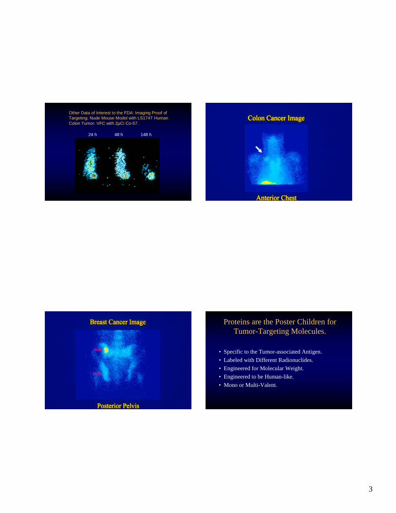
Other Data of Interest to the FDA: Imaging Proof ofTargeting; Nude Mouse Model with LS174T HumanColon Tumor. VFC with 2µCi Co-57.
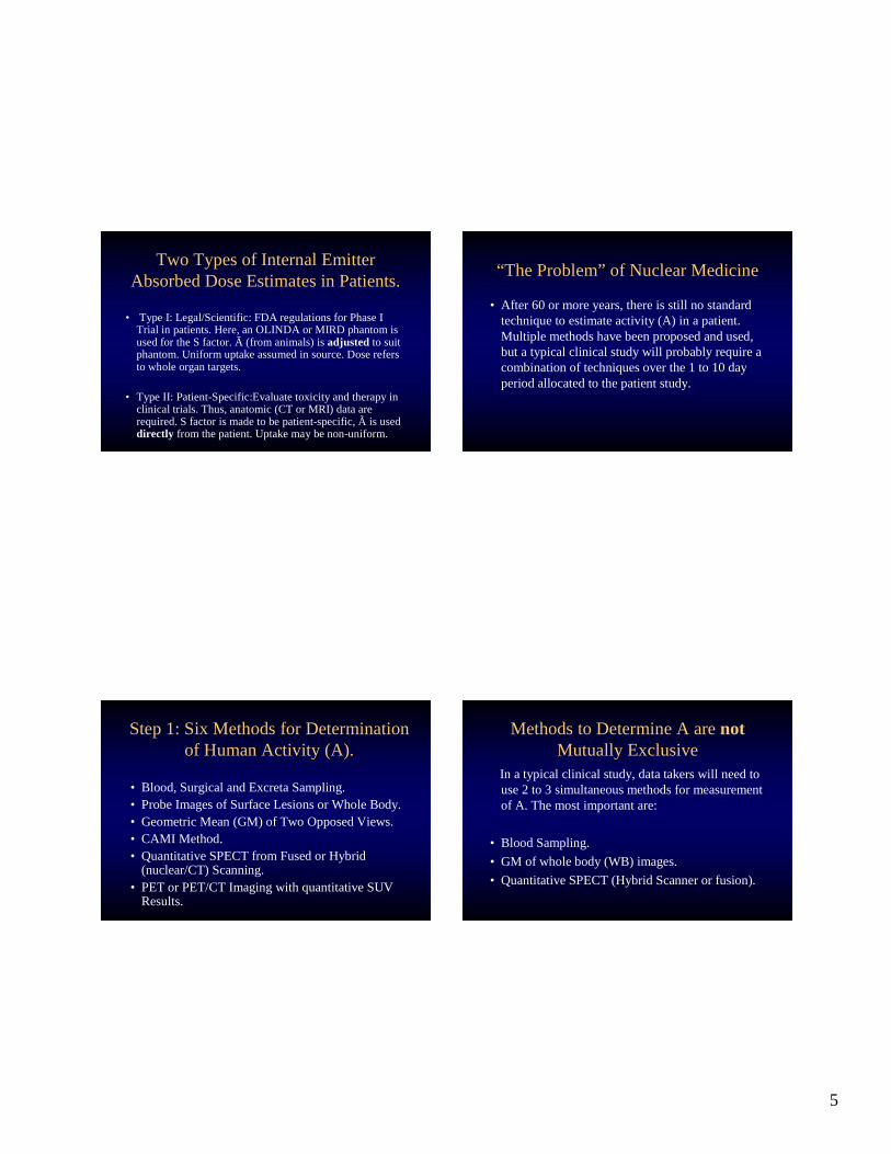
Theaboveagentsrely on catheterplacementof agent.UseTc-99mMAA to definelungtoxicity.
• BexxarTositumomab(I-131)for Lymphoma.
• ZevalinIbritumomab(Y-90) for Lymphoma.
Theseagents areinjectedIV and circulate.
InternalEmitter DoseEstimation.
In orderof decreasingdifficult y theprocesshasthreesteps.
1. Most difficult: Determination of activity (A) in tissues ofinterestat varioustimes(t). Many methods.
2. Next most difficult: Integration of A(t) over very longtimes (∞) time to form Ã. Varioustechniques.
3. Leastdifficult (usually):Convertingà to dose(D) via thematrix transformation D = S * Ã. However,S mayneedto be verydifferent from OLIND A or MIRD standardphantomvalues.UseCT or MRI data.
5
Two Typesof Internal EmitterAbsorbedDoseEstimatesin Patients.
• TypeI: Legal/Scientific:FDA regulationsfor PhaseITrial in patients.Here,anOLIN DA or MIRD phantom isusedfor theS factor. Ã (from animals) is adjusted to suitphantom. Uniform uptakeassumedin source.Doserefersto whole organtargets.
• TypeII : Patient-Specific:Evaluatetoxicity andtherapyinclinical trials.Thus,anatomic(CT or MRI) dataarerequired.S factor is madeto bepatient-specific, Ã is useddir ectly from thepatient.Uptake maybenon-uniform.
“TheProblem” of NuclearMedicine
• After 60 or more years, thereis still no standardtechniqueto estimateactivity (A) in apatient.Multiple methodshavebeenproposedandused,but a typical clinical studywill probablyrequireacombination of techniquesoverthe1 to 10 dayperiod allocatedto thepatientstudy.
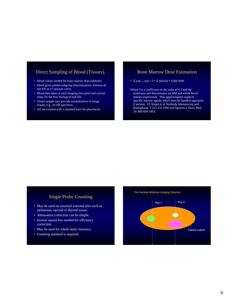
Step1: Six Methods for Determinationof HumanActivity (A).
• Blood,SurgicalandExcreta Sampling.• ProbeImagesof Surface Lesionsor WholeBody.• Geometric Mean(GM) of Two OpposedViews.• CAMI Method.• QuantitativeSPECTfrom Fusedor Hybrid
• All arecounted with a standard from thepharmacist.
BoneMarrow DoseEstimation
• Ã (rm→ rm) = f * Ã (blood)* 1500/5000
Where f is a coefficient on theorderof 0.3andthenumeratorand denominator are RM andwholebloodmassesrespectively. This approximationneglectsspecific marrowuptakewhich mustbehandledseparatelyif present. Cf. Siegel et al Antibody Immunoconj andRadiopharm. 3 213-2331990andSgouros J.Nucl. Med.34: 689-6941993.
SingleProbeCounting
• May beusedon essential externalsitessuchasmelanoma,sarcoidor thyroid tissue.
• Typically usesanterior-posterior projection.• Tissueattenuationis correctedwith CT, MRI or
direct measurement(externalsource).• Shouldhavestandard sourcein thefield of view.• Suffersfrom possibleorganand tumor overlap.• May alsosuffer from observerconfusion; hot
spotanterior image≠ hot spotposteriorimage.• Typical errorsare+/- 30 % (literature).
CAMI Method
• UsesCT data to correctattenuationalongrays ofinterestthru thepatient’s majororgansystems.
• May beusedfroma singlewholebodyscan.• Problemof activi ty becomesa setof activity
to the BetaEmitter (Therapy)Label.For example,goingfromIn-111-Antibodyto Y-90-Antibody.
10
Blood
UrineFeces
Residual Liver
RKrb
Kbr
Kbl
Klb
Krf Klu
Kd
Kd
Kd
Kd
Kd
5 Compartment PharmacokineticHuman Data Model
Step3: Methods to DetermineAbsorbedDose(D = S*Ã)
• OLINDA, MIRDOSE3or MIRDOSE2Programs;S dependsupona given phantom.Traditional Method; favoredby regulatoryagenciesand mostusers of radioactivity.
• Voxel-BasedCalculation (MAVSK) ; S is local.
• Point-SourceKernels; S is local.
• CompleteMonteCarlo Analysis; no useof S (!).
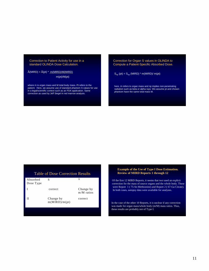
Two Correctionsto OLINDAEstimationsof AbsorbedDose.
• Correctà (patient) to Al low Substitution intoStandardProgram.TypeI Estimate.
• CorrectS (OLINDA or MIRD) to Allow Patient-Specific Estimation of AbsorbedDose.TypeIIEstimate.
11
Correction to Patient Activity for use in astandard OLINDA Dose Calculation.
Ã(MIRD) = Ã(pt) * m(MIRD)/M(MIRD)
m(pt)/M(pt)
where m is organ mass and M total body mass. Pt refers to thepatient. Here, we assume use of standard phantom S values for usein a legal/scientific context such as an FDA application. Samecorrection as used by Jeff Siegel in red marrow analysis.
Correction for Organ S values in OLINDA toCompute a Patient-Specific Absorbed Dose.
Snp (pt) = Snp (MIRD) * m(MIRD)/ m(pt)
here, m refers to organ mass and np implies non-penetratingradiation such as beta or alpha rays. We assume pt and chosenphantom have the same total mass M.
Tableof DoseCorrection ResultsA bsorbedDoseType
S Ã
I correct Changebym/M ratios
I I Changebym(MIRD)/m(pt)
correct
Example of the Useof Type I DoseEstimation.Reviewof MIRD Reports 1 through 12
Of the fi rst 12 MIRD Reports, it seemsthattwo usedanexplicitcorrection for themass of source organsandthewhole body. ThesewereReport 1 ( 75-Se-Methionine)andReport2 ( 67-GaCitrate).In bothcases,autopsydatawere available for analyses.
In thecaseof theother10 Reports,it is unclearif anycorrectionwasmadefor organmass/wholebody(m/M) massratios. Thus,theseresultsareprobablynot of TypeI.
12
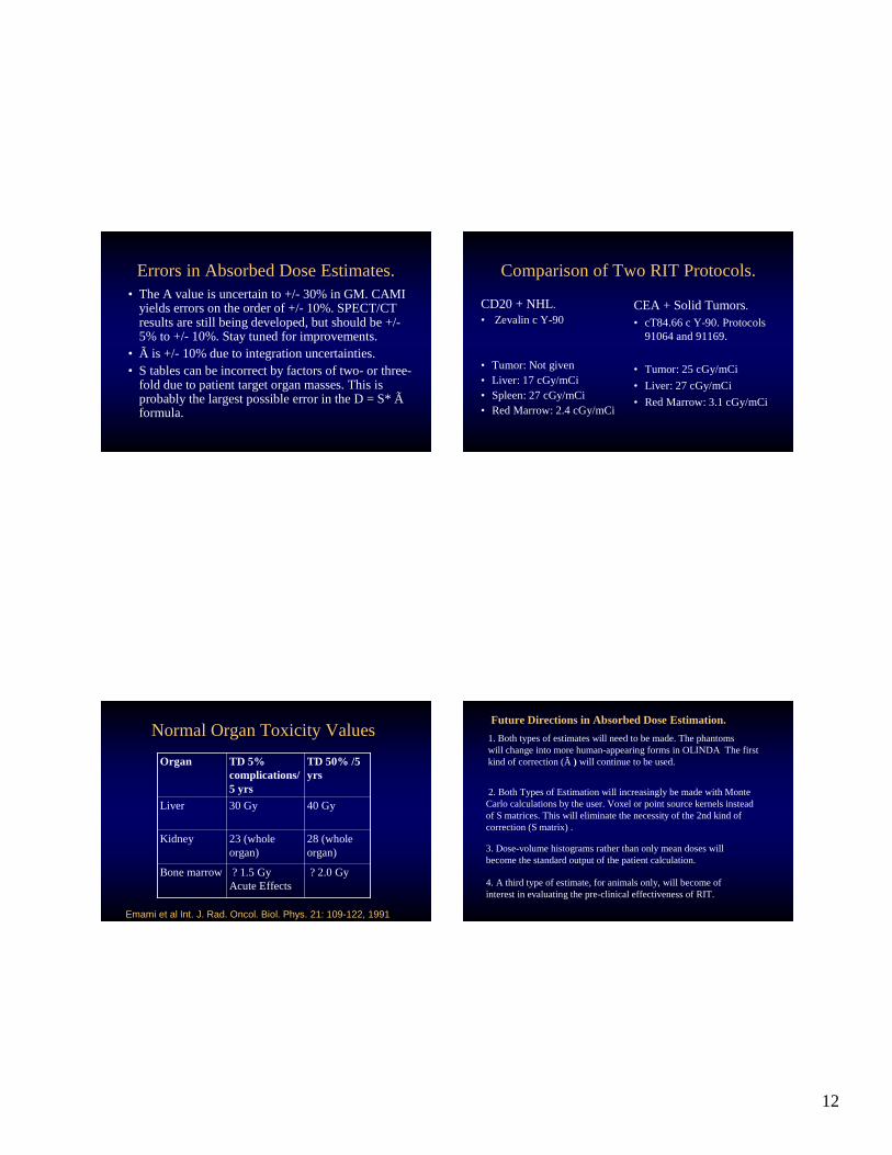
Errorsin Absorbed DoseEstimates.• The A valueis uncertain to +/- 30%in GM. CAMI
yields errorson theorder of +/- 10%.SPECT/CTresultsarestill beingdeveloped,but shouldbe+/-5% to +/- 10%.Staytunedfor improvements.
• Ã is +/- 10%dueto integration uncertainties.• S tablescanbeincorrect by factorsof two- or three-
Emami et al Int. J. Rad. Oncol. Biol. Phys. 21: 109-122, 1991
Future Directions in Absorbed DoseEstimation.
1. Both typesof estimateswill needto bemade.Thephantomswill changeinto morehuman-appearingforms in OLINDA Thefirstkind of correction(Ã ) will continueto beused.
2. Both Typesof Estimationwill increasingly bemadewith MonteCarlocalculationsby theuser.Voxel or point sourcekernels insteadof Smatrices.This will eliminatethenecessity of the2ndkind ofcorrection (Smatrix) .
3. Dose-volumehistogramsratherthanonly meandoseswillbecomethestandardoutput of the patientcalculation.
4. A third typeof estimate,for animalsonly, wil l become ofinterestin evaluating thepre-clinical effectivenessof RIT.
13
SomeReferencesfor Internal EmitterDoseEstimation
• ThePrimer.AAPM ReportNo. 71,2001.RIT.
• Stabin et al. JNM 46: 1023-1027, 2005. OLINDA.
• Siegel et al. Antibod. Immunoconj. Radiopharm. 3:213-233, 1990.BoneMarrow DoseEstimates.