Page 1
ORIGINAL PAPER
Evaporative respiratory cooling augments pit organ thermaldetection in rattlesnakes
Viviana Cadena • Denis V. Andrade •
Rafael P. Bovo • Glenn J. Tattersall
Received: 17 June 2013 / Revised: 23 August 2013 / Accepted: 25 August 2013 / Published online: 5 September 2013
� Springer-Verlag Berlin Heidelberg 2013
Abstract Rattlesnakes use their facial pit organs to sense
external thermal fluctuations. A temperature decrease in
the heat-sensing membrane of the pit organ has the
potential to enhance heat flux between their endothermic
prey and the thermal sensors, affect the optimal functioning
of thermal sensors in the pit membrane and reduce the
formation of thermal ‘‘afterimages’’, improving thermal
detection. We examined the potential for respiratory
cooling to improve strike behaviour, capture, and con-
sumption of endothermic prey in the South American rat-
tlesnake, as behavioural indicators of thermal detection.
Snakes with a higher degree of rostral cooling were more
accurate during the strike, attacking warmer regions of
their prey, and relocated and consumed their prey faster.
These findings reveal that by cooling their pit organs, rat-
tlesnakes increase their ability to detect endothermic prey;
disabling the pit organs caused these differences to disap-
pear. Rattlesnakes also modify the degree of rostral cooling
by altering their breathing pattern in response to biologi-
cally relevant stimuli, such as a mouse odour. Our findings
reveal that low humidity increases their ability to detect
endothermic prey, suggesting that habitat and ambush site
selection in the wild may be influenced by external
humidity levels as well as temperature.
Keywords Thermosensation � Pit organ � Thermal
imaging � Respiratory cooling � Heat detection
Introduction
Pit vipers possess one heat-sensitive pit organ on each side
of the head, located between the eye and the nostril. Neural
information from the pit organs is integrated with their
visual system (Chiszar et al. 1977; de Cock Buning 1983a),
and although this sensory system can aid snakes in ori-
enting toward thermally favourable environments (Kroch-
mal and Bakken 2003; Krochmal et al. 2004), endothermic
prey detection and capture remain the hallmark features of
thermal sensing in many snakes, particularly pit vipers (de
Cock Buning et al. 1981; de Cock Buning 1983a). Indeed,
on moonless nights when rattlesnakes are most active
(Clarke et al. 1996) or in darkened burrows, where visual
cues are unavailable, thermal radiation is the main source
of information about the location of live prey. Moreover, in
the absence of visual cues, rattlesnakes and pythons are
capable of striking prey based solely on thermal detection,
with no effects on the accuracy or efficacy of the strike
(Kardong and Mackessy 1991; Kardong 1992; Grace et al.
2001).
The pit organs of Crotalinae snakes provide the highest
level of thermal sensitivity ever described for a biological
system (Molenaar 1992; Ebert and Westhoff 2006; Ebert
et al. 2007). In the case of pit vipers, the sensitive region of
the pit organ is a membranous structure located within the
pit. The heat sensors in the membrane appear to operate as
thermal receptors, as opposed to photonic infrared (IR)
V. Cadena � G. J. Tattersall (&)
Department of Biological Sciences, Brock University,
St. Catharines, ON L2S 3A1, Canada
e-mail: [email protected]
D. V. Andrade � R. P. Bovo
Departamento de Zoologia, Universidade Estadual
Paulista, Rio Claro, SP 13506-900, Brazil
D. V. Andrade � G. J. Tattersall
Instituto Nacional de Ciencia e Tecnologia em Fisiologia
Comparada, Rio Claro, SP, Brazil
123
J Comp Physiol A (2013) 199:1093–1104
DOI 10.1007/s00359-013-0852-4
Author's personal copy
Page 2
receptors, such that temperature changes in the membrane
of the pit organ evoke changes in the discharge frequency
of the heat-sensitive units (Bullock and Diecke 1956; de
Cock Buning et al. 1981; Pappas et al. 2004). IR radiation
reaches the membrane and induces miniscule changes in
membrane temperature, which are sensed and transduced
by metabolically active (i.e., mitochondria-rich) neural
mast cells (Bleichmar and de Robertis 1962; Ebert et al.
2006). Therefore, the pit organ detects changes in heat flux
from the stimulus which in turn, depends on pit membrane
temperature (de Cock Buning 1983b). Furthermore, pit
membrane temperature will affect the opening probability
of the TRPA1 ion channels located on the pit membrane,
which have been implicated in the molecular sensation of
heat flux (Gracheva et al. 2010). It has also been suggested
that rapid heat removal from the membrane is important to
reduce thermal ‘‘afterimages’’ by allowing the thermal
receptors to return to their resting state after activation
from a thermal stimulus, thereby increasing spatial or
temporal resolution (Amemiya et al. 1999; Goris et al.
2003). Cooling of the pit membrane via evaporation may
enhance thermal detection via any of these mechanisms.
We hypothesized that a decrease in rostral temperature
(primarily around the nose or snout), and, therefore, in the
pit organs, leads to enhanced temperature-based prey
detection in rattlesnakes. Moreover, we also tested whether
rattlesnakes are capable of modifying pit organ temperature
via respiratory cooling, and whether they are capable of
doing it rapidly enough to influence thermal detection and
translate into a higher efficiency of predatory behaviours.
To accomplish these objectives, we manipulated pit organ
temperature by changing ambient humidity, thereby
affecting the potential for respiratory evaporative cooling.
In all cases, the effectiveness of this approach was vali-
dated by simultaneously tracking rostral and body surface
temperatures with infrared thermography. Then, we tested
predatory efficiency during staged predatory encounters
with endothermic prey under conditions allowing for
maximal (i.e., low relative humidity; RH) and minimal
(i.e., high RH) respiratory cooling. Finally, we investigated
whether South American rattlesnakes can rapidly lower pit
organ temperature by changing their respiratory pattern as
an alertness response to relevant stimuli, such as visual,
mechanical, and chemosensory (prey odours) cues.
Materials and methods
Animals
South American Rattlesnakes (N = 25; mass: 741 ± 137 g,
mean ± SD; snout vent length: 98.6 ± 5.3 cm), Crotalus
durissus, were collected from Sao Paulo state, Brazil, and
were housed for a period of 2 or 3 months at *28 ± 2 �C
(daily range) ambient temperature (Ta) in individual wooden
cages (25 9 26 9 26 cm; height 9 width 9 length) with a
sliding glass door. Snakes were fed every other week with
live mice and given free access to water. Two weeks prior to
experimentation, all animals were fasted. Only non-ecdysial
individuals were used in experimental procedures. Animals
were obtained from snakes rescued from nearby sugar
plantations, and research was performed at the Jacarezario
Laboratory, which runs a conservation program for reptiles
in captivity under the Brazilian Institute for Environment
and Natural Renewable Resources (IBAMA; license number
1/35/94/1088-8). Approval to perform the animal studies
involving feeding live prey to the rattlesnakes was issued by
the Commission for Ethics in Animal Experimentation from
University of Sao Paulo State (UNESP), SP, Brazil, Protocol
Numbers: 03/08-CEEA and 021/2010.
Experimental procedures
All experiments were performed inside the snakes’ home
cages under normal housing conditions and as part of their
normal care and feeding schedule. We used this setting
rather than a larger experimental arena since more con-
sistent behaviours could be elicited and preliminary
experiments indicated that transferring the snakes to a
novel test environment interfered with their normal pred-
atory behaviour, precluding snakes from striking at prey for
hours following the transfer (data not shown). The cages
were devoid of foreign objects (e.g. water dish) starting
24 h before the beginning of each experiment. During all
experiments cameras (thermal imaging camera and an
infrared night vision camera; see details below) were
located in front of the cage. Each measurement period
started at least 5 min after the sliding glass door of the cage
was gently removed. We also observed extreme care in
minimizing disturbance from the presence of the experi-
menter; the observer was outside the snake’s field of view,
movements were slow and minimal and silence was kept
all times to avoid disturbing the rodent prey.
Series I: Feeding trials: intact pit organs
The purpose of these experiments was to evaluate the effect
of rostral cooling on the predatory efficiency of rattle-
snakes. Each snake was tested on two separate days (at
least 2 days apart) at low (LH; 40–45 % RH) and high
humidity (HH; 90–95 % RH), presented in random order.
Low humidity was obtained by fitting the room with
dehumidifiers while high humidity was achieved with open
source water heaters and water vapourisers (Ta was con-
stant between treatments). RH and Ta were measured with
a digital humidity/temperature meter (Model HHF81,
1094 J Comp Physiol A (2013) 199:1093–1104
123
Author's personal copy
Page 3
Omega Engineering Inc., Laval, Canada). To eliminate
visual cues that could influence the snakes’ predatory
ability, the rattlesnakes were kept in complete darkness
throughout the feeding. At the beginning of each experi-
ment, a mouse (Strain CD-1; mass: 38 ± 6 g) was gently
placed at the corner of the cage furthest from the snake.
The snake was then allowed to strike, search, and attempt
feeding on the mouse.
Predatory behaviour in venomous snakes is usually
structured into the following identifiable stereotyped phases
(Chiszar et al. 1977; de Cock Buning 1983a): (1) the strike:
this is defined as a rapid projection of the snake’s head
followed by physical contact between the mouth of the
snake with the mouse, resulting in its envenomation; (2)
prey relocation: since rattlesnakes usually release their prey
after envenomation, they subsequently must initiate a post-
strike search (de Cock Buning 1983a); for our purposes, a
snake was considered to have relocated the prey when its
rostrum was within *1 cm of the mouse; (3) attempt to eat
after prey inspection: after relocation of the prey, snakes
spend considerable time inspecting it before attempting to
eat it. To determine predatory effectiveness relative to time
0 (time when the mouse was placed inside the cage), the
latency (in seconds) to strike (S), relocate (R), and eat
(E) the prey were scored from the video recordings. This
was done by re-playing the videos frame by frame and using
the time-stamp to determine the duration (to the nearest
second) of each of the behaviours of interest.
We also determined the accuracy of the strike (i.e. how
many snakes struck the warmer head vs. the cooler body of
the mouse), and the snakes’ propensity to strike (whether the
snake struck within 15 min). We also calculated the distance
from the head of the snake to the mouse immediately before
the strike and the beginning of the relocation phase (obtained
from videos) to test for possible differences in these dis-
tances between humidity treatments. Tongue flick frequency
was also recorded; tongue flicks were visible from the video
recordings (see description below) and were counted man-
ually and binned across 10-s intervals. These were then
averaged over the 10 s prior to a strike, the 60 s following a
strike, and the 60 s prior to finding the prey.
Once the snake had made an attempt to eat, the mouse
was removed from the cage. Snakes were not allowed to
feed until the completion of the study, to maintain moti-
vational state, and thus allow the same individuals to be
used at both humidity levels. Since some snakes struck and
held onto mice, data on search and eat times were not
available for all animals (Table 1). Since we were interested
in the latency between the strike and relocation and eating
of the prey, we were unable to use these data. For this
reason, and to maintain a repeated measures design, only
snakes that struck and released prey in both humidity con-
ditions were formally analysed for behavioural latencies.
Series II: Feeding trials: blocked pits
To account for potential effects of vomerolfactory senses
being influenced by humidity, and thus, driving any dif-
ferences in predatory performance between humidity lev-
els, we repeated the experiments from Series I, but in
animals devoid of pit organ thermosensation. To accom-
plish this we blocked the pit organs of snakes (Krochmal
and Bakken 2003) using dental amalgam (3M ESPE,
ImpregnumTM F) covered with a small piece of aluminium
duct tape. Snakes were anaesthetized briefly with CO2
(Andrade et al. 2004), while the amalgam mix was inserted
into the pit organs. Mass and snout vent length of each
snake were also recorded at this time. The entire procedure
took\5 min and at least 17 h were allowed to pass before
the beginning of the first experiment. Due to their reluc-
tance to strike without thermal detection, snakes with
blocked pits were allowed to strike in a lit environment.
Lights were turned off immediately after the strike, and
since the remaining portion of the experiments was con-
ducted in complete darkness, the primary sense available to
search and attempt to eat the prey was vomerolfaction.
Animals that did not strike within 15 min of being pre-
sented with a mouse, or experiments in which the snake
held onto the mouse after having struck, or in which the pit
plugs fell out (three animals) were not included in the
analysis. As a result, out of 20 snakes of which the pit
organs were blocked, only 10 were used in the analysis
(N = 5 per humidity level, not paired).
Series III: Disturbance trials
The objective of these experiments was to quantify tran-
sient changes in rostral and body surface temperature in
response to disturbance. We tracked dynamic changes in
Table 1 Number of snakes exhibiting different strike parameters
during feeding trials at low (LH) and high humidity (HH)
Intact pits Blocked pits
HH LH HH LH
Strikea 23 23 7 6
No strikea 2 2b 7 4
Struck and held onto 7 9 2 1
Struck and released 16 14 5 5
Nosed live preyc 2 0 1 0
Attempted to swallow live prey 1 0 1 2
a Propensity to strike (whether the snake struck or not within a period
of 15 min) was not significantly affected by humidityb One of these two snakes struck the prey but was unsuccessful at
envenomating itc Refers to events in which, after having struck, the snake immedi-
ately re-approached and started touching the mouse with its nose
J Comp Physiol A (2013) 199:1093–1104 1095
123
Author's personal copy
Page 4
rostral (Tr) and body surface temperatures (Ts) of seven
snakes, at each humidity level using infrared thermography
(details below). Five minutes after removing the glass door
of the cage the first disturbance was applied. This first
disturbance consisted of simply turning the lights off in the
room; after a 4- to 5-min period of darkness, a stream of air
was forced over the snake for 10 s using a hand held
electrical fan (diameter 10 cm), followed by another
4–5 min of no disturbance. Tongue flick frequency was
recorded as in Series I.
Series IV: Olfactory stimulation
To determine whether a mouse odour would induce
respiratory cooling similar to those observed with light and
airflow disturbances, the respiratory cooling responses to a
mouse odour was tested on a subset of snakes (N = 15)
under total darkness (at *50–60 % RH). A plastic tube
was connected to the rear of the snake’s cage, which was
fed back to an air pump in another room (5 m away with
mechanical and auditory isolation) and a three-way valve,
and subsequently returned to the front of the snake’s cage,
approximately 20 cm from the snake. Once the lights were
turned off and the glass covering the cage was removed,
the snakes were left undisturbed for a minimum of 5 min
before applying the olfactory stimulus. Air was drawn from
the snake’s cage at approximately 200 mL/min and
allowed to flow back toward the snake for approximately
5 min before measurements began. A subsequent control
olfactory (snake smell) stimulation lasted 30 s, after which
the valve was switched, allowing the air to pass through a
chamber (*2 L) containing a live mouse before it was
directed back to the snake’s cage. To eliminate mechanical
stimuli from air vibrations, the air flow was diffused by
cotton gauze attached to the end of the outflow. The mouse
odour was delivered continuously for a period of 3 min
after which the valve was switched back allowing for the
snake’s own cage air smell to be re-circulated for a further
4 min. Because of the tubing dead space, there was a 30-s
delay from the moment the valve was turned and the
stimulus was delivered to the snake’s cage. Throughout the
procedure, changes in body and rostral surface temperature
were monitored by infrared thermography as described
below.
Thermal imaging of surface temperatures and video
capture
Thermographic images were collected using an IR thermal
imaging camera (Mikron Instruments, Model 7515, Vista,
California for all experiments except Series IV in which a
Model SC640, FLIR Systems, Wilsonville, Oregon was
used) connected continuously to a video acquisition
program (MikrospecRT, Mikron Instruments or Therma-
cam Researcher Pro). Images were collected at 4 (Series I)
and 1 frames s-1 (Series II–IV). The camera was calibrated
prior to every experiment against an internal thermocouple
(NIST standard), and in all cases emissivity was assumed
to be 0.95 (Tattersall et al. 2004). Using specialized soft-
ware (MikroSpec RT, Mikron Instruments or Thermacam
Researcher Pro), regions of interest on the snake’s body
and the background were digitally ‘drawn’ on each frame
to obtain the average surface body (Ts) and rostral (Tr)
temperatures (corresponding to the surface along the body
axis, and the region around the nares and surrounding the
pit organs, respectively), as well as ambient temperature
(Ta). Similarly, average mouse surface temperatures (Tm)
were obtained for Series I. Maximum mouse head (Tmh)
and mouse body (Tmb) for Series I were also compared
before the strike. Simultaneously with the thermal imaging
camera, snakes were recorded during experimental Series
I–III using a digital IR (night vision) video camera (Model
DCR-HC90 Handycam, Sony Corporation, Tokyo, Japan)
at 30 frames�s-1.
Statistical analysis
Series I and II Latencies to strike, relocate, and eat were
analysed for the influence of humidity using one-way
analysis of variance (ANOVA) with humidity treated as a
between-subjects factor when appropriate. The propensity
to strike (i.e., whether the snake struck within a 15-min
period) between blocked vs. intact pit organs was com-
pared with a v2 test. The location accuracy of the strike in
Series I and II was compared between humidities with v2
tests. Ta, Ts and Tr for Series I were compared between
humidities using paired t-tests. Ts and Tr were assessed
from the thermal video frames 5 s prior to strike (-S), 30 s
after strike (?S), and 30 s prior to relocating the mouse
(-R). The influence of humidity on Tr- Ts was then analysed
using two-way repeated measures ANOVA (RMANOVA;
humidity and predatory event as factors). Tm from Series I
at -S, ?S, and R were analysed with two-way RMANO-
VA (humidity and predatory event as factors). Maximum
mouse head vs. body surface temperatures were compared
with a two-way RMANOVA with humidity and body
region (i.e, head vs. body) as factors. The distance between
the snake and the mouse immediately before the strike and
before initiating the relocation phase was compared using
two-way RMANOVA.
Series III Ts and Tr were assessed for the 60-s imme-
diately before the beginning of darkness (no disturbance,
ND), and for the 90 s after turning the lights off (LO) and
the air flow (AF) disturbances. Tr - Ts was analysed for
the influence of humidity using a two-way RMANOVA
(with humidity and disturbance as factors).
1096 J Comp Physiol A (2013) 199:1093–1104
123
Author's personal copy
Page 5
Series IV Tr - Ts were calculated and averaged over
30 s immediately preceding the olfactory stimulus (NS),
for 60 s during exposure to the mouse odour (OS), and for
the 60 s following removal of the stimulus (PS). Tr - Ts
values before, during and after the stimulus were compared
using RMANOVA.
In all cases, normality was evaluated by residual ana-
lysis using the D’Agostino-Pearson test. If log transfor-
mation could not improve the residual distribution,
statistical tests were performed on ranked data. Post hoc
multiple comparisons were made using the Holm-Sidak
test. Statistical tests were considered significant at
a = 0.05 and Bonferroni corrections were applied where
multiple variables were tested within the same experi-
mental series. Data are presented as mean ± SD.
Results
Series I and II: Feeding trials
Respiratory cooling during feeding: effects of humidity
Humidity proved to be an effective way of manipulating
the degree of respiratory evaporation and, therefore, the
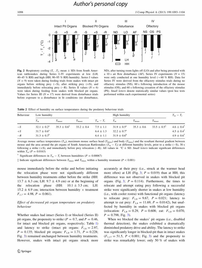
temperature around the pit organ (t6 = 7.18, P = 0.0037;
Fig. 1). However, Ta and Ts remained unchanged between
humidity levels (Ta: t6 = -1.196, P = 0.277; Ts:
t6 = 0.47, P = 0.652) and, therefore, the Tr -Ts decreased
(i.e, was more negative) at low humidity (Fig. 2). This was
true whether snakes had intact (F1,12 = 81.96,
P = 0.0001) or blocked pit organs (F1,27 = 106.33,
P \ 0.001). However, there was no change in Tr - Ts
between behavioural events (i.e., -S, ?S and -R) in either
the intact (Series I) or the blocked pit (Series II) treatment
(F2,12 = 0.65, P = 0.538 and F2,27 = 0.40, P = 0.676,
respectively; Fig. 2).
We also observed a larger thermal gradient between the
rodent prey and the area surrounding the pit organs at low
than at high humidity (F1,12 = 24.01, P = 0.003; Table 2).
This was caused by the decrease in rostral temperature of
the snake since Tm was unaffected by humidity
(F1,12 = 1.77, P = 0.239; Table 2). However, Tm fell
significantly from the time immediately before the strike to
the time when snakes attempted feeding (F2,12 = 7.036,
P = 0.00951; Table 2), but was not affected by humidity.
Maximum mice head surface temperature was *2 �C
higher than maximum body surface temperature before the
strike (Tmh = 35.3; Tmb = 33.3; F1,7, P \ 0.001; Table 2).
There were no significant differences in either maximum
head or body temperatures between humidity treatments
(P = 0.387). The distance between the snake and the
a b
c d
27.6
26.8
26.0
27.2
26.4
25.6
24.0
24.4
24.8
25.2
28.0
°C
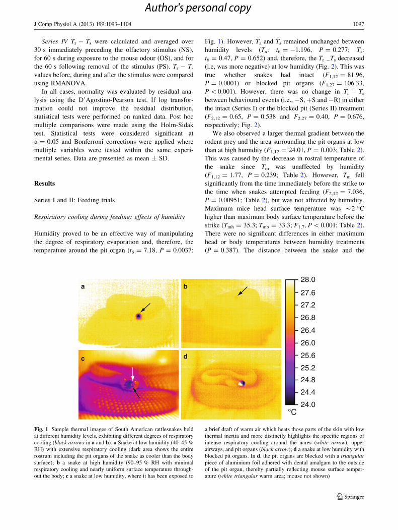
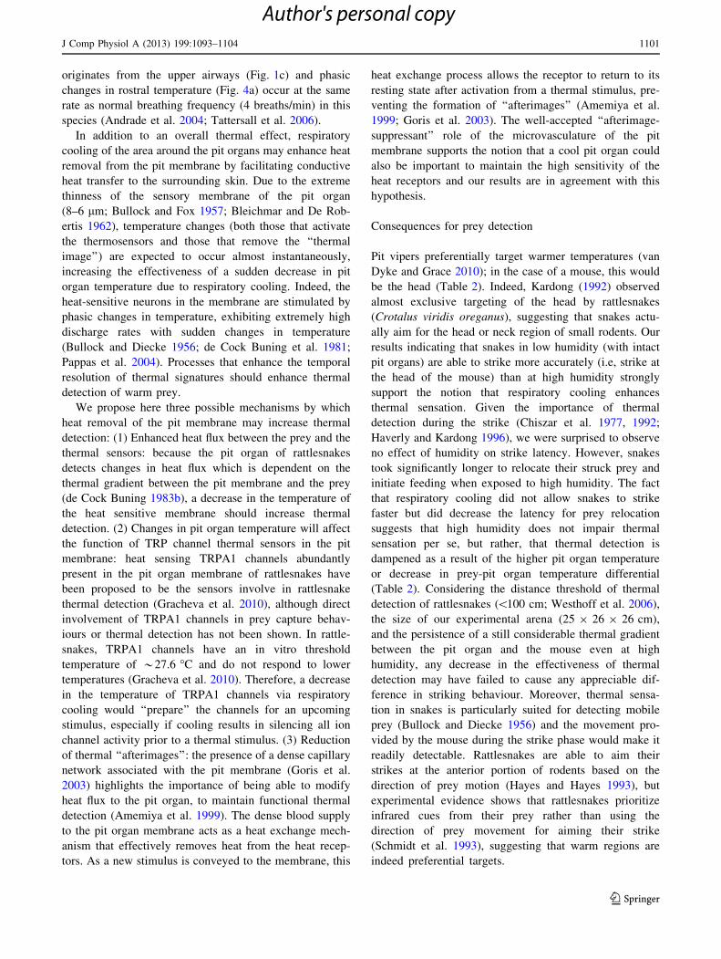
Fig. 1 Sample thermal images of South American rattlesnakes held
at different humidity levels, exhibiting different degrees of respiratory
cooling (black arrows in a and b). a Snake at low humidity (40–45 %
RH) with extensive respiratory cooling (dark area shows the entire
rostrum including the pit organs of the snake as cooler than the body
surface); b a snake at high humidity (90–95 % RH with minimal
respiratory cooling and nearly uniform surface temperature through-
out the body; c a snake at low humidity, where it has been exposed to
a brief draft of warm air which heats those parts of the skin with low
thermal inertia and more distinctly highlights the specific regions of
intense respiratory cooling around the nares (white arrow), upper
airways, and pit organs (black arrow); d a snake at low humidity with
blocked pit organs. In d, the pit organs are blocked with a triangular
piece of aluminium foil adhered with dental amalgam to the outside
of the pit organ, thereby partially reflecting mouse surface temper-
ature (white triangular warm area; mouse not shown)
J Comp Physiol A (2013) 199:1093–1104 1097
123
Author's personal copy
Page 6
mouse immediately before the strike and before initiating
the relocation phase were not significantly different
between humidity treatments either before the strike (HH:
13.7 ± 6.3 cm; LH: 9.7 ± 4.9 cm) or at the beginning of
the relocation phase (HH: 10.1 ± 3.5 cm; LH:
15.2 ± 6.9 cm; interaction between humidity 9 treatment
F1,7 = 4.98, P = 0.061).
Effect of decreased pit organ temperature on predatory
behaviour
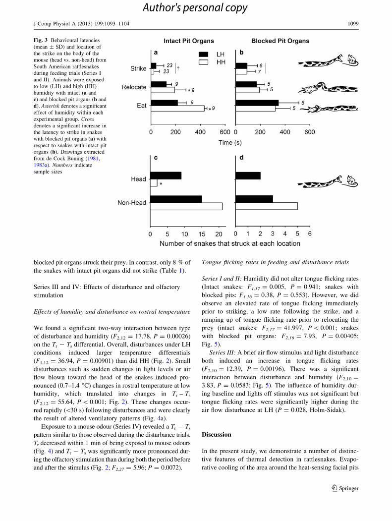
Whether snakes had intact (Series I) or blocked (Series II)
pit organs, the propensity to strike (P = 0.7, and P = 0.46,
for intact and blocked pit organs, respectively; Table 1)
and latency to strike (intact pit organs: F1,6 = 2.97,
P = 0.135; blocked pit organs: F1,8 = 1.71, P = 0.228;
Fig. 3) remained unchanged between humidity treatments.
However, snakes with intact pit organs struck more
accurately at their prey (i.e., struck at the warmer head
more often) at LH (Fig. 3; P = 0.019) than at HH; this
difference was not observed in snakes with blocked pit
organs (Fig. 3; P = 0.114). Furthermore, the times to
relocate and attempt eating prey following a successful
strike were significantly shorter in snakes at low humidity
(i.e., with cooler rostra) with functional pit organs (latency
to relocate prey: F1,6 = 9.67, P = 0.021; latency to
attempt to eat prey: F1,6 = 11.69, P = 0.0142), but unaf-
fected by humidity in snakes with blocked pit organs
(relocation: F1,8 = 0.29, P = 0.608; eat: F1,8 = 0.070,
P = 0.798; Fig. 3).
When we blocked the snakes’ pit organs (i.e., disabled
thermal detection), the snakes exhibited a dramatically
diminished predatory drive and ability. The latency to strike
was significantly longer in blocked-pit than in intact snakes
(F1,17 = 51.5, P \ 0.001; Fig. 3) and the propensity to
strike was remarkably lower; only 50 % of snakes with
Tr -
Ts (
oC
)
-4
-3
-2
-1
0
aa a
b b b
Intact Pit Organs Blocked Pit Organs
cc c
d d d
LH HH
e
Disturbance
f g
h h h
NS OS PS
Olfactory
ij
i
-S +S -R -S +S -R
I II III IV
ND LO AF
Fig. 2 Respiratory cooling (Tr -Ts; mean ± SD) from South Amer-
ican rattlesnakes during Series I–IV experiments at low (LH;
40–45 % RH) and high (HH; 90–95 % RH) humidity. Series I values
(N = 9) were taken during feeding trials from snakes with intact pit
organs before striking prey (-S), after striking prey (?S), and
immediately before relocating prey (-R). Series II values (N = 6)
were taken during feeding from snakes with blocked pit organs.
Values for Series III (N = 17) were derived from disturbance trials
before exposure to a disturbance in lit conditions (no disturbance,
ND), after turning room lights off (LO) and after being presented with
a 10 s air flow disturbance (AF). Series IV experiments (N = 15)
were only conducted at one humidity level (*60 % RH). Data for
Series IV were derived from the olfactory stimulus trials during no
olfactory stimulus (NS), 60 s following introduction of the mouse
stimulus (OS), and 60 s following cessation of the olfactory stimulus
(PS). Small letters denote statistically similar values (post hoc tests
performed within each experimental series)
Table 2 Effect of humidity on surface temperatures during the predatory behaviour trials
Behaviour Low humidity High humidity Tm - Tr
Tm Thmax Tbmax Tm - Tr Tm Thmax Tbmax
-S 32.1 ± 0.2a 35.3 ± 0.6� 33.2 ± 0.4 7.5 ± 1.1 31.9 ± 0.5a 35.3 ± 0.6 33.5 ± 0.5� 4.6 ± 0.4�
?S 31.7 ± 0.6a 6.4 ± 1.3 32.2 ± 0.7a 4.5 ± 0.4�
-R 31.3 ± 0.3b 6.4 ± 1.1 31.9 ± 0.6b 4.9 ± 0.6�
Average mouse surface temperatures (Tm), maximum mouse surface head (Thmax) and body (Tbmax) and the resultant thermal gradient between
mouse and the area around the pit organs of South American Rattlesnakes (Tm - Tr) at different humidity levels, prior to a strike (-S), 30 s
following a strike (?S), and immediately before prey relocation (-R). All values in �C ± SD. Small letters indicate significant differences
within Tm (P = 0.0163)� Significant differences in Tm - Tr between humidities (P = 0.00067)
� Indicate significant differences between Thmax and Tbmax within a humidity treatment (P \ 0.001)
1098 J Comp Physiol A (2013) 199:1093–1104
123
Author's personal copy
Page 7
blocked pit organs struck their prey. In contrast, only 8 % of
the snakes with intact pit organs did not strike (Table 1).
Series III and IV: Effects of disturbance and olfactory
stimulation
Effects of humidity and disturbance on rostral temperature
We found a significant two-way interaction between type
of disturbance and humidity (F2,12 = 17.78, P = 0.00026)
on the Tr - Ts differential. Overall, disturbances under LH
conditions induced larger temperature differentials
(F1,12 = 36.94, P = 0.00901) than did HH (Fig. 2). Small
disturbances such as sudden changes in light levels or air
flow blown toward the head of the snakes induced pro-
nounced (0.7–1.4 �C) changes in rostral temperature at low
humidity, which translated into changes in Tr – Ts
(F2,12 = 55.64, P \ 0.001; Fig. 2). These changes occur-
red rapidly (\30 s) following disturbances and were clearly
the result of altered ventilatory patterns (Fig. 4a).
Exposure to a mouse odour (Series IV) revealed a Tr - Ts
pattern similar to those observed during the disturbance trials.
Tr decreased within 1 min of being exposed to mouse odours
(Fig. 4) and Tr - Ts was significantly more pronounced dur-
ing the olfactory stimulation than during both the period before
and after the stimulus (Fig. 2; F2,27 = 5.96; P = 0.0072).
Tongue flicking rates in feeding and disturbance trials
Series I and II: Humidity did not alter tongue flicking rates
(Intact snakes: F1,17 = 0.005, P = 0.941; snakes with
blocked pits: F1,16 = 0.38, P = 0.553). However, we did
observe an elevated rate of tongue flicking immediately
prior to striking, a low rate following the strike, and a
ramping up of tongue flicking rate prior to relocating the
prey (intact snakes: F2,17 = 41.997, P \ 0.001; snakes
with blocked pit organs: F2,16 = 7.93, P = 0.00405;
Fig. 5).
Series III: A brief air flow stimulus and light disturbance
both induced an increase in tongue flicking rates
(F2,10 = 12.39, P = 0.00196). There was a significant
interaction between disturbance and humidity (F2,10 =
3.83, P = 0.0583; Fig. 5). The influence of humidity dur-
ing baseline and lights off stimulus was not significant but
tongue flicking rates were significantly higher during the
air flow disturbance at LH (P = 0.028, Holm-Sidak).
Discussion
In the present study, we demonstrate a number of distinc-
tive features of thermal detection in rattlesnakes. Evapo-
rative cooling of the area around the heat-sensing facial pits
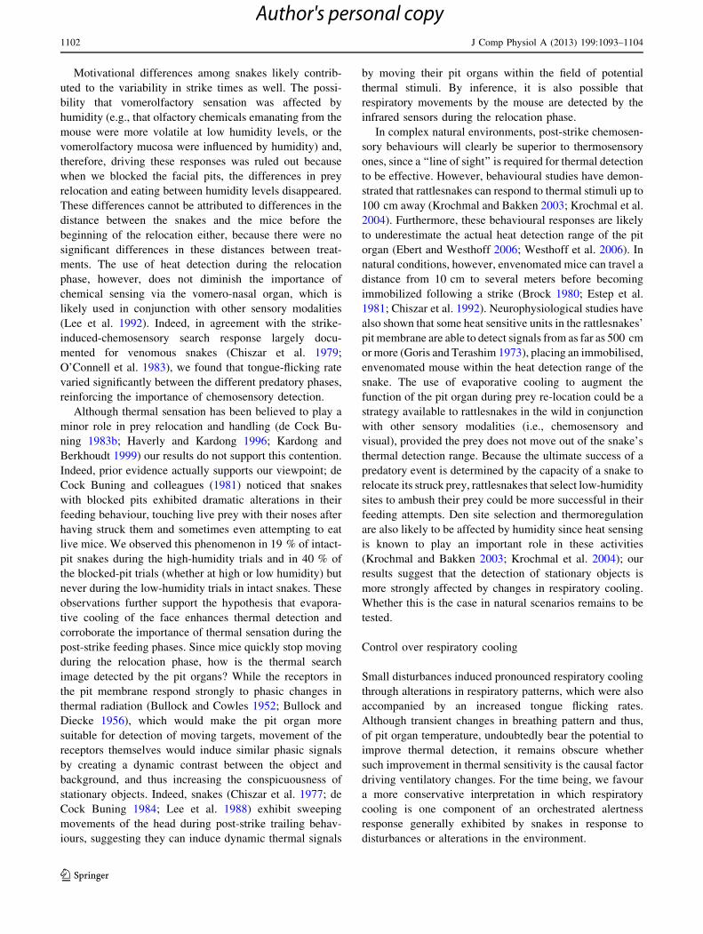
Fig. 3 Behavioural latencies
(mean ± SD) and location of
the strike on the body of the
mouse (head vs. non-head) from
South American rattlesnakes
during feeding trials (Series I
and II). Animals were exposed
to low (LH) and high (HH)
humidity with intact (a and
c) and blocked pit organs (b and
d). Asterisk denotes a significant
effect of humidity within each
experimental group. Cross
denotes a significant increase in
the latency to strike in snakes
with blocked pit organs (a) with
respect to snakes with intact pit
organs (b). Drawings extracted
from de Cock Buning (1981,
1983a). Numbers indicate
sample sizes
J Comp Physiol A (2013) 199:1093–1104 1099
123
Author's personal copy
Page 8
enhances some aspects of endothermic prey detection, and
by inference, thermal detection. This was evident from the
more accurate strikes and from the shorter latency of the
post-strike behaviours in snakes with increased rostral
cooling (i.e., in the area around the pit organs). It follows,
then, that thermal sensation, a sense that is integrated with
the visual system, may contribute to the post-strike
searching behaviour of stationary prey objects, a behaviour
that has traditionally been thought to be mediated primarily
by vomerolfactory sensation (Chiszar et al. 1977; Chiszar
et al. 1992; Haverly and Kardong 1996). We have provided
evidence that C. durissus alters the degree of respiratory
cooling by changing the depth or duration of breathing, as
part of a behavioural ‘alertness’ response; this response
provides for greater cooling of the region around the pit
organs and may augment or at least influence the animal’s
thermal detection. In this manner, South American rattle-
snakes are able to capitalize upon a phenomenon (i.e.,
respiratory cooling) that is common among vertebrates
(Robertshaw 2006; Tattersall et al. 2006) to increase their
prey capture ability.
Effect of rostral cooling on thermal sensation
Our results demonstrate that the area around the upper
airways and the pit organs can be cooled up to 2.6 �C
below the surface temperature of the rest of the body. This
was clearly the result of respiratory evaporation (Borrell
et al. 2005; Tattersall et al. 2006) because an increase in
environmental humidity diminished the magnitude of ros-
tral cooling (Figs. 1, 2, 3, 4). Furthermore, evaporation
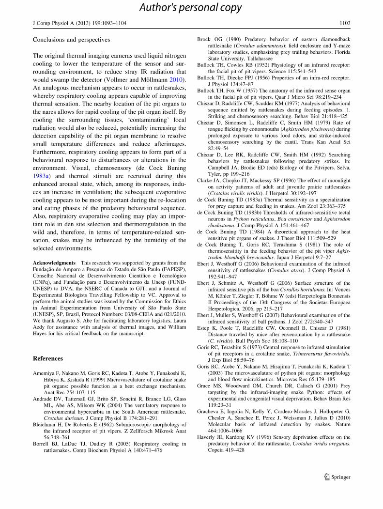
0 30 60 90 120
Tr -
Ts (
oC
)
-3.0
-2.5
-2.0
-1.5
-1.0
-0.5HH
LH
0 100 200 300 400 500 600
Tem
pera
ture
(oC
)
24
25
26
27
28
a
b
Ts
Tr
Air flowMouse
Lights off
Time (s)0 60 120 180 240 300 360
Tem
pera
ture
(oC
)
23
24
25
26
c
Ts
Tn
LO AF
Introductionof mouse odour
Removal of mouse odour
Fig. 4 Time course changes in rostral temperature of South Amer-
ican rattlesnakes held at low (LH) and high (HH) humidity during
disturbance and olfactory stimulation trials. a Sample trace from one
snake at low humidity during Series III (disturbance trials) showing
body surface (Ts; black line) and rostral surface temperature (Tr; dark
grey line) fluctuating in synchrony with inspiration (arrows), and
decreasing moderately when room light was turned off (LO; dotted
vertical line at 0 s) and quite severely when a brief (10 s) air flow
stimulus was presented (AF; dashed vertical line at 260 s). b Average
(N = 17) respiratory cooling responses (Tr - Ts) for snakes when the
room light was turned off (black line), when an air flow stimulus was
presented (dark grey line), and during the feeding trials when a mouse
was presented (light grey line). c Average (N = 15) time course
changes in nose temperature (Tr; grey line) and body surface
temperature (Ts; black line) of South American rattlesnakes during
Series III experiments before, during (0 s) and after removal of a
mouse olfactory stimulus (180 s)
ND LO AF
Ton
gue
flick
rat
e (
s-1)
0.0
0.2
0.4
0.6
0.8
1.0
1.2
1.4
-S +S -R0.0
0.2
0.4
0.6
0.8
1.0
1.2
1.4
-S +S -R0.0
0.2
0.4
0.6
0.8
1.0
1.2
1.4
y
x
x
y
x x
x
y
xx
y
x
Time (s)-40 -20 R20 40 60-10 -5 S
Ton
gue
flick
rat
e (
s-1)
0.0
0.2
0.4
0.6
0.8
1.0
1.2
1.4
ba
dc
xx x x
y
x
LH
HH
Fig. 5 Tongue flicking rates in South American rattlesnakes exposed
to low (LH) and high (HH) humidity. Snakes with intact pit organs
(Series I; N = 17) are shown in a and c (expanded time course;
S denotes the time of strike and R denotes time of prey relocation),
before striking (-S), after striking (?S), and before finding prey
(-R). Snakes with blocked pit organs (Series II; N = 5) are shown in
b. d Shows tongue flicking rates for snakes during the disturbance
trials (Series III) under control lighted conditions (LO), following
lights off (LO), and following an air flow stimulus (AF). Small letters
denote statistically similar effects of humidity within an experimental
series
1100 J Comp Physiol A (2013) 199:1093–1104
123
Author's personal copy
Page 9
originates from the upper airways (Fig. 1c) and phasic
changes in rostral temperature (Fig. 4a) occur at the same
rate as normal breathing frequency (4 breaths/min) in this
species (Andrade et al. 2004; Tattersall et al. 2006).
In addition to an overall thermal effect, respiratory
cooling of the area around the pit organs may enhance heat
removal from the pit membrane by facilitating conductive
heat transfer to the surrounding skin. Due to the extreme
thinness of the sensory membrane of the pit organ
(8–6 lm; Bullock and Fox 1957; Bleichmar and De Rob-
ertis 1962), temperature changes (both those that activate
the thermosensors and those that remove the ‘‘thermal
image’’) are expected to occur almost instantaneously,
increasing the effectiveness of a sudden decrease in pit
organ temperature due to respiratory cooling. Indeed, the
heat-sensitive neurons in the membrane are stimulated by
phasic changes in temperature, exhibiting extremely high
discharge rates with sudden changes in temperature
(Bullock and Diecke 1956; de Cock Buning et al. 1981;
Pappas et al. 2004). Processes that enhance the temporal
resolution of thermal signatures should enhance thermal
detection of warm prey.
We propose here three possible mechanisms by which
heat removal of the pit membrane may increase thermal
detection: (1) Enhanced heat flux between the prey and the
thermal sensors: because the pit organ of rattlesnakes
detects changes in heat flux which is dependent on the
thermal gradient between the pit membrane and the prey
(de Cock Buning 1983b), a decrease in the temperature of
the heat sensitive membrane should increase thermal
detection. (2) Changes in pit organ temperature will affect
the function of TRP channel thermal sensors in the pit
membrane: heat sensing TRPA1 channels abundantly
present in the pit organ membrane of rattlesnakes have
been proposed to be the sensors involve in rattlesnake
thermal detection (Gracheva et al. 2010), although direct
involvement of TRPA1 channels in prey capture behav-
iours or thermal detection has not been shown. In rattle-
snakes, TRPA1 channels have an in vitro threshold
temperature of *27.6 �C and do not respond to lower
temperatures (Gracheva et al. 2010). Therefore, a decrease
in the temperature of TRPA1 channels via respiratory
cooling would ‘‘prepare’’ the channels for an upcoming
stimulus, especially if cooling results in silencing all ion
channel activity prior to a thermal stimulus. (3) Reduction
of thermal ‘‘afterimages’’: the presence of a dense capillary
network associated with the pit membrane (Goris et al.
2003) highlights the importance of being able to modify
heat flux to the pit organ, to maintain functional thermal
detection (Amemiya et al. 1999). The dense blood supply
to the pit organ membrane acts as a heat exchange mech-
anism that effectively removes heat from the heat recep-
tors. As a new stimulus is conveyed to the membrane, this
heat exchange process allows the receptor to return to its
resting state after activation from a thermal stimulus, pre-
venting the formation of ‘‘afterimages’’ (Amemiya et al.
1999; Goris et al. 2003). The well-accepted ‘‘afterimage-
suppressant’’ role of the microvasculature of the pit
membrane supports the notion that a cool pit organ could
also be important to maintain the high sensitivity of the
heat receptors and our results are in agreement with this
hypothesis.
Consequences for prey detection
Pit vipers preferentially target warmer temperatures (van
Dyke and Grace 2010); in the case of a mouse, this would
be the head (Table 2). Indeed, Kardong (1992) observed
almost exclusive targeting of the head by rattlesnakes
(Crotalus viridis oreganus), suggesting that snakes actu-
ally aim for the head or neck region of small rodents. Our
results indicating that snakes in low humidity (with intact
pit organs) are able to strike more accurately (i.e, strike at
the head of the mouse) than at high humidity strongly
support the notion that respiratory cooling enhances
thermal sensation. Given the importance of thermal
detection during the strike (Chiszar et al. 1977, 1992;
Haverly and Kardong 1996), we were surprised to observe
no effect of humidity on strike latency. However, snakes
took significantly longer to relocate their struck prey and
initiate feeding when exposed to high humidity. The fact
that respiratory cooling did not allow snakes to strike
faster but did decrease the latency for prey relocation
suggests that high humidity does not impair thermal
sensation per se, but rather, that thermal detection is
dampened as a result of the higher pit organ temperature
or decrease in prey-pit organ temperature differential
(Table 2). Considering the distance threshold of thermal
detection of rattlesnakes (\100 cm; Westhoff et al. 2006),
the size of our experimental arena (25 9 26 9 26 cm),
and the persistence of a still considerable thermal gradient
between the pit organ and the mouse even at high
humidity, any decrease in the effectiveness of thermal
detection may have failed to cause any appreciable dif-
ference in striking behaviour. Moreover, thermal sensa-
tion in snakes is particularly suited for detecting mobile
prey (Bullock and Diecke 1956) and the movement pro-
vided by the mouse during the strike phase would make it
readily detectable. Rattlesnakes are able to aim their
strikes at the anterior portion of rodents based on the
direction of prey motion (Hayes and Hayes 1993), but
experimental evidence shows that rattlesnakes prioritize
infrared cues from their prey rather than using the
direction of prey movement for aiming their strike
(Schmidt et al. 1993), suggesting that warm regions are
indeed preferential targets.
J Comp Physiol A (2013) 199:1093–1104 1101
123
Author's personal copy
Page 10
Motivational differences among snakes likely contrib-
uted to the variability in strike times as well. The possi-
bility that vomerolfactory sensation was affected by
humidity (e.g., that olfactory chemicals emanating from the
mouse were more volatile at low humidity levels, or the
vomerolfactory mucosa were influenced by humidity) and,
therefore, driving these responses was ruled out because
when we blocked the facial pits, the differences in prey
relocation and eating between humidity levels disappeared.
These differences cannot be attributed to differences in the
distance between the snakes and the mice before the
beginning of the relocation either, because there were no
significant differences in these distances between treat-
ments. The use of heat detection during the relocation
phase, however, does not diminish the importance of
chemical sensing via the vomero-nasal organ, which is
likely used in conjunction with other sensory modalities
(Lee et al. 1992). Indeed, in agreement with the strike-
induced-chemosensory search response largely docu-
mented for venomous snakes (Chiszar et al. 1979;
O’Connell et al. 1983), we found that tongue-flicking rate
varied significantly between the different predatory phases,
reinforcing the importance of chemosensory detection.
Although thermal sensation has been believed to play a
minor role in prey relocation and handling (de Cock Bu-
ning 1983b; Haverly and Kardong 1996; Kardong and
Berkhoudt 1999) our results do not support this contention.
Indeed, prior evidence actually supports our viewpoint; de
Cock Buning and colleagues (1981) noticed that snakes
with blocked pits exhibited dramatic alterations in their
feeding behaviour, touching live prey with their noses after
having struck them and sometimes even attempting to eat
live mice. We observed this phenomenon in 19 % of intact-
pit snakes during the high-humidity trials and in 40 % of
the blocked-pit trials (whether at high or low humidity) but
never during the low-humidity trials in intact snakes. These
observations further support the hypothesis that evapora-
tive cooling of the face enhances thermal detection and
corroborate the importance of thermal sensation during the
post-strike feeding phases. Since mice quickly stop moving
during the relocation phase, how is the thermal search
image detected by the pit organs? While the receptors in
the pit membrane respond strongly to phasic changes in
thermal radiation (Bullock and Cowles 1952; Bullock and
Diecke 1956), which would make the pit organ more
suitable for detection of moving targets, movement of the
receptors themselves would induce similar phasic signals
by creating a dynamic contrast between the object and
background, and thus increasing the conspicuousness of
stationary objects. Indeed, snakes (Chiszar et al. 1977; de
Cock Buning 1984; Lee et al. 1988) exhibit sweeping
movements of the head during post-strike trailing behav-
iours, suggesting they can induce dynamic thermal signals
by moving their pit organs within the field of potential
thermal stimuli. By inference, it is also possible that
respiratory movements by the mouse are detected by the
infrared sensors during the relocation phase.
In complex natural environments, post-strike chemosen-
sory behaviours will clearly be superior to thermosensory
ones, since a ‘‘line of sight’’ is required for thermal detection
to be effective. However, behavioural studies have demon-
strated that rattlesnakes can respond to thermal stimuli up to
100 cm away (Krochmal and Bakken 2003; Krochmal et al.
2004). Furthermore, these behavioural responses are likely
to underestimate the actual heat detection range of the pit
organ (Ebert and Westhoff 2006; Westhoff et al. 2006). In
natural conditions, however, envenomated mice can travel a
distance from 10 cm to several meters before becoming
immobilized following a strike (Brock 1980; Estep et al.
1981; Chiszar et al. 1992). Neurophysiological studies have
also shown that some heat sensitive units in the rattlesnakes’
pit membrane are able to detect signals from as far as 500 cm
or more (Goris and Terashim 1973), placing an immobilised,
envenomated mouse within the heat detection range of the
snake. The use of evaporative cooling to augment the
function of the pit organ during prey re-location could be a
strategy available to rattlesnakes in the wild in conjunction
with other sensory modalities (i.e., chemosensory and
visual), provided the prey does not move out of the snake’s
thermal detection range. Because the ultimate success of a
predatory event is determined by the capacity of a snake to
relocate its struck prey, rattlesnakes that select low-humidity
sites to ambush their prey could be more successful in their
feeding attempts. Den site selection and thermoregulation
are also likely to be affected by humidity since heat sensing
is known to play an important role in these activities
(Krochmal and Bakken 2003; Krochmal et al. 2004); our
results suggest that the detection of stationary objects is
more strongly affected by changes in respiratory cooling.
Whether this is the case in natural scenarios remains to be
tested.
Control over respiratory cooling
Small disturbances induced pronounced respiratory cooling
through alterations in respiratory patterns, which were also
accompanied by an increased tongue flicking rates.
Although transient changes in breathing pattern and thus,
of pit organ temperature, undoubtedly bear the potential to
improve thermal detection, it remains obscure whether
such improvement in thermal sensitivity is the causal factor
driving ventilatory changes. For the time being, we favour
a more conservative interpretation in which respiratory
cooling is one component of an orchestrated alertness
response generally exhibited by snakes in response to
disturbances or alterations in the environment.
1102 J Comp Physiol A (2013) 199:1093–1104
123
Author's personal copy
Page 11
Conclusions and perspectives
The original thermal imaging cameras used liquid nitrogen
cooling to lower the temperature of the sensor and sur-
rounding environment, to reduce stray IR radiation that
would swamp the detector (Vollmer and Mollmann 2010).
An analogous mechanism appears to occur in rattlesnakes,
whereby respiratory cooling appears capable of improving
thermal sensation. The nearby location of the pit organs to
the nares allows for rapid cooling of the pit organ itself. By
cooling the surrounding tissues, ‘contaminating’ local
radiation would also be reduced, potentially increasing the
detection capability of the pit organ membrane to resolve
small temperature differences and reduce afterimages.
Furthermore, respiratory cooling appears to form part of a
behavioural response to disturbances or alterations in the
environment. Visual, chemosensory (de Cock Buning
1983a) and thermal stimuli are recruited during this
enhanced arousal state, which, among its responses, indu-
ces an increase in ventilation; the subsequent evaporative
cooling appears to be most important during the re-location
and eating phases of the predatory behavioural sequence.
Also, respiratory evaporative cooling may play an impor-
tant role in den site selection and thermoregulation in the
wild and, therefore, in terms of temperature-related sen-
sation, snakes may be influenced by the humidity of the
selected environments.
Acknowledgments This research was supported by grants from the
Fundacao de Amparo a Pesquisa do Estado de Sao Paulo (FAPESP),
Conselho Nacional de Desenvolvimento Cientıfico e Tecnologico
(CNPq), and Fundacao para o Desenvolvimento da Unesp (FUND-
UNESP) to DVA, the NSERC of Canada to GJT, and a Journal of
Experimental Biologists Travelling Fellowship to VC. Approval to
perform the animal studies was issued by the Commission for Ethics
in Animal Experimentation from University of Sao Paulo State
(UNESP), SP, Brazil, Protocol Numbers: 03/08-CEEA and 021/2010.
We thank Augusto S. Abe for facilitating laboratory logistics, Laura
Aedy for assistance with analysis of thermal images, and William
Hayes for his critical feedback on the manuscript.
References
Amemiya F, Nakano M, Goris RC, Kadota T, Atobe Y, Funakoshi K,
Hibiya K, Kishida R (1999) Microvasculature of crotaline snake
pit organs: possible function as a heat exchange mechanism.
Anat Rec 254:107–115
Andrade DV, Tattersall GJ, Brito SP, Soncini R, Branco LG, Glass
ML, Abe AS, Milsom WK (2004) The ventilatory response to
environmental hypercarbia in the South American rattlesnake,
Crotalus durissus. J Comp Physiol B 174:281–291
Bleichmar H, De Robertis E (1962) Submicroscopic morphology of
the infrared receptor of pit vipers. Z Zellforsch Mikrosk Anat
56:748–761
Borrell BJ, LaDuc TJ, Dudley R (2005) Respiratory cooling in
rattlesnakes. Comp Biochem Physiol A 140:471–476
Brock OG (1980) Predatory behavior of eastern diamondback
rattlesnake (Crotalus adamanteus): field enclosure and Y-maze
laboratory studies, emphasizing prey trailing behaviors. Florida
State University, Tallahassee
Bullock TH, Cowles RB (1952) Physiology of an infrared receptor:
the facial pit of pit vipers. Science 115:541–543
Bullock TH, Diecke FPJ (1956) Properties of an infra-red receptor.
J Physiol 134:47–87
Bullock TH, Fox W (1957) The anatomy of the infra-red sense organ
in the facial pit of pit vipers. Quar J Micro Sci 98:219–234
Chiszar D, Radcliffe CW, Scudder KM (1977) Analysis of behavioral
sequence emitted by rattlesnakes during feeding episodes. 1.
Striking and chemosensory searching. Behav Biol 21:418–425
Chiszar D, Simonsen L, Radcliffe C, Smith HM (1979) Rate of
tongue flicking by cottonmouths (Agkistrodon piscivorus) during
prolonged exposure to various food odors, and strike-induced
chemosensory searching by the cantil. Trans Kan Acad Sci
82:49–54
Chiszar D, Lee RK, Radcliffe CW, Smith HM (1992) Searching
behaviors by rattlesnakes following predatory strikes. In:
Campbell JA, Brodie ED (eds) Biology of the Pitvipers. Selva,
Tyler, pp 199–216
Clarke JA, Chopko JT, Mackessy SP (1996) The effect of moonlight
on activity patterns of adult and juvenile prairie rattlesnakes
(Crotalus viridis viridis). J Herpetol 30:192–197
de Cock Buning TD (1983a) Thermal sensitivity as a specialization
for prey capture and feeding in snakes. Am Zool 23:363–375
de Cock Buning TD (1983b) Thresholds of infrared-sensititive tectal
neurons in Python reticulatus, Boa constrictor and Agkistrodon
rhodostoma. J Comp Physiol A 151:461–467
de Cock Buning TD (1984) A theoretical approach to the heat
sensitive pit organs of snakes. J Theor Biol 111:509–529
de Cock Buning T, Goris RC, Terashima S (1981) The role of
thermosensitity in the feeding behavior of the pit viper Agkis-
trodon blomhoffi brevicaudus. Japan J Herpetol 9:7–27
Ebert J, Westhoff G (2006) Behavioural examination of the infrared
sensitivity of rattlesnakes (Crotalus atrox). J Comp Physiol A
192:941–947
Ebert J, Schmitz A, Westhoff G (2006) Surface structure of the
infrared sensitive pits of the boa Corallus hortulanus. In: Vences
M, Kohler T, Ziegler T, Bohme W (eds) Herpetologia Bonnensis
II Proceedings of the 13th Congress of the Societas Europaea
Herpetologica, 2006, pp 215–217
Ebert J, Muller S, Westhoff G (2007) Behavioural examination of the
infrared sensitivity of ball pythons. J Zool 272:340–347
Estep K, Poole T, Radcliffe CW, Oconnell B, Chiszar D (1981)
Distance traveled by mice after envenomation by a rattlesnake
(C. viridis). Bull Psych Soc 18:108–110
Goris RC, Terashim S (1973) Central response to infrared stimulation
of pit receptors in a crotaline snake, Trimeresurus flavoviridis.
J Exp Biol 58:59–76
Goris RC, Atobe Y, Nakano M, Hisajima T, Funakoshi K, Kadota T
(2003) The microvasculature of python pit organs: morphology
and blood flow microkinetics. Microvas Res 65:179–185
Grace MS, Woodward OM, Church DR, Calisch G (2001) Prey
targeting by the infrared-imaging snake Python: effects of
experimental and congenital visual deprivation. Behav Brain Res
119:23–31
Gracheva E, Ingolia N, Kelly Y, Cordero-Morales J, Hollopeter G,
Chesler A, Sanchez E, Perez J, Weissman J, Julius D (2010)
Molecular basis of infrared detection by snakes. Nature
464:1006–1066
Haverly JE, Kardong KV (1996) Sensory deprivation effects on the
predatory behavior of the rattlesnake, Crotalus viridis oreganus.
Copeia 419–428
J Comp Physiol A (2013) 199:1093–1104 1103
123
Author's personal copy
Page 12
Hayes WK, Hayes DM (1993) Stimuli influencing the release and aim
of predatory strikes of the Northern Pacific Rattlesnake, Crotalus
v. viridis. Northwestern Naturalist 74:1–9
Kardong KV (1992) Proximate factors affecting guidance of the
rattlesnake strike. Zool Jahrb Anat 122:233–244
Kardong KV, Berkhoudt H (1999) Rattlesnake hunting behavior:
correlations between plasticity of predatory performance and
neuroanatomy. Brain Behav Evol 53:20–28
Kardong KV, Mackessy SP (1991) The strike behavior of a
congenitally blind rattlesnake. J Herpetol 25:208–211
Krochmal AR, Bakken GS (2003) Thermoregulation is the pits: use of
thermal radiation for retreat site selection by rattlesnakes. J Exp
Biol 206:2539–2545
Krochmal AR, Bakken GS, LaDuc TJ (2004) Heat in evolution’s
kitchen: evolutionary perspectives on the functions and origin of
the facial pit of pitvipers (Viperidae:Crotalinae). J Exp Biol
207:4231–4238
Lee RKK, Chiszar DA, Smith HM (1988) Post-strike orientation of
the prairie rattlesnake facilitates location of envenomated prey.
J Ethol 6:129–134
Lee RKK, Chiszar DA, Smith HM, Kandler K (1992) Chemical and
orientational cues mediate selection of prey trails by Prairie
Rattlesnakes (Crotalus viridis). J Herpetol 26:95–98
Molenaar GJ (1992) Anatomy and physiology of infrared sensitivity
of snakes. In: Gans C, Ulinski PS (eds) Biology of the Reptilia,
vol 17. University of Chicago Press, Chicago, pp 367–453
O’Connell B, Chiszar D, Smith HM (1983) Strike-induced chemo-
sensory searching in prairie rattlesnakes (Crotalus viridis) during
daytime and at night. J Herpetol 17:193–196
Pappas TC, Motamedi M, Christensen BN (2004) Unique tempera-
ture-activated neurons from pit viper thermosensors. Am J
Physiol Cell Physiol 287:C1219–C1228
Robertshaw D (2006) Mechanisms for the control of respiratory
evaporative heat loss in panting animals. J Appl Physiol
101:664–668
Schmidt DF, Hayes WK, Hayes FE (1993) The influence of prey
movement on the aim of predatory strikes in the rattlesnake,
Crotalus viridis. Great Basin Naturalist 53:203–206
Tattersall GJ, Milsom WK, Abe AS, Brito SP, Andrade DV (2004)
The thermogenesis of digestion in rattlesnakes. J Exp Biol
207:579–585
Tattersall GJ, Cadena V, Skinner MC (2006) Respiratory cooling and
thermoregulatory coupling in reptiles. Resp Physiol Neurobiol
154:302–318
Van Dyke JU, Grace MS (2010) The role of thermal contrast in
infrared-based defensive targeting by the copperhead, Agkistro-
don contortrix. Anim Behav 79:993–999
Vollmer M, Mollmann K-P (2010) Infrared thermal imaging funda-
mentals, research and applications. Wiley-VCH, Weinheim
Westhoff G, Morsch M, Ebert J (2006) Infrared detection in the
rattlesnake Crotalus atrox—from behavioural studies to mid-
brain recordings. In: Vences M, Kohler T, Ziegler T, Bohme W
(eds) Herpetologia Bonnensis II Proceedings of the 13th
Congress of the Societas Europaea Herpetologica, pp 225–228
1104 J Comp Physiol A (2013) 199:1093–1104
123
Author's personal copy