52
iMaging 5.0: Our Newest Radiology Operating System Unleashed Kenneth L. Pierce, M.D. Associate Professor Stritch School of Medicine
| Date post: | 16-Dec-2015 |
| Category: |
Documents |
| Upload: | elmer-dickerson |
| View: | 214 times |
| Download: | 0 times |

iMaging 5.0: Our Newest Radiology Operating System
Unleashed
Kenneth L. Pierce, M.D.
Associate Professor
Stritch School of Medicine

iMaging 5.0: What’s New?
•PACS
•Consultants
•Modalities
•Protocols
•HIPAA

PACS•picture archiving and
communication systems
•replaces hard-copy based means of managing medical images
•‘filmless’
•off-site viewing/interpretation
•data storage vs. fileroom


PACS•Workstations in the main department
•3MP resolution
•In OR
•Web-based browsers
•On PCs throughout hospital/clinics
•Available on home PC thru VPN
•CDs of studies are available in file room

PACS vs Film
•Advantages/ Disadvantages
•Storage
•Access
•Cost
•Security

Physics
• Xray imaging
• Shoot electrons at tungsten target
• Emit xrays (photons)
• Directed at object/ detector

Physics
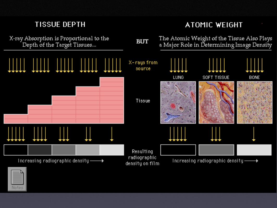
• Some of the photons absorbed by patient
• Photons that penetrate patient strike detector
• Different tissues have different xray absorption - contrast

Helical CT

MR Basics• Hydrogen proton
imaging
• Observe behavior of protons in magnet after application of RF signal
• Unsurpassed contrast resolution, spatial resolution limited
• Time consuming, costly
• Contraindications?

Ultrasound•1 to 10 MHz frequency/ 1.5mm
wavelength
•speed determined by tissue
•different tissues(impedance)->different speed->reflection
•time for echo to travel back to probe used to calculate depth of tissue interface causing echo

Doppler Ultrasound
• apparent change in frequency or wavelength of a wave that is perceived by an observer moving relative to the source of the waves
•rbc’s move away or towards the transducer
•measuring frequency shift of a particular sample blood volume determines speed and direction

Nuclear Medicine
• uses unsealed radioactive substances in diagnosis and therapy
• differ from most other imaging modalities in that the tests show the function of the system being investigated as opposed to the anatomy
• majority of diagnostic tests involve formation of an image using gamma camera
• Most diagnostic radionuclides emit gamma rays

Nuclear Medicine• The most commonly used radionuclides
are:
• technetium-99m
• iodine-123 and 131
• thallium-201
• gallium-67
• PET - metabolically active molecule (sugar)
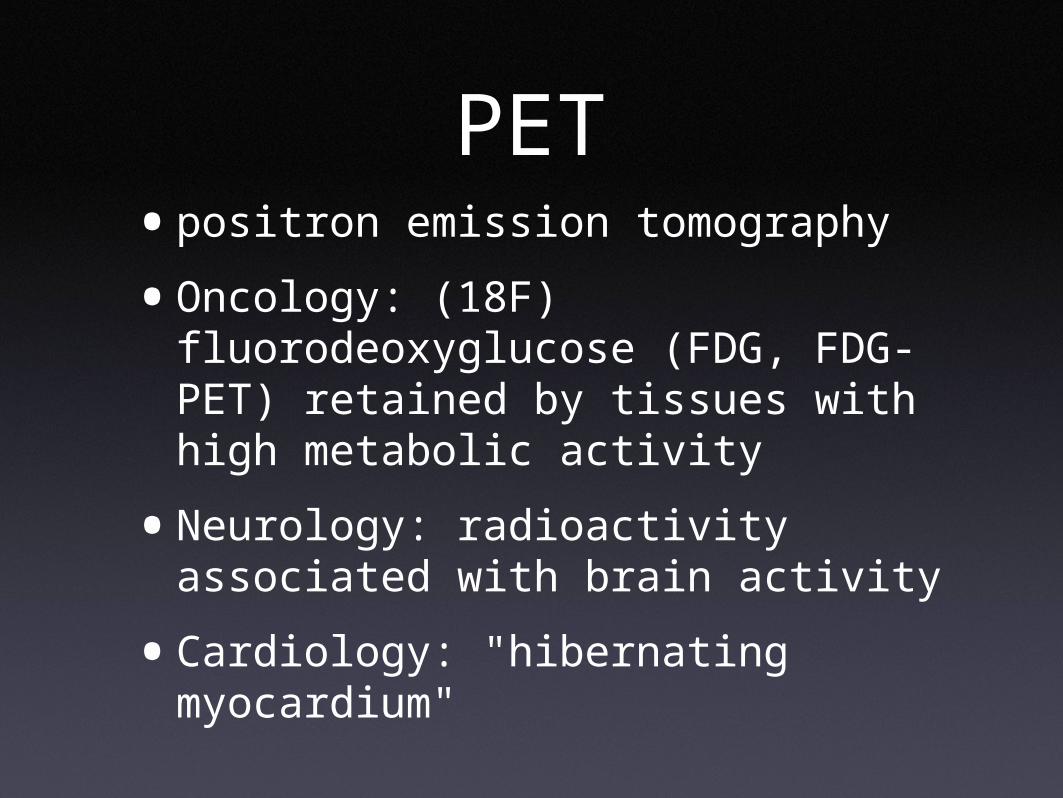
PET•positron emission tomography
•Oncology: (18F) fluorodeoxyglucose (FDG, FDG-PET) retained by tissues with high metabolic activity
•Neurology: radioactivity associated with brain activity
•Cardiology: "hibernating myocardium"

Interventional Radiology
•Vascular Diagnosis- Arteriography- Venography- Lymphangiography
•Vascular Intervention- Angioplasty/stents- Embolization- Filters- Chemoembo
• Venous access
•Non Vascular Intervention- Regional tumor therapy- Biopsy- Drainage- Biliary
• Urological

Radiologist as Consultant
•We’re not ‘technologists’
•Offer advice re:
•Exam indication
•Procedures
•Interpretation
•Conferences

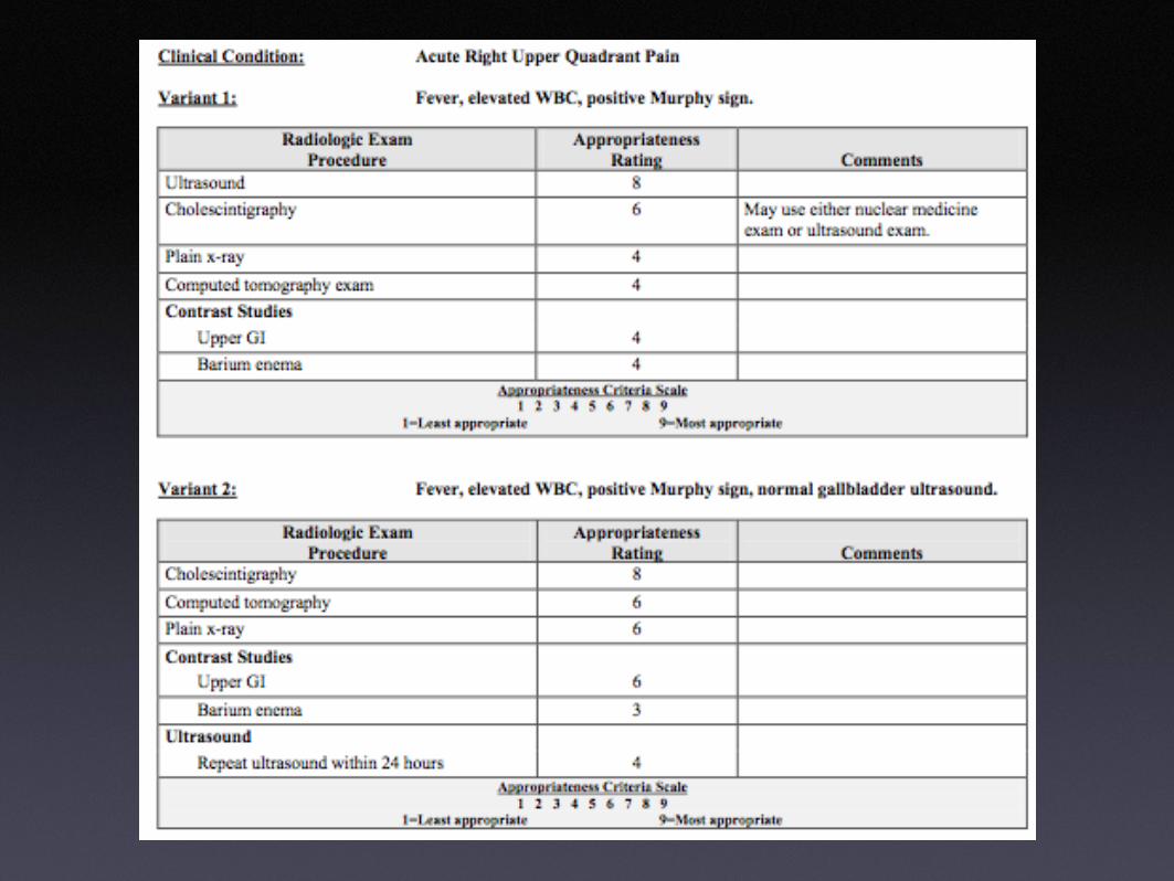
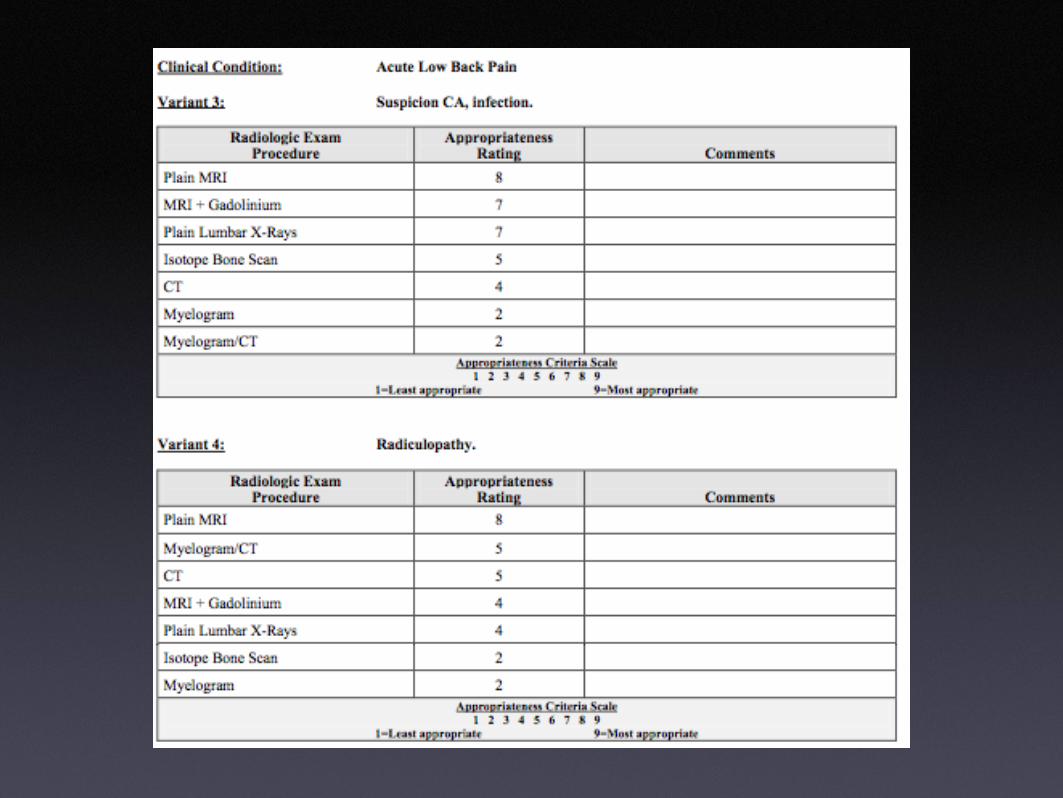
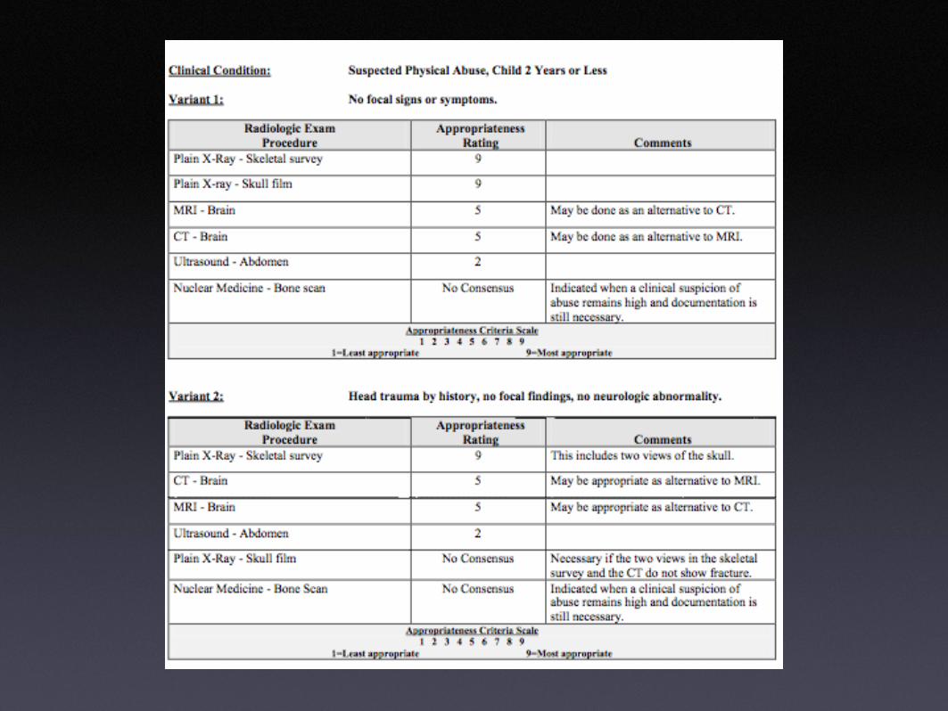
PROTOCOL
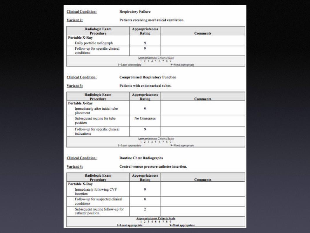
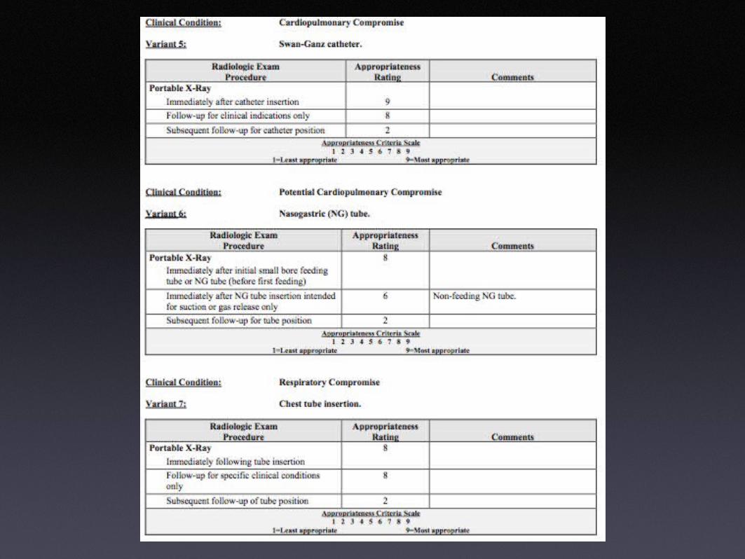
•ACR appropriateness criteria
•Available at acr.org
•Not perfect, but helpful
•Not followed


Risk Management
•Radiation safety
•Allergic reactions
•Medical emergencies and treatment issues
•Diagnostic issues
•Competency

HIPAA
• Health Insurance Portability and Accountability Act
• communications networks that link radiology information systems, billing software, and image transmission technology (PACS/teleradiology). hospital demographic downloads, electronic claims submission and remittance, and remote referring physician (reports and images) or patient access (billing records) to information via a web site.

Contrast Reaction•not caused by iodine
•not related to shellfish
•not true allergy (no drug-antibody)
•mechanism remains unknown
•unpredictable
•dose independent
•prevalence 1-2% (0.04 - 0.22% severe)
•fatal 1 in 75,000

Contrast Reaction - Premedication
•Prednisone 50 mg P.O, 13 hours before test
•Prednisone 50mg P.O, 7 hours before test
•Prednisone 50mg P.O, 1 hour before test plus Benadryl 50 mg P.O, 1 hour before test.

Renal Toxicity • serum creatinine up more than 25% or >
0.5 mg%
• Risk Factors
• 5 - 10 fold increase with pre-existing renal insufficiency (increased creatinine)
• Dehydration
• CHF
• Age > 70
• nephrotoxic drugs

Renal Toxicity
•direct relationship between serum creatinine and likelihood nephrotoxicity
•Hydrate 100 ml/hr Normal saline 4 hrs prior to procedure, continue for 24 hours
•Those on hemodialysis do not need extra seesions or dialysis immediately following contrast administration

Renal Toxicity•Metformin (Glucophage)
•oral diabetic agent
•patients with renal insufficiency may develop lactic acidosis
•withhold drug for 48 hrs after contrast administration in all patients taking this drug - restart if Cr back to baseline

iMaging 5.0
•Ready for primetime
•Easily accessible
•Integrates well with clinical work
•Free iPod for every 3rd year student
•See Dr Gruener after this lecture


