Veterinary World, EISSN: 2231-0916 664 Veterinary World, EISSN: 2231-0916 Available at www.veterinaryworld.org/Vol.12/May-2019/6.pdf RESEARCH ARTICLE Open Access Isolation and molecular characterization of Mycoplasma spp. in sheep and goats in Egypt Mounier M. Abdel Halium 1 , Fayez A. Salib 1 , S. A. Marouf 2 and Emil S. Abdel Massieh 1 1. Department of Medicine and Infectious Diseases, Faculty of Veterinary Medicine, Cairo University, Giza, Egypt; 2. Department of Microbiology and Mycology, Faculty of Veterinary Medicine, Cairo University, Giza, Egypt. Corresponding author: Emil S. Abdel Massieh, e-mail: [email protected]Co-authors: MMAH: [email protected], FAS: [email protected], SAM: [email protected]Received: 04-12-2018, Accepted: 15-03-2019, Published online: 13-05-2019 doi: 10.14202/vetworld.2019.664-670 How to cite this article: Abdel Halium MM, Salib FA, Marouf SA, Abdel Massieh ES (2019) Isolation and molecular characterization of Mycoplasma spp. in sheep and goats in Egypt, Veterinary World, 12(5): 664-670. Abstract Background and Aim: Different species of Mycoplasma are associated with many pathological problems in small ruminants including respiratory manifestation, this problem results in significant losses, especially in African countries. This study aimed to (I) study some epidemiological aspects of Mycoplasma species infections in Egyptian sheep and goats at Giza Governorate, (II) diagnosis of Mycoplasma species affections using bacterial isolation and identification, (III) apply the polymerase chain reaction (PCR) for typing of different Mycoplasma species, and (IV) illustrate the phylogenetic tree for the isolated Mycoplasma species and other species from GenBank using the purified PCR product. Materials and Methods: A total of 335 samples were collected from sheep and goats from Giza Governorate in Egypt as 142 nasal swabs from clinically affected animals, 167 pneumonic lungs, 18 samples from tracheal bifurcation, and 8 samples by bronchial wash were cultured on pleuropneumonia-like organisms (PPLOs) media for cultivation of Mycoplasma species. PCR and sequencing and phylogenetic analysis were adopted to identify and classify the isolated Mycoplasma species. Results: A total of 24 Mycoplasma isolates were isolated on PPLO media, identified by biochemical tests, and confirmed and typed by PCR using specific primers. 10 isolates were confirmed as Mycoplasma arginini, four isolates as Mycoplasma ovipneumoniae by PCR, and 10 isolates as undifferentiated Mycoplasma species. A purified isolate of M. arginini and M. ovipneumoniae was sequenced and phylogenetic analysis was illustrated. Conclusion: M. arginini and M. ovipneumoniae are prevalent in Egyptian sheep and goats. Further studies on M. arginini are required due to its high frequency of isolation from pneumonic sheep and goats and also from animals suffer from different respiratory manifestations. Keywords: goats, Mycoplasma, polymerase chain reaction, sequencing and phylogenetic analysis, sheep. Introduction Mycoplasma belongs to a group of bacteria named Mollicutes which characterized by its min- ute genome size and perpetually devoid of the cell wall [1]. Different Mycoplasma species are accompa- nying by many diseases and problems in both mam- malian and avian species [2], sheep and goats (poor man’s cow) are economically critical in many coun- tries including Egypt as consumed for meat, wool, and milk production, more than 10% of meat production in Egypt is from sheep and goats [3]. Mycoplasma infections leading to a great eco- nomic loss due to it cause high morbidity and mor- tality rates in sheep and goats populations in African countries including Egypt, European countries, and India [4]. Mycoplasma causes various clinical man- ifestations as pneumonia, conjunctivitis, arthritis, and mastitis [5]. Mycoplasma species commonly associated with pneumonia in small ruminants are Mycoplasma ovipneumoniae, Mycoplasma arginini, Mycoplasma capri, Mycoplasma capripneumoniae, and Mycoplasma capricolum [6]. M. arginini is fre- quently isolated with M. ovipneumoniae from cases of atypical pneumonia in sheep and goats [5] and other cases with lung consolidation [7]. M. arginini is also isolated from other locations such as genital organs, eyes, and ears [8]; in addition, isolation of M. arginini from pathological cases in human [9,10] gives suspi- cion that M. arginini may have zoonotic importance. In Egypt, different Mycoplasma species have been isolated including M. arginini, M. ovipneumoniae, and Mycoplasma agalactiae [11]. The respiratory problems are considered as mul- tifactorial disease where interaction between different microbial agents such as bacteria (Pasteurella and Mycoplasma), viruses (PI3, reovirus, and adenovi- rus), and fungi and also other factors as host defense mechanism and environmental factors including cli- matic conditions and stress of transportations lead to increase the incidence of those problems [12]. Mycoplasma is highly fastidious microorganism required very precise media to develop in vitro, the Copyright: Abdel Halium, et al. Open Access. This article is distributed under the terms of the Creative Commons Attribution 4.0 International License (http://creativecommons.org/licenses/ by/4.0/), which permits unrestricted use, distribution, and reproduction in any medium, provided you give appropriate credit to the original author(s) and the source, provide a link to the Creative Commons license, and indicate if changes were made. The Creative Commons Public Domain Dedication waiver (http:// creativecommons.org/publicdomain/zero/1.0/) applies to the data made available in this article, unless otherwise stated.
Transcript
Veterinary World, EISSN: 2231-0916 664
Veterinary World, EISSN: 2231-0916Available at www.veterinaryworld.org/Vol.12/May-2019/6.pdf
RESEARCH ARTICLEOpen Access
Isolation and molecular characterization of Mycoplasma spp. in sheep and goats in Egypt
Mounier M. Abdel Halium1, Fayez A. Salib1, S. A. Marouf2 and Emil S. Abdel Massieh1
1. Department of Medicine and Infectious Diseases, Faculty of Veterinary Medicine, Cairo University, Giza, Egypt;2. Department of Microbiology and Mycology, Faculty of Veterinary Medicine, Cairo University, Giza, Egypt.
Received: 04-12-2018, Accepted: 15-03-2019, Published online: 13-05-2019
doi: 10.14202/vetworld.2019.664-670 How to cite this article: Abdel Halium MM, Salib FA, Marouf SA, Abdel Massieh ES (2019) Isolation and molecular characterization of Mycoplasma spp. in sheep and goats in Egypt, Veterinary World, 12(5): 664-670.
AbstractBackground and Aim: Different species of Mycoplasma are associated with many pathological problems in small ruminants including respiratory manifestation, this problem results in significant losses, especially in African countries. This study aimed to (I) study some epidemiological aspects of Mycoplasma species infections in Egyptian sheep and goats at Giza Governorate, (II) diagnosis of Mycoplasma species affections using bacterial isolation and identification, (III) apply the polymerase chain reaction (PCR) for typing of different Mycoplasma species, and (IV) illustrate the phylogenetic tree for the isolated Mycoplasma species and other species from GenBank using the purified PCR product.
Materials and Methods: A total of 335 samples were collected from sheep and goats from Giza Governorate in Egypt as 142 nasal swabs from clinically affected animals, 167 pneumonic lungs, 18 samples from tracheal bifurcation, and 8 samples by bronchial wash were cultured on pleuropneumonia-like organisms (PPLOs) media for cultivation of Mycoplasma species. PCR and sequencing and phylogenetic analysis were adopted to identify and classify the isolated Mycoplasma species.
Results: A total of 24 Mycoplasma isolates were isolated on PPLO media, identified by biochemical tests, and confirmed and typed by PCR using specific primers. 10 isolates were confirmed as Mycoplasma arginini, four isolates as Mycoplasma ovipneumoniae by PCR, and 10 isolates as undifferentiated Mycoplasma species. A purified isolate of M. arginini and M. ovipneumoniae was sequenced and phylogenetic analysis was illustrated.
Conclusion: M. arginini and M. ovipneumoniae are prevalent in Egyptian sheep and goats. Further studies on M. arginini are required due to its high frequency of isolation from pneumonic sheep and goats and also from animals suffer from different respiratory manifestations.
Mycoplasma belongs to a group of bacteria named Mollicutes which characterized by its min-ute genome size and perpetually devoid of the cell wall [1]. Different Mycoplasma species are accompa-nying by many diseases and problems in both mam-malian and avian species [2], sheep and goats (poor man’s cow) are economically critical in many coun-tries including Egypt as consumed for meat, wool, and milk production, more than 10% of meat production in Egypt is from sheep and goats [3].
Mycoplasma infections leading to a great eco-nomic loss due to it cause high morbidity and mor-tality rates in sheep and goats populations in African countries including Egypt, European countries, and India [4]. Mycoplasma causes various clinical man-ifestations as pneumonia, conjunctivitis, arthritis,
and mastitis [5]. Mycoplasma species commonly associated with pneumonia in small ruminants are Mycoplasma ovipneumoniae, Mycoplasma arginini, Mycoplasma capri, Mycoplasma capripneumoniae, and Mycoplasma capricolum [6]. M. arginini is fre-quently isolated with M. ovipneumoniae from cases of atypical pneumonia in sheep and goats [5] and other cases with lung consolidation [7]. M. arginini is also isolated from other locations such as genital organs, eyes, and ears [8]; in addition, isolation of M. arginini from pathological cases in human [9,10] gives suspi-cion that M. arginini may have zoonotic importance. In Egypt, different Mycoplasma species have been isolated including M. arginini, M. ovipneumoniae, and Mycoplasma agalactiae [11].
The respiratory problems are considered as mul-tifactorial disease where interaction between different microbial agents such as bacteria (Pasteurella and Mycoplasma), viruses (PI3, reovirus, and adenovi-rus), and fungi and also other factors as host defense mechanism and environmental factors including cli-matic conditions and stress of transportations lead to increase the incidence of those problems [12].
Mycoplasma is highly fastidious microorganism required very precise media to develop in vitro, the
Copyright: Abdel Halium, et al. Open Access. This article is distributed under the terms of the Creative Commons Attribution 4.0 International License (http://creativecommons.org/licenses/by/4.0/), which permits unrestricted use, distribution, and reproduction in any medium, provided you give appropriate credit to the original author(s) and the source, provide a link to the Creative Commons license, and indicate if changes were made. The Creative Commons Public Domain Dedication waiver (http://creativecommons.org/publicdomain/zero/1.0/) applies to the data made available in this article, unless otherwise stated.
Veterinary World, EISSN: 2231-0916 665
Available at www.veterinaryworld.org/Vol.12/May-2019/6.pdf
low ability of Mycoplasma to form macromolecules needed for their growth refers to their evolutionary development from other bacteria; it is highly sus-pected that many Mycoplasmas exist in nature, how-ever, have not been isolated due to their hard growth in vitro on artificial media [1].
Lately, the high development of polymerase chain reaction (PCR) technique makes the detection of different species of Mycoplasma using specific primers much simpler, till now, PCR remains the most valuable and rapid method for detecting specific spe-cies of Mycoplasma [13]. Moreover, the amplified purified PCR products of many genes from differ-ent strains can be sequenced, offering the chance for whole or partial typing of various species and strains at a more accurate level allowing molecular epidemi-ological studies and research.
The molecular epidemiological analysis allows the genotyping of different strains and has used in the tracing, control, and prevention of some diseases; also, molecular typing of some strains such as M. arginini would help in strain differentiation and then know the suspected pathological importance of these strains [8].This study aimed to (I) study some epidemiological aspects of Mycoplasma species infections in Egyptian sheep and goats at Giza Governorate, (II) diagnosis of Mycoplasma species affections using bacterial isola-tion and identification, (III) apply the PCR for typing of different Mycoplasma species, and (IV) illustrate the phylogenetic tree for the isolated Mycoplasma species and other species from GenBank using the purified PCR product.Materials and MethodsEthical approval
Animal ethical approval was obtained from the Institutional Animal Care and Use Committee, Cairo University (II F 23 18).Animals and samples
A total of 246 sheep and 89 goats of different ages, sexes, and breeds suffering from respiratory manifes-tations were examined clinically and bacteriology for isolation and identification of Mycoplasma species.
The present investigation was carried out between December 2015 and November 2016.
A total of 142 nasal swabs (58 sheep and 84 goats) and eight bronchial washes (seven sheep and one goat) were collected from sheep and goats suffer-ing from respiratory manifestations in different local-ities at Giza Governorate. A total of 167 lung tissues (165 sheep and two goats) and 18 tracheal bifurcations (16 sheep and two goats) were collected from slaugh-tered sheep and goats at Monieb abattoir.Clinical examination
Clinical signs of different respiratory manifes-tations in examined sheep and goats were recorded, a general examination of affected animals includ-ing body temperature was measured, cough test was applied, and chest auscultation was also adopted [14].
Postmortem examinationLung tissues, tracheas, and tracheal bifurcation
of slaughtered sheep and goats were examined to detect and record different pathological changes [15].Epidemiological investigation
The prevalence of isolation of different Mycoplasma species in the examined sheep and goats was recorded according to species, ages, sexes, and season of the year [16].Bacterial isolation and identification of Mycoplasma species
For nasal swab from living sheep and goats with respiratory manifestation, sterile cotton swabs were used to collect nasal swabs from nostrils of affected sheep and goats, and they were taken on pleuro-pneumonia-like organisms (PPLOs) broth placed in an icebox and submitted directly to the laboratory. Serial dilutions from nasal swab were formed and incubated on PPLO broth of pH 7.4-7.6 with 15% heat-inactivated swine serum, 10% fresh yeast extract, and 0.0005 g/ml thallium acetate at 37°C for 3-7 days, then cultured on solid PPLO media by running drop technique [17].
For bronchial washes, approximately 1 cm long skin incision was done with a surgical scalpel over the midpoint of the trachea under local anesthesia of 2% lidocaine with epinephrine, a 16-gauge hypodermic needle was inserted into the tracheal lumen. A catheter equipped with two-way lumen connect valve was then inserted through the needle up to bronchial bifurca-tion; approximately 20-50 ml of Hank’s balanced salt solution was injected through the catheter and then immediately aspirated with syringe. The debris was then removed by low-speed centrifugation; and then, samples were cultured on PPLO broth for 3-7 days at 37°C and then cultured on solid PPLO by running drop technique [18,19].
For lung tissues and tracheal bifurcations from slaughtered sheep and goats, 167 samples from pneu-monic areas and 18 samples from tracheal bifurcation were aseptically taken and placed in sterile plates kept in an icebox and were submitted to the laboratory. The outer surface of the pneumonic lungs was first seared with a heated spatula before cutting the inner surface of the lungs. Samples were taken either from bron-chioles of each lobe of cut lung sections with micro tipped swabs or from lung homogenates by blending lung tissue with Hank’s balanced salt solution fol-lowed by removal of debris by low-speed centrifu-gation., Samples were then cultured on PPLO broth for 3-7 days at 37°C and then cultured on solid PPLO by running drop technique; filter syringe of 0.45 nm pore diameter was used for filtration of cultured broth media before culturing on solid media.
Mild turbidity in liquid media of cultured sam-ples and fried egg appearance after examination of plates under the microscope indicates mullicates growth [20].
Veterinary World, EISSN: 2231-0916 666
Available at www.veterinaryworld.org/Vol.12/May-2019/6.pdf
PCRFor detection and typing of Mycoplasma spe-
cies using PCR, positive cultures in liquid broth were centrifuged at 15,000 rpm for 10 min, and the pellet was resuspended in 300 µl of sterile distilled water, 300 µl TNES buffer (20 mM Tris, pH 8.0, 150 mM NaCl, 10 mM Tris-ethylenediamine tetra-acetic acid, and 0.2% sodium dodecyl sulfate) and proteinase K (200 µg/ml) were then added to the bacterial suspen-sion and kept at 56°C for 1 h. The suspension was heated at 95°C for 10 min to inactivate proteinase K using a heat block method [20], the primer sequences, conditions of PCR, and the amplicon size with refer-ences are illustrated in Table-1 [2,5,10,20].DNA sequencing and phylogenetic analysis
For PCR product purification and sequencing, QIAquick PCR Product Extraction Kit (Qiagen Inc., Valencia, CA) was used for purification of the PCR product directly while a purified PCR product was sequenced in the forward and/or reverse directions on an Applied Biosystems 3130 automated DNA Sequencer (ABI, 3130, USA), using already avail-able BigDye Terminator V3.1 sequencing kit (Perkin-Elmer/Applied Biosystems, Foster City, CA) [21].
A BLAST® analysis (Basic Local Alignment Search Tool) was initially performed to establish sequence identity to GenBank accessions. A com-parative analysis of sequences was performed using the CLUSTAL W multiple sequence alignment pro-gram, version 1.83 of Meg Align module of Lasergene DNAStar software pairwise, which was designed by Thompson et al. [22] and phylogenetic analyses were done using maximum likelihood, neighbor-joining, and maximum parsimony in MEGA6 [23].ResultsClinical examination
Living animals were examined clinically; ani-mals were suffered from different signs of respira-tory manifestations including fever 40-42°C with depression, nasal and ocular discharges (mucoid,
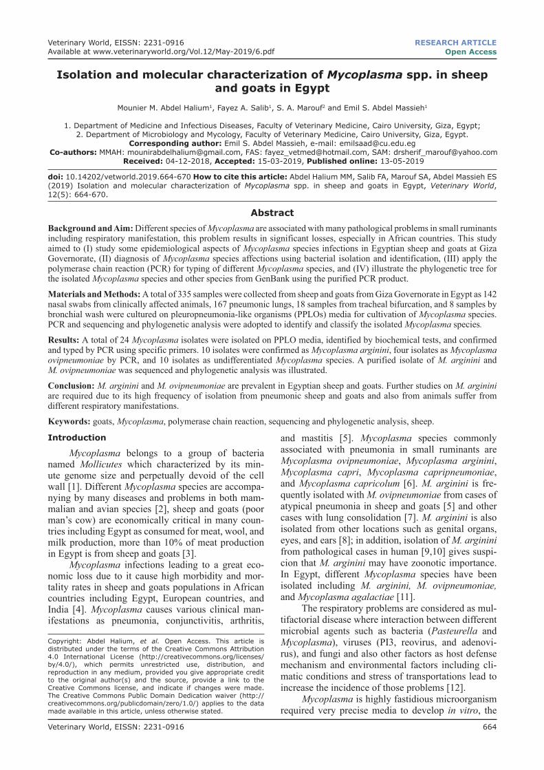
mucopurulent, and purulent), cough, and abnormal chest sound with auscultation (Figure-1a and b).Postmortem examination
The examined lungs, tracheas, and tracheal bifur-cation showed different pathological lesions including congestion and edema with red and gray hepatization, hemorrhages in the trachea and tracheal bifurcation, and different degrees of pleurisy (Figure-1c).Some epidemiological data
The results of isolation of different Mycoplasma species in the examined sheep and goats in relation to sex, season, and age are illustrated in Tables-2 and 3.Bacterial isolation and identification of Mycoplasma species
The collected samples were considered positive to Mycoplasma species when bacterial growth was observed, and it was indicated by turbidity in PPLO broth and fried egg appearance colonies in the agar
Table-1: PCR primers, conditions, and amplicon size.
Species Primers Annealing temperature
Amplicon size
References
Mycoplasma genus GPO3F: 5’-TGGGGAGCAAACAGGATTAGATACC-3’MGSO: 5’- TGCACCATCTGTCACTCTGTTAACCTC-3
At 53°C for 15 s 278 [20]
M. arginini MAGF: 5’-GCA TGG AAT CGCATG ATT CCT-3’ GP4R: 5’-GGT GTT CTT CCTTAT ATC TAC GC-3’
M. agalactiae Mag-F: 5’-CCTTTTAGATTGGGATAGCGGATG-3’ Mag-R: 5’-CCGTCAAGGTAGCGTCATTTCCTAC-3’
60°C for 60 s 360 [5]
PCR=Polymerase chain reaction, M. arginine=Mycoplasma arginine, M. agalactiae=Mycoplasma agalactiae, Mycoplasma ovipneumoniae=M. ovipneumoniae
Figure-1: (a) Buck suffers from different respiratory manifestations with nasal and ocular discharges. (b) Goats suffer from severe respiratory distress with nasal and ocular discharge (corneal opacity). (c) Pneumonic sheep lung with areas of red hepatization, especially at the cranial lobe. (d) Colonies of Mycoplasma appear as a fried egg under the microscope.
a b
c d
Veterinary World, EISSN: 2231-0916 667
Available at www.veterinaryworld.org/Vol.12/May-2019/6.pdf
plates (Figure-1d). Bacterial growth was observed in 24 samples (15 from sheep and nine from goats). Biochemically, all the 24 samples showed zones of growth inhibition around digitonin discs 5 mm in diameter or more, 14 were glucose fermentation test positive, and 10 samples were arginine hydrolysis positive.PCR
The total of 24 culture-positive isolates was confirmed as belonging to the Mycoplasma genus (Figure-2a) by the group-specific PCR, which pro-duced specific bands with the molecular size of 278 bp.
In M. arginini-specific PCR amplification of the samples, six of the sheep isolates and four of the goat iso-lates produced positive products with the approximate molecular size of 525 bp (Figure-2b). The isolation per-centages of M. arginini were, therefore, calculated as 40% (6/15) in sheep and 44.4 (4/9) in goats.
In M. ovipneumoniae-specific PCR amplifica-tion of the samples, three of the sheep isolates and one of the goat isolates produced positive products with the approximate molecular size of 418 bp (Figure-2c). The isolation percentages of M. ovipneumoniae in related to total Mycoplasma isolates were, therefore, calculated as 20% (3/15) in sheep and 11.11 (1/9) in goats.
The other 10 isolates which were positive with the general primer of Mycoplasma genus were tested against (M. capricolum subspecies capripneumoniae,
M. capricolum subspecies capricolum, Mycoplasma mycoides subspecies capri, and M. agalactiae) using species-specific primers and were nega-tive, the 10 samples considered as undifferentiated Mycoplasma species (Table-4).
The high-frequency rate of isolation of M. argi-nini from sheep and goats suffering from respiratory manifestations and pneumonic lungs makes us more interested, amplified purified PCR product of one isolate was subjected to sequencing analysis and the sequence then was submitted to NCBI GenBank tak-ing accession number of MH685445 and also, purified PCR product of one isolate of M. ovipneumoniae was subjected to sequencing analysis and the sequence then submitted to NCBI GenBank taking accession number of MK045665.Phylogenetic analysis
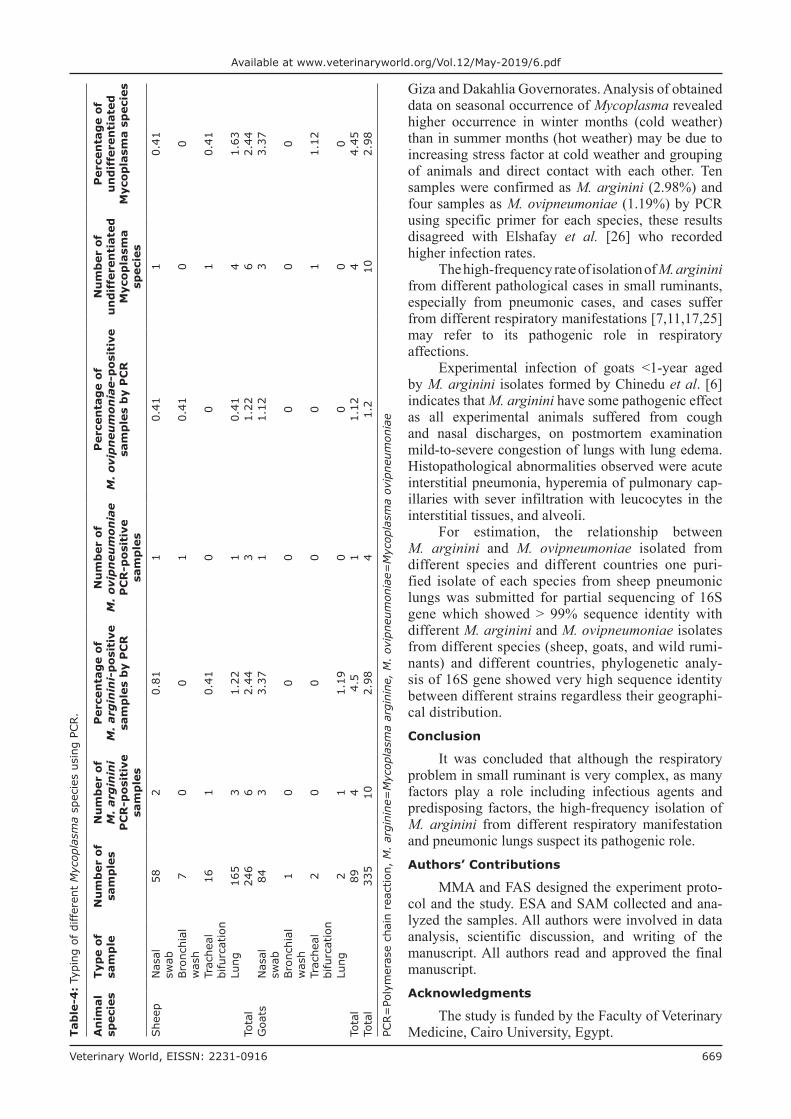
A phylogenetic tree (Figure-3a and b) was formed based on 16S gene sequence of one purified strain of each species from the Egyptian isolates and other isolates of M. arginini and M. ovipneumoniae isolated from different countries and different species (from GenBank). Sequencing of 16S genes of M. argi-nini and M. ovipneumoniae Egyptian isolate and other isolates from other countries showed no significant difference between them.Discussion
Mycoplasmas of different species have been implicated in causing various diseases in sheep and
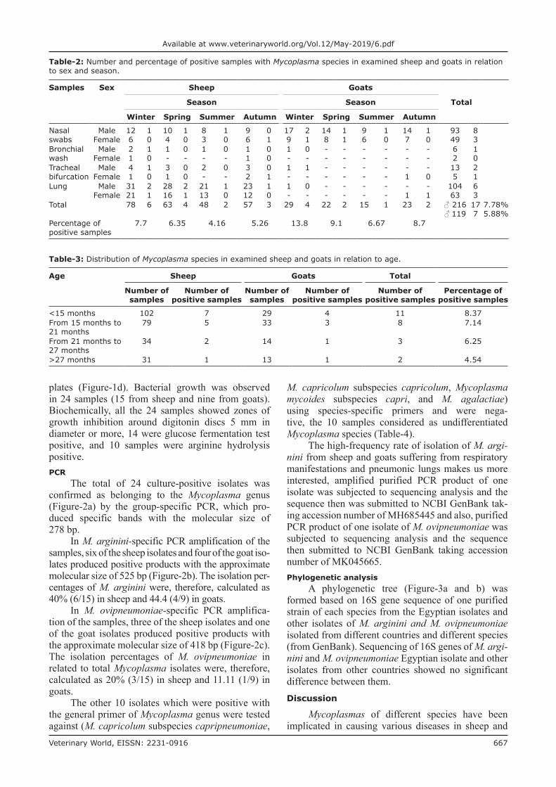
Table-2: Number and percentage of positive samples with Mycoplasma species in examined sheep and goats in relation to sex and season.
Samples Sex Sheep Goats
TotalSeason Season
Winter Spring Summer Autumn Winter Spring Summer Autumn
Available at www.veterinaryworld.org/Vol.12/May-2019/6.pdf
goats including different respiratory manifestations, joint affection, conjunctivitis, and mastitis. The most common species isolated from small ruminants are M. mycoides cluster as Mycoplasma capricolum sub-species capripneumoniae the causative agent of conta-gious caprine pleuropneumonia, other species isolated from sheep and goats including Mycoplasma ovipneu-moniae [24]; however, M. arginini has been isolated from a different pathological condition in small rumi-nants including pneumonia [17,25].
In this study, 335 samples including lung tis-sue, nasal swab, tracheal bifurcation, and bronchial wash were collected from sheep and goats in Egypt
to isolation, identification, and classification of dif-ferent Mycoplasmas species; Mycoplasma were iso-lated from 24 samples identified biochemically using digitonin, confirmed using PCR as Mycoplasma using Mycoplasma genus-specific primers.
The results obtained in this study showed that the prevalence rate of Mycoplasma species was higher in goats than sheep. In Egypt, similar finding has been reported by Ammar et al. [11]; the occurrence of Mycoplasma was more frequently in young ani-mals than adult; these findings are closely related to Elshafay et al. [26] who found higher percent of isola-tion in young animals than adult, especially in goats in
Figure-2: (a) Agarose gel of conventional polymerase chain reaction for detection of 16S gene using Mycoplasma genus-specific primer at amplicon size 278 bp. Lane M (Molecular weight marker, 100-1000 bp); Lanes 1-14, positive samples except for lane 3 negative control. (b) Agarose gel of conventional polymerase chain reaction for detection of Mycoplasma arginini species at amplicon size 525 bp. Lane M (Molecular weight marker, 100-1000 bp); Lanes 1-10 positive. (c) Agarose gel of conventional polymerase chain reaction for detection of Mycoplasma ovipneumoniae at amplicon size 418 bp. Lane M, (Molecular weight marker, 100-1000 bp); lanes 1-4 positive samples.
a b
c d
Figure-3: (a) Phylogenetic analysis of Mycoplasma arginini isolates based on 16S gene sequences, MH685445 isolate under study, KP972459 and KP972459 strains isolated from goats in Egypt, HQ661830 and HQ661829 strains from sheep in South Africa, LC158832 from sheep in Japan, and KX2304770 from goat in China. (b) Phylogenetic analysis of M. ovipneumoniae isolates based on 16S gene sequence, KU870650 and KU870647 strains isolated from goats in China, JN257120, EF687778, DQ0000588, and JN257121 from sheep in China, NR_025989 from wild sheep (Ovis aries) in the USA and EU265779 from bighorn sheep in the USA and KJ433280 from Norwegian Muskov in Norway.
a
b
Veterinary World, EISSN: 2231-0916 669
Available at www.veterinaryworld.org/Vol.12/May-2019/6.pdf
Giza and Dakahlia Governorates. Analysis of obtained data on seasonal occurrence of Mycoplasma revealed higher occurrence in winter months (cold weather) than in summer months (hot weather) may be due to increasing stress factor at cold weather and grouping of animals and direct contact with each other. Ten samples were confirmed as M. arginini (2.98%) and four samples as M. ovipneumoniae (1.19%) by PCR using specific primer for each species, these results disagreed with Elshafay et al. [26] who recorded higher infection rates.
The high-frequency rate of isolation of M. arginini from different pathological cases in small ruminants, especially from pneumonic cases, and cases suffer from different respiratory manifestations [7,11,17,25] may refer to its pathogenic role in respiratory affections.
Experimental infection of goats <1-year aged by M. arginini isolates formed by Chinedu et al. [6] indicates that M. arginini have some pathogenic effect as all experimental animals suffered from cough and nasal discharges, on postmortem examination mild-to-severe congestion of lungs with lung edema. Histopathological abnormalities observed were acute interstitial pneumonia, hyperemia of pulmonary cap-illaries with sever infiltration with leucocytes in the interstitial tissues, and alveoli.
For estimation, the relationship between M. arginini and M. ovipneumoniae isolated from different species and different countries one puri-fied isolate of each species from sheep pneumonic lungs was submitted for partial sequencing of 16S gene which showed ˃ 99% sequence identity with different M. arginini and M. ovipneumoniae isolates from different species (sheep, goats, and wild rumi-nants) and different countries, phylogenetic analy-sis of 16S gene showed very high sequence identity between different strains regardless their geographi-cal distribution.Conclusion
It was concluded that although the respiratory problem in small ruminant is very complex, as many factors play a role including infectious agents and predisposing factors, the high-frequency isolation of M. arginini from different respiratory manifestation and pneumonic lungs suspect its pathogenic role.Authors’ Contributions
MMA and FAS designed the experiment proto-col and the study. ESA and SAM collected and ana-lyzed the samples. All authors were involved in data analysis, scientific discussion, and writing of the manuscript. All authors read and approved the final manuscript.Acknowledgments
The study is funded by the Faculty of Veterinary Medicine, Cairo University, Egypt.Ta
ble
-4:
Typi
ng o
f di
ffer
ent
Myc
opla
sma
spec
ies
usin
g PC
R.
An
imal
sp
ecie
sTy
pe
of
sam
ple
Nu
mb
er o
f sa
mp
les
Nu
mb
er o
f M
. ar
gin
ini
PC
R-p
osit
ive
sam
ple
s
Per
cen
tag
e of
M
. ar
gin
ini-
pos
itiv
e sa
mp
les
by
PC
R
Nu
mb
er o
f M
. ov
ipn
eum
onia
e P
CR
-pos
itiv
e sa
mp
les
Per
cen
tag
e of
M
. ovi
pn
eum
onia
e-p
osit
ive
sam
ple
s b
y P
CR
Nu
mb
er o
f u
nd
iffe
ren
tiat
ed
Myc
opla
sma
spec
ies
Per
cen
tag
e of
u
nd
iffe
ren
tiat
ed
Myc
opla
sma
spec
ies
She
ep
Nas
al
swab
582
0.81
10.
411
0.41
Bro
nchi
al
was
h7
00
10.
410
0
Trac
heal
bi
furc
atio
n16
10.
410
01
0.41
Lung
165
31.
221
0.41
41.
63To
tal
246
62.
443
1.22
62.
44G
oats
Nas
al
swab
843
3.37
11.
123
3.37
Bro
nchi
al
was
h1
00
00
00
Trac
heal
bi
furc
atio
n2
00
00
11.
12
Lung
21
1.19
00
00
Tota
l89
44.
51
1.12
44.
45To
tal
335
102.
984
1.2
102.
98
PCR=
Poly
mer
ase
chai
n re
actio
n, M
. ar
gini
ne=
Myc
opla
sma
argi
nine
, M
. ov
ipne
umon
iae=
Myc
opla
sma
ovip
neum
onia
e
Veterinary World, EISSN: 2231-0916 670
Available at www.veterinaryworld.org/Vol.12/May-2019/6.pdf
Competing Interests
The authors declare that they have no competing interests.Publisher’s Note
Veterinary World remains neutral with regard to jurisdictional claims in published institutional affiliation.References1. Nicholas, R., Ayling, R. and McAuliffe, L. (2008)
Respiratory diseases of small ruminants. In: Mycoplasma Diseases of Ruminants. 1st ed. CABI International, Oxfordshire, UK. p169-198.
2. Glen, C.W., Mark, L.D., Frances, E.C. and Alton, C.S.W. (2012) Detection of Mycoplasma ovipneumoniae and M. arginini in bighorn sheep using enrichment culture coupled with genus and species-specific polymerase chain reaction. J. Wildl. Dis., 48(2): 449-453.
3. Hakim, A.S., Bakry, M.A., Nagwa, S.S.A. and Mona, S.Z. (2014) Role of molecular techniques in the characteriza-tion of bacteria causing pneumonia in small ruminants. Life Sci. J., 11(6): 147-153.
4. Sandip, C., Amit, K., Ruchi, T., Anu, R., Yash, M., Kuldeep, D., Amar, P. and Minakshi, P. (2014) Advances in the diagnosis of respiratory diseases of small ruminants. Vet. Med. Int., 2014: 508304.
5. Kumar, V., Rajneesh, R., Somya, M. and Pramod, K.R. (2013) Isolation and characterization of Mycoplasma mycoides subspecies capri from milk of natural goat masti-tis cases. ISRN Vet. Sci., 2013: 593029.
6. Chinedu, A.A., Kennedy, F.C., Stephen, I.O. and Jude, S.R. (2016) Pathogenicity of local isolates of Mycoplasma ovi-pneumoniae and Mycoplasma arginini in experimental West African dwarf goats. J. Adv. Vet. Anim. Res., 3(3): 242-251.
7. Fernández, S., Galapero, J., Rey, J., Pérez, C.J., Ramos, A., Rosales, R., Ayling, R., Alonso, J.M., Gómez, L. (2016) Investigations into the seasonal presence of Mycoplasma species in fattening lambs. Vet. J., 212 : 80-82.
8. Olusola, M.O., Anna, K., Stuart, R.B., Kelly, A.T., Philip, F.M., Marc, S.M. and Glenn, F.B. (2015) Genetic diversity of Mycoplasma arginini isolates based on multilo-cus sequence typing. Vet. Microbiol., 180(1-2): 123-128.
9. Mayumi, W., Shigemi, H., Miki, G. and Yuichi, H. (2017) Bloodstream infection due to Mycoplasma arginini in an immunocompromised patient. J. Clin. Microbiol., 50(9): 3133-3135.
10. Pálma, S., Dóra, P, Eszter, O., Mercedes, M., Norbert, W., Katinka, P., Dmitriy, V.V., Vladimir, E.C., Susan, S., Laszlo, S. and Sarolta, K. (2018) Eosinophilic fasciitis associated with Mycoplasma arginini infection. J. Clin. Microbiol., 50(3): 1113-1117.
11. Ammar, A.M., Eissa, S.I., El-Hamid, M.I.A., Ahmed, H.A. and ElAziz, A.E.E. (2008) Advanced study on Mycoplasmas isolated from sheep. Zag. Vet. J., 36: 128-137.
12. Azizi, S., Tajbakhsh, E., Rezaii, A., Nekouei, S.H. and Namjoo, A.R. (2011) The role of Mycoplasma ovipneu-moniae and Mycoplasma arginini in pneumonic lungs of
Masroor, A.B., Mohammad, A.A., Zafar, A., Nadeem, R., Majed, R. and Mohammad, S. (2012) Prevalence of Mycoplasma species by polymerase chain reaction (PCR) directly from the nasal swab samples of goats. Pak. J. Life Soc. Sci., 10(1): 5-12.
14. Jackson, P.G.G. and Cockcroft, P.D. (2008) Clinical Examination of Farm Animals. Blackwell Publishing, Oxford, UK.
15. Wäsle, K., Pospischil, A., Hässig, M., Gerspach, C. and Hilbe, M. (2017) The post-mortem examination in rumi-nants and its possible benefit to ruminant clinical medicine. J. Comp. Pathol., 156(2-3): 202-216.
16. Thrusfield, M.V. and Robert, C. (2018) Veterinary Epidemiology. 4th ed. Blackwell Publishing, Oxford, UK.
17. Valsala, R., Rana, R., Remesh, A.T. and Singh, V.P. (2017) Mycoplasma arginini: High frequency involvement in goat pneumonia. Turk. J. Vet. Anim. Sci., 41(1): 393-399.
18. Voigt, K., Brugmann, M., Huber, K., Dewar, P., Cousens, C., Hall, M., Sharp, J.M. and Ganter, M. (2007) PCR examina-tion of bronchoalveolar lavage samples is a useful tool in pre-clinical diagnosis of ovine pulmonary adenocarcinoma (Jaagsiekte). Res. Vet. Sci., 83(3): 419-427.
19. Ponnusamy, P., Samuel, B.M.R., Ranjith, K.M. and Manickam, R. (2017) Isolation, identification and anti-biogram of Mannheimia haemolytica associated with cap-rine pneumonia in the Cauvery delta region of Tamil Nadu, India. Int. J. Curr. Microbiol. Appl. Sci., 6(9): 3118-3122.
20. Çetinkaya, B., Kalin, R., Karahan, M., Atil, E., Manso-Silván, L. and Thiaucourt, F. (2009) Detection of conta-gious caprine pleuropneumonia in East Turkey. Rev. Sci. Tech., 28(3): 1037-1044.
21. Altschul, S.F., Gish, W., Miller, W., Myers, E.W. and Lipmanl, D.J. (1990) Basic local alignment search tool. J. Mol. Biol., 215(3): 403-410.
22. Thompson, J.D., Higgins, D.G. and Gibson, T.J. (1994) CLUSTAL W: Improving the sensitivity of progressive multiple sequence alignment through sequence weighting, position-specific gap penalties and weight matrix choice. Nucleic Acids Res., 22(22): 4673-4680.
23. Tamura, K., Stecher, G., Peterson, D., Filipski, A. and Kumar, S. (2013) MEGA6: Molecular evolutionary genetics analysis version 6.0. Mol. Biol. Evol., 30(12): 2725-2729.
24. Yatoo, M.I., Oveas, R.P., Masood, S.M., Sabiya, Q., Zahid, A.K., Mir, N., Mujeeb, U.R.F., Noor, A.T., Maheshwar, S.K., Sandip, C., Kuldeep, D. and Rajneesh, R. (2018) Mycoplasmosis in small ruminants in India. J. Exp. Biol. Agric. Sci., 6(2): 264-281.
25. Andrea, D.P., Daniela, A., Katiuscia, Z., Giuseppe, M. and Massimo, S. (2017) Mycoplasma pneumonia in small rumi-nants: A ten-year long retrospective survey. Small Rumin. Res., 153: 103-106.
26. Elshafay, D.Y., Nermin, A., Yasser, F.E. and Emad, E.Y. (2016) Isolation and Molecular Characterization of Mycolplasma arginini Isolated from Sheep and Goats with Respiratory Manifestation in Dakahilia Governorate. 3rd Conference of the Scientific Research Institute of Animal Society. Vol. 3. Modern Trends Health for the Advancement of Animal Health, Egypt, Giza.