Page 1
Journal of Stress Physiology & Biochemistry, Vol. 9 No. 1 2013, pp. 184-208 ISSN 1997-0838Original Text Copyright © 2013 by Kapoor, Tripathi and Shrivastava
ORIGINAL ARTICLE
Isolation and Purification of Heterotetrameric Catalase from a
Desiccation Tolerant Cyanobacterium Lyngbya arboricola
Shivali Kapoor* , S. N. Tripathi, Alpana Shrivastava
Department of Botany, Banaras Hindu University, Varanasi 221005, India
*E-Mail: [email protected]
Received October 25, 2012
The desiccation tolerant cyanobacterium Lyngbya arboricola, isolated from bark surfaces of Mangifera indica, possessed up to four stable isoforms of catalase in addition to other antioxidative enzymes, for several years under a dry state. Purification of the two most persistent isoforms of catalase (Cat) has been undertaken by employing acetone precipitation, ethanol: chloroform treatment, gel filtration and ion exchange chromatography. The two isoforms of catalase remained almost unchanged on varying matric and osmotic hydration levels of mats of the cyanobacterium. The purification procedures resulted in a 1.3 % yield of purified single isoform (0.22 mg mL-1 protein) with 709 Units mg-1 specific activity and a purity index of 0.83. Five millimolar of dithiothreitol (DTT) was observed to be pertinent in maintaining the optimum redox state of the enzyme. The purification procedures additionally facilitated the simultaneous elimination and procurement of phycoerythrins (PE) and mycosporine-like amino acids (MAA). Each purified isoform gave a single band (~45kDa) upon SDS-PAGE and denaturing urea isoelectric focusing (IEF) depicted the presence of 2 subunits each of CatA and CatB. The monoisotopic mass and pI value of CatA and CatB as revealed by LC-MS analysis and internal amino acid sequencing was 78.96, 5.89 and 80.77, 5.92, respectively, showing resemblance with CatA of Erysiphe graminis subs. hordei and CatB of Ajellomyces capsulata. The heterotetrameric monofunctional catalase (~320 kDa), due to its stability in the form of resistance to ethanol: chloroform, its thermoalkaliphilic nature and the presence of innumerable hydrophobic amino acid residues (~40%), thus exhibited its potential for biotechnological applications.
Key words: Chromatography; Denaturing isoelectric focusing; Desiccation; Electrophoresis; Lyngbya arboricola; Stable Catalase
Abbreviations: Cat, Catalase; DTT, dithiothreitol; IEF, isoelectric focusing; MAA, mycosporine like amino acids ; PE, phycoerythrins; KPB, potassium phosphate buffer; PMSF, phenylmethane-sulphonyl fluoride; ß-ME, beta mercaptoethanol
JOURNAL OF STRESS PHYSIOLOGY & BIOCHEMISTRY Vol. 9 No. 1 2013
Page 2
Isolation and Purification of Heterotetrameric Catalase...
ORIGINAL ARTICLE
Isolation and Purification of Heterotetrameric Catalase from a
Desiccation Tolerant Cyanobacterium Lyngbya arboricola
Shivali Kapoor* , S. N. Tripathi, Alpana Shrivastava
Department of Botany, Banaras Hindu University, Varanasi 221005, India
*E-Mail: [email protected]
Received October 25, 2012
The desiccation tolerant cyanobacterium Lyngbya arboricola, isolated from bark surfaces of Mangifera indica, possessed up to four stable isoforms of catalase in addition to other antioxidative enzymes, for several years under a dry state. Purification of the two most persistent isoforms of catalase (Cat) has been undertaken by employing acetone precipitation, ethanol: chloroform treatment, gel filtration and ion exchange chromatography. The two isoforms of catalase remained almost unchanged on varying matric and osmotic hydration levels of mats of the cyanobacterium. The purification procedures resulted in a 1.3 % yield of purified single isoform (0.22 mg mL-1 protein) with 709 Units mg-1 specific activity and a purity index of 0.83. Five millimolar of dithiothreitol (DTT) was observed to be pertinent in maintaining the optimum redox state of the enzyme. The purification procedures additionally facilitated the simultaneous elimination and procurement of phycoerythrins (PE) and mycosporine-like amino acids (MAA). Each purified isoform gave a single band (~45kDa) upon SDS-PAGE and denaturing urea isoelectric focusing (IEF) depicted the presence of 2 subunits each of CatA and CatB. The monoisotopic mass and pI value of CatA and CatB as revealed by LC-MS analysis and internal amino acid sequencing was 78.96, 5.89 and 80.77, 5.92, respectively, showing resemblance with CatA of Erysiphe graminis subs. hordei and CatB of Ajellomyces capsulata. The heterotetrameric monofunctional catalase (~320 kDa), due to its stability in the form of resistance to ethanol: chloroform, its thermoalkaliphilic nature and the presence of innumerable hydrophobic amino acid residues (~40%), thus exhibited its potential for biotechnological applications.
Key words: Chromatography; Denaturing isoelectric focusing; Desiccation; Electrophoresis; Lyngbya arboricola; Stable Catalase
Abbreviations: Cat, Catalase; DTT, dithiothreitol; IEF, isoelectric focusing; MAA, mycosporine like amino acids ; PE, phycoerythrins; KPB, potassium phosphate buffer; PMSF, phenylmethane-sulphonyl fluoride; ß-ME, beta mercaptoethanol
Metabolic disorders in an oxygen-enriched
environment often result in the generation of
reactive oxygen species (ROS) such as superoxide,
hydroxyl radical, and hydrogen peroxide leading to
oxidation of cellular biomolecules causing
breakdown of normal cellular, membrane, and
JOURNAL OF STRESS PHYSIOLOGY & BIOCHEMISTRY Vol. 9 No. 1 2013
158170185
Page 3
Kapoor et al
reproductive functions (Kranner and Birtic, 2005;
Abele, 2002; Marx, 1985). In order to maintain
normal growth and function, as well as to neutralize
the lethal effects of ROS, organisms have evolved
specific antioxidant defenses. Catalase (H2O: H2O
oxidoreductase; EC 1.11.1.6), a ubiquitous enzyme
that was one of the first enzymes isolated in a state
of high purity (Schonbaum and Chance, 1976),
efficiently catalyzes the decomposition of hydrogen
peroxide into oxygen and water and thus is a
significant component of antioxidative defense
systems in aerobic organisms. Potential applications
of catalases in medicine and industry have led to
numerous attempts to engineering this protein
(Shaked and Wolfe, 1988). Catalase has also gained
attention in recent years because of its link to
cancer, ageing in humans and animals (Melov et al.,
2000; Preston et al., 2001; Turdi et al., 2007). Also
the application of catalases for the elimination of
H2O2 in textile bleaching effluents would not only
allow the recycling of enormous amount of water
used for dyeing but would also have advantage over
the use of unfavorable high quantities of salt
concentrations such as sodium bisulfite and act as a
more environmentally friendly alternative to
existing chemical treatments (Thompson et al.,
2003; Tzanov et al., 2001; Gujelj et al., 2001). Most
of the commercial catalases available in the market
are optimally active at 20-50oC and at neutral pH.
This necessititates the requirement of new
thermoalkalistable catalases which can act at
temperatures above 50oC and pH values above 9.0.
Catalases are not restricted to one protein type
structurally, functionally, or by sequence. In
general, there are three classes of catalase: the
typical or monofunctional catalases, the catalase-
peroxidases and the Mn-catalases or
pseudocatalases (Zamocky and Koller 1999,
Thompson et al., 2003). Additionally the
monofunctional catalases are divided into three
phylogenetic clades arising from a minimum of two
genes duplication - two distint clades or
subgroupings of small subunit enzymes and one
clade of large subunit enzymes (Chelikani et al.,
2004; Klotz et al., 1997). It has been demonstrated
that catalases belonging to clade 2 mainly exhibit
unusual resistances to physical and chemical
denaturation. Catalase HPII was found to show
thermal as well as pH stability by showing
considerable activity till 60oC which declined after
80oC with a Tm value 83oC (Goldberg and Hochman
1989; Switala et al., 1999). The thermal stability as
well as resistance property of catalase to treatment
with chloroform and ethanol has been widely
exploited for purification of the enzyme (Weiting et
al., 1990).
Cyanobacteria, one of the most primitive
oxygenic phototrophs were probably the first
organisms to develop elaborate mechanisms for the
detoxification of partially reduced oxygen species.
Desiccation tolerant terrestrial cyanobacteria,
mainly exhibiting subaerial growth, due to oxygen
rich atmosphere are supposed to generate
considerable ROS and also to possess defense
mechanisms against damages expected due to
generation of ROS mainly during the phases of dry-
down (dehydration) followed by desiccation (dry)
and then subsequent recovery of metabolism
(rehydration) at their natural habitats (Potts, 2001;
Tripathi and Maurya, 2001). Cyanobacteria are
mostly known to possess bifunctional catalase
peroxidase (KatG), Mn-catalases (MnCat), and
peroxiredoxins (Zamocky et al., 2008). However, in
spite of being the largest group of H2O2 degrading
enzymes, typical catalases are very uncommon in
cyanobacteria. Until now the only one complete
JOURNAL OF STRESS PHYSIOLOGY & BIOCHEMISTRY Vol. 9 No. 1 2013
186
Page 4
Isolation and Purification of Heterotetrameric Catalase...
and nonfused gene in which all essential amino
acids of typical catalase is conserved is found in
Nostoc punctiforme PCC73102. Phylogenetic studies
have futher revealed that it belongs to clade 3 of
small subunit catalases that contain haem b as the
active site and use NADPH as a second active
cofactor (Bernroitner et al., 2009). Also incomplete
catalase genes have been reported in Cyanothece
sp. ATCC51142 and Synechococcus elongatus
PCC7942.
The genus Lyngbya has a prominent ecological
role in marine ecosystems and is a group rich in
bioactive secondary metabolites (Gerwick et al.,
2008; Hoffman, 1994). Recently the whole genome
analysis of Lyngba majuscula 3L was done to gain
insight into potential microbial interactions and
gauge the natural product synthesis of this genus
(Jones et al., 2011). Also there are reports of
purification and isolation of a catalase and its gene
from other microbes like Aspergillus species
(Chandrashekar, 2011) and Bacillus sp. (Wang et al.,
2011) but none have reported catalase from any
cyanobacterial species yet. Tripathi and Srivastava
in 2001 were the first to report the presence of
active catalases, and dismutases in desiccation
tolerant cyanobacterium Lyngbya arboricola.
Catalases are known to retain its stability for
longer duration, being active in freeze-dried
permafrost samples (Gilichinsky et al., 1992) over a
period of million years. But the presence of stable
and active catalase in cells of this desiccation
tolerant terrestrial cyanobacterium Lyngbya
arboricola stored in desiccated state for two years
is not only reflective of the ability of the
cyanobacterium to maintain structural and
functional integrity of their macromolecules
including proteins under extremes of desiccation,
but also signifies for better availability of stable
catalase in dry mats of this cyanobacterium.
Due to the low structural stability of most of
catalases much work in recent times has been
devoted to increase their stability. This makes this
cyanobacterium excellent source material for future
exploitation of such enzymes for bioengineering
and scientific research. This study will also be one
of the first reports of presence and purification of
catalases in a dessication tolerant cyanonbacteria
which may serve as potential sources of
thermophilic industrial catalases.
Unfortunately, structural, functional and
molecular basis of stability of catalases from the
cyanobacterium have not been explored at the
desired level. In order to exploit this activity in the
future, an understanding of the characteristics of
the enzyme is required, necessitating the
development of procedures for purification of
catalase from the cyanobacterium.
Besides cyanobacteria being useful potential
sources for antioxidants and antioxidative enzymes,
they also contain significant amounts of unusual
phycobiliproteins (PBPs), especially (PEs) (Tripathi
et al., 2007) and UV-absorbing pigments MAAs and
scytonemins (Garcia-Pichel et al., 1993) that are
important in biotechnology but pose great
problems for the purification of catalases. Thus, this
work also describes a process of recovering these
water soluble pigments as by-products of the
catalase purification process. In addition we have
also developed procedure(s) to maintain the
pertinent level of redox state of the cell-free extract
to minimize denaturation of the enzymes, i.e.,
catalases.
This method of purification and isolation of
antioxidant enzyme catalase with simultaneous
JOURNAL OF STRESS PHYSIOLOGY & BIOCHEMISTRY Vol. 9 No. 1 2013
187
Page 5
Kapoor et al
recovery of biotechnologically important PEs and
UV screening MAAs, may also save the individual
efforts and costs to extract these pigments from
this cyanobacterium.
MATERIALS AND METHODS
(i) Cyanobacterial material: L. arboricola
inhabiting bark surfaces of Mangifera indica from
the campus of Banaras Hindu University faces 8-9
dry months during the hot summer and cold winter
seasons due to a considerably low amount of
rainfall (2-12%). However, 3-4 months of a warm
and moist rainy season with torrential rainfall (90%)
and intermittent breaks result in uncertain wetting
and drying and provide relatively better conditions
for growth of the cyanobacterium (Tripathi et al.,
1990/91). Collection of the cyanobacterial samples
was performed by scraping cyanobacterial mats
from bark at the end of the rainy season (during the
last week of September); after removing any
adherent soil particles, the intact mats were used
for further analysis.
Under natural habitat, rehydration (wetting) of
dry mats of L. arboricola is on direct availability of
water; whereas, dehydration (drying) results due to
loss of water from wet mats to the atmosphere. In
other words, it can be said that under natural
habitats, rehydration and dehydration of the
cyanobacterium can be controlled by regulating the
osmotic and matric availability of water,
respectively. So, osmotic water potential of the
cyanobacterial mat was controlled by incubating
the mat to the solutes of the medium (water
potential solutions) on filter papers; whereas,
matric water potential of the mats was controlled
by equilibrating the mats in the atmosphere of a
solution of defined water potential (isopiestic
control, Harris et al., 1970; Potts and Friedman
1981).
In order to obtain the cyanobacterium under
growing conditions, the dry cyanobacterial mats
were placed over filter paper pre-soaked with
double distilled water at 0 MPa, for 72 h at 25oC
and 72 μmol photon m-2s-1 light intensity. To
maintain hydration levels, the growing and dry
natural mats of L. arboricola were incubated at
different osmotic or matric water potentials by
equilibrating the mats over sodium chloride
solutions and saturated solutions of different salts
equivalent to different water potentials (0, -2.8,
-21, -39, -167, and -355 MPa) at 25oC as described
by Brock (1975) and Tripathi and Maurya (2001).
For rehydration of dry mat, the mat was incubated
on the filter paper soaked with the required
osmotic water potential solutions for the required
incubation period; whereas, for dehydration the 72
h grown mat at 0 MPa osmotic water potential was
in the atmosphere of the required matric water
potential solution for the required time period.
(ii) Application of reducing agents: The impact of
some reducing agents in optimization of redox state
of the enzyme was studied by incubating catalase
(0.05 mg protein per lane) with varying
concentrations (0-20 mM) of cysteine, DTT and ß-
ME, respectively, for 30 min at 4oC and monitoring
changes in the enzyme activity by native-PAGE gel.
The optimum redox state of the enzyme was
evaluated by monitoring the concentration of
reductant at which an optimum enzyme activity
could be recorded. The levels of sharpness and
intensity on native-PAGE gel were related to the
state of the enzyme.
(iii) Purification of catalase: Purification of the
enzyme was achieved by employing acetone
precipitation, ethanol-chloroform treatment, gel
filtration, ion-exchange chromatography and
electroelution processes. These procedures were
JOURNAL OF STRESS PHYSIOLOGY & BIOCHEMISTRY Vol. 9 No. 1 2013
188
Page 6
Isolation and Purification of Heterotetrameric Catalase...
carried out at 4oC unless otherwise stated, and the
purity of catalase at each step of purification was
determined using spectroscopy and PAGE.
(a) Preparation of cell-free extract: The cell-free
extract used for purification of the enzyme was
obtained by crushing cyanobacterial mats (25 g) to
powder in liquid nitrogen and homogenizing the
powder with 110 mL extraction buffer containing
100 mM potassium phosphate buffer (KPB, pH 7.5),
1 mM Na-EDTA and 1 mM phenylmethane sulfonyl
fluoride (PMSF) at 4oC. The homogenate was
sonicated at 130 W, 20 kHz for 5 min at 50%
amplitude using an ultrasonicator (Sonic &
Materials, USA) and centrifuged at 25,000 × g at 4oC
for 1 h. The supernatant (cell-free extract) was
filtered with 0.45 μm cellulose-acetate membrane
filter (Millipore, USA) and was used to obtain
catalase enriched fraction of the extract.
(b) Sequential acetone precipitation: The
catalase enriched fraction was acquired by addition
of acetone (20-60%) to the respective supernatants
obtained after stepwise addition of acetone to cell
free extract. The cell-free extract was mixed with
chilled acetone (20%), kept overnight at 4oC for
precipitation and the supernatant and precipitate
were obtained by centrifuging the mixture at
17,000 × g for 30 min at 4oC. The supernatant was
lyophilized, re-dissolved in extraction buffer and
was further undertaken for second and third
acetone fractionation (40& 60%) successively as
before. Catalase enzyme activity was measured in
the supernatants and pellets obtained at each step
of acetone treatments.
(c) Ethanol: chloroform treatment: The enzyme-
enriched fractions collected after acetone
precipitations were mixed with cold ethanol (95%)
and chloroform in a ratio of 10:5:3 (v/v). After
vortexing vigorously for 2-5 min, the mixture was
centrifuged at 17,000 × g at 4oC for 30 min. Out of
the three layers formed, the top aqueous layer
displaying high catalase activity was collected and
further purified; the dense solid middle layer
containing denatured proteins and some pigments
was used for purification of PBPs.
(d) Column chromatography: Gel filtration - One
milliliter of the enzyme extract (the top aqueous
layer after lyophilization) containing 2 mg protein
mL-1 was filtered with a 0.22 µm filter and loaded
onto a Hi-Prep Sephacryl (16/60) S-300 HR column
(Akta Prime Plus, GE Healthcare) that had been
washed and pre-equilibrated with ~ 200 mL 100
mM KPB (pH 7.5) and extraction buffer,
respectively. The enzyme was eluted at a flow rate
of 30 mL h-1. Two-milliliter fractions with maximum
enzyme activity were pooled and concentrated
using dialysis sacks (12 kDa cut off, Sigma, USA).
After dialysis overnight against 10 mM Tris-HCl (pH
8.0) at 4oC, the pooled fraction was applied to a
DEAE-Sephadex A-50 column.
Ion exchange chromatography - One milliliter of
dialyzed sample (1.5 mg protein mL-1) obtained
after gel-filtration was applied to a DEAE-Sephadex
A-50 column (20 × 1.5 cm) that was pre-
equilibrated and was thoroughly washed with 10
mM Tris (pH 8.0) at a flow rate of 24 mL h-1 to
remove any unbound proteins (mainly, MAA);
bound proteins were eluted using a linear gradient
of NaCl (0-1.0 M) in 10 mM Tris-HCl (pH 8.0). Ten
milliliters (each fraction of 2.5 mL) was collected
and desalted overnight at 4oC. Fractions showing
high catalase activity at each gradient were pooled,
concentrated using 10 kDa cut-off filters (Ultrafree-
MC, Millipore, USA) and used for further analysis.
(iv) Determination of purity index: The purity
index for catalase (PIcatalase) at each step of
purification was expressed as A405/A280, where A405
JOURNAL OF STRESS PHYSIOLOGY & BIOCHEMISTRY Vol. 9 No. 1 2013
189
Page 7
Kapoor et al
and A280 stand for absorbance at 405 & 280 nm,
respectively. Also, the ratios of A565/A280 and of
A320/A280 corresponding to PE and to MAA,
respectively, were determined in order to monitor
the level of pigment impurity persisting at each step
of purification.
Protein concentration was also determined
using the Lowry method, and lysozyme (1 mg
protein mL-1) was used as the standard (Lowry et
al., 1951).
(v) Native PAGE: The cell-free extract and
samples obtained at each step of purification were
evaluated by native- PAGE for catalase activity using
a vertical slab gel apparatus (Miniprotein-II, Bio-
Rad, USA) on 8% polyacrylamide gel supported with
glycerol following the method of Davis (1964) with
modifications as described by Tripathi and
Srivastava (2001).
(vi) Electroelution of individual isoforms:
Individual isoforms of the enzyme were
electroeluted from the native-PAGE of the enzyme
by adopting the procedure (with minor
modifications) developed by Harrington (1990). The
native-PAGE was carried out using the enzyme
fraction obtained after 60% acetone precipitation.
Packing of the gel slices in the electroeluter was
performed by using loading buffer containing 20
mM Tris-HCl (pH 8.0), 100 μM DTT, 1 mM PMSF, 1
mM EDTA and 10 % (v/v) glycerol. The eluates were
collected at 8-10 mA per tube for 3-4 h at 4˚C in
elution buffer containing 20 mM Tris-HCl (pH 8.0)
and 5 mM DTT and were further concentrated with
a 10 kDa cut-off filter (Ultrafree-MC, Millipore USA)
to the desired protein concentration.
(vii) Enzyme characterization:
(a) SDS PAGE: 15% polyacrylamide resolving gel
supported with 0.1 % SDS and 5% stacking gel) was
used for further analysis of purity and estimation of
molecular weight of the isoforms of catalase
obtained in the eluate of DEAE-Sephadex column or
after electroelution from native PAGE following the
method of Sambrook (Sambrook et al., 1989). The
proteins on gels were visualized by staining with
0.25% (w/v) Coomassie Brilliant Blue R-250 in 45%
methanol and 10% acetic acid and destained with
45% (v/v) methanol and 7% (v/v) glacial acetic acid.
Molecular weights were analyzed using low
molecular weight markers as standards (MW-SDS-
70, Sigma, USA).
(b) IEF: The isoform(s) of catalase purified by
ion-exchange chromatography and by
electroelution were further evaluated by
denaturing urea-IEF on a 10% polyacrylamide gel
with 2% (v/v) Ampholine (pH 3.5-10.0, Sigma, USA)
(Robertson et al., 1987) in 8 M urea using a vertical
slab gel apparatus (Miniprotein-II, Bio-Rad, USA).
Samples (10-30 µg protein/lane) were incubated
with equal volumes of urea-IEF sample solution [8
M-urea, ampholytes (2%), β-ME- 50 μL,
bromophenol blue- 50 μg in water] for 5 min at
room temperature, applied to bottom of wells and
overlaid with a 1% ampholyte, 15 % glycerol
solution. Focusing was performed using 50 mM
NaOH and 20 mM acetic acid as catholyte and
anolyte solutions, respectively, and the gel was run
at 250 V for 30 min followed by 450 V (constant
current) for another 2 h at 4˚C. After
electrophoretic separation, the gel was fixed for 20-
30 min with constant shaking at room temperature
in the IEF fixing solution [Trichloroacetic acid (12%
w/v) and Sulfosalicyclic acid (4 % w/v)] in deionized
water. Afterward, it was rinsed in destaining
solution (the same as that used for SDS-PAGE gels)
for 10 min. The protein bands in IEF gels were
JOURNAL OF STRESS PHYSIOLOGY & BIOCHEMISTRY Vol. 9 No. 1 2013
190
Page 8
Isolation and Purification of Heterotetrameric Catalase...
visualized by Coomassie Blue staining as described
for SDS-PAGE gels.
(c) Amino acid sequence analysis: The pI and
molecular weights of individual subunits and
internal amino acid sequence analyses were
outsourced to The Centre for Genomic Application
(TCGA), New Delhi, India. The internal amino acid
sequences of catalases were analyzed using LC-MS
by excising the gel bands after denaturing-IEF,
performing tryptic digestion of the protein, and
separating and fragmenting them on a reverse
phase column. The data were analyzed by matching
with Xcalibur/database/uniprot.fasta, and the
deduced amino acid compositions of the catalases
used as reference (with GenBank gene sequence
primary accession numbers in parentheses) were
CATA_ERYGR (Q8X1P0) EC.1.11.1.6) and
CATB_AJECA (Q9Y7C2) EC 1.11.1.6).
(d) Enzyme assay: Catalase activity in enzyme
samples (cell-free extract or purified) was
determined by monitoring the decrease in
absorbance of H2O2 as described by Tripathi and
Srivastava (2001) with certain modifications. The
reaction was initiated by addition of 4.4 mM H2O2 to
a 3 mL reaction mixture containing 2.86 mL of KPB
(50 mM, pH 7.5) and 100 µL of enzyme extract (1.4
mg protein mL-1), and catalase activity was
measured by recording the amount of H2O2
(extinction coefficient (ε) of H2O2=43.6 M-1cm-1)
consumed. One unit (U) of catalase activity was
defined as the amount of enzyme required to
degrade 1 μmol of H2O2 min-1 at 25°C. The kinetic
parameters were determined with the catalase
solution using standard assay with varying substrate
concentrations in the range of 3-460 mM. The
apparent Km value and Vmax for the enzyme was
estimated by analysis of data by Michaelis-Menten/
Lineweaver-Burk plots.
(e) Effect of pH and temperature: The effect of
pH on catalase activity was evaluated in the range
of pH 3.5-11.0 by employing different buffers, 50
mM sodium acetate buffer (pH 3.6-5.6), 50 mM K-
phosphate buffer (pH 5.7-8.0) and 50 mM Glycine-
NaOH buffer (pH 8.6-10.6). 100 µL enzyme solutions
(1.4mg mL-1) was mixed with 2.86 mL of buffer of
desired pH and incubated at 25˚C for 1 h and
enzyme activity was measured using the standard
assay. Also, the effect of temperature (20-80˚C) on
catalase activity was evaluated by incubating the
enzyme solution in standard reaction mixtures at
each temperature for 1 h. The effect of all the
above was expressed as Enzyme Relative activity
(%) by taking the pH and temperature at which
maximum enzyme activity was recorded as
optimum (100%).
The effect of different concentrations of
reducing agent DTT (0 -20 mM) on catalase activity
was determined by incubating the reaction mixture
containing 100 µl of the enzyme solution along with
DTT at 25˚C for varying time periods. However,
relative enzyme activity (%) was calculated by taking
the enzyme activity recorded in the native enzyme
without the presence of any additive compound
taken as control (100%).
RESULTS
(i) Selection of the catalase isoform(s) for
purification: An apparent variability in the number
of catalase isoforms from four in the present study,
to three, reported earlier by Tripathi and Srivastava
in 2001, in the mats of L. arboricola harvested from
the same natural habitat poses a problem for
selection of the isoform(s) to be selected for
purification. Native PAGE reflecting CAT activity in
the natural mats grown at 0 MPa osmotic water
potential for 72 h when incubated matrically
JOURNAL OF STRESS PHYSIOLOGY & BIOCHEMISTRY Vol. 9 No. 1 2013
191
Page 9
Kapoor et al
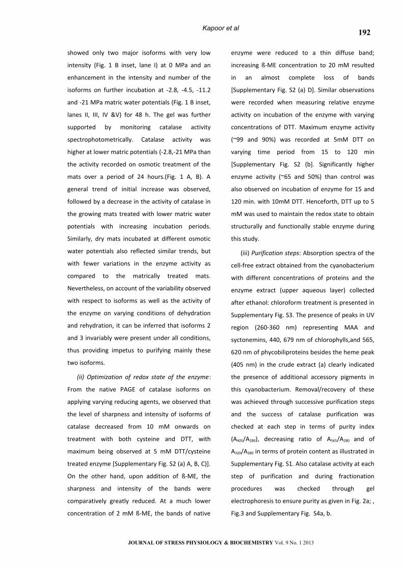
showed only two major isoforms with very low
intensity (Fig. 1 B inset, lane I) at 0 MPa and an
enhancement in the intensity and number of the
isoforms on further incubation at -2.8, -4.5, -11.2
and -21 MPa matric water potentials (Fig. 1 B inset,
lanes II, III, IV &V) for 48 h. The gel was further
supported by monitoring catalase activity
spectrophotometrically. Catalase activity was
higher at lower matric potentials (-2.8,-21 MPa than
the activity recorded on osmotic treatment of the
mats over a period of 24 hours.(Fig. 1 A, B). A
general trend of initial increase was observed,
followed by a decrease in the activity of catalase in
the growing mats treated with lower matric water
potentials with increasing incubation periods.
Similarly, dry mats incubated at different osmotic
water potentials also reflected similar trends, but
with fewer variations in the enzyme activity as
compared to the matrically treated mats.
Nevertheless, on account of the variability observed
with respect to isoforms as well as the activity of
the enzyme on varying conditions of dehydration
and rehydration, it can be inferred that isoforms 2
and 3 invariably were present under all conditions,
thus providing impetus to purifying mainly these
two isoforms.
(ii) Optimization of redox state of the enzyme:
From the native PAGE of catalase isoforms on
applying varying reducing agents, we observed that
the level of sharpness and intensity of isoforms of
catalase decreased from 10 mM onwards on
treatment with both cysteine and DTT, with
maximum being observed at 5 mM DTT/cysteine
treated enzyme [Supplementary Fig. S2 (a) A, B, C)].
On the other hand, upon addition of ß-ME, the
sharpness and intensity of the bands were
comparatively greatly reduced. At a much lower
concentration of 2 mM ß-ME, the bands of native
enzyme were reduced to a thin diffuse band;
increasing ß-ME concentration to 20 mM resulted
in an almost complete loss of bands
[Supplementary Fig. S2 (a) D]. Similar observations
were recorded when measuring relative enzyme
activity on incubation of the enzyme with varying
concentrations of DTT. Maximum enzyme activity
(~99 and 90%) was recorded at 5mM DTT on
varying time period from 15 to 120 min
[Supplementary Fig. S2 (b]. Significantly higher
enzyme activity (~65 and 50%) than control was
also observed on incubation of enzyme for 15 and
120 min. with 10mM DTT. Henceforth, DTT up to 5
mM was used to maintain the redox state to obtain
structurally and functionally stable enzyme during
this study.
(iii) Purification steps: Absorption spectra of the
cell-free extract obtained from the cyanobacterium
with different concentrations of proteins and the
enzyme extract (upper aqueous layer) collected
after ethanol: chloroform treatment is presented in
Supplementary Fig. S3. The presence of peaks in UV
region (260-360 nm) representing MAA and
syctonemins, 440, 679 nm of chlorophylls,and 565,
620 nm of phycobiliproteins besides the heme peak
(405 nm) in the crude extract (a) clearly indicated
the presence of additional accessory pigments in
this cyanobacterium. Removal/recovery of these
was achieved through successive purification steps
and the success of catalase purification was
checked at each step in terms of purity index
(A405/A280), decreasing ratio of A565/A280 and of
A320/A280 in terms of protein content as illustrated in
Supplementary Fig. S1. Also catalase activity at each
step of purification and during fractionation
procedures was checked through gel
electrophoresis to ensure purity as given in Fig. 2a; ,
Fig.3 and Supplementary Fig. S4a, b.
JOURNAL OF STRESS PHYSIOLOGY & BIOCHEMISTRY Vol. 9 No. 1 2013
192
Page 10
Isolation and Purification of Heterotetrameric Catalase...
The data of purification of catalase from
Lyngbya arboricola are summarized in Table 1. The
purified catalase was stored as a concentrated
solution at -20oC without any significant loss in
enzyme activity. Purification of the two most
persistent isoforms [2 and 3; See Fig. 2 (a), E] of
catalase with purity index of 0.83 and 0.6,
respectively, contributing to 45 % and 40% of the
total proteins and yield of 1.3 % was achieved by
this procedure. Fig. 3 shows the SDS-PAGE profile of
the subsequent purification steps of through which
successful procurement of two major isoforms (2
and 3) of catalase was achieved. However
electroelution of catalase isoforms from the native
PAGE of the partially purified enzyme (after 60%
acetone precipitation) supported the isolation of all
the four individual isoforms separately (Fig. 2b) if
required.
(iv) Characterization of catalase:
(a) Electrophoresis: The purity of the each of the
isoforms isolated by precipitation steps and by
electroelution was verified by presence of a single
band at ~45kDa in SDS-PAGE (Fig. 4a). Urea-IEF and
internal amino acid sequencing of subunits of
individual isoforms obtained after ion-exchange
chromatography and electroelution revealed the
presence of 4 subunits in a single isoform, with the
fourth subunit overlapping the third (Fig. 4c). The
subunits 1 & 4 and 2 & 3, when analyzed for their
internal amino acid sequences, molecular weight
and pI revealed greatest homology and sequence
matching with CatB (80.77 kD, pI:5.92) of
Ajellomyces capsulata and CatA (78.96 kD,pI:5.89)
of Erysiphe graminis subs. hordei, respectively (Fig.
5). Thus, an individual isoform of the catalase
enzyme is heterotetrameric (MW equivalent to
approximately 320 kDa), and each of its dimers is
composed of two heteromeric subunits, where
each one resembles either CatA or CatB, as
depicted by the line diagram in Supplementary Fig.
S5.
(b) Kinetic Properties: Catalase activity recorded
with varying H2O2 concentration indicated a gradual
but steady increase in the enzyme activity till 150
mM attaining almost a stationary state at 200 mM.
Nevertheless, on further increasing the
concentration from 200 mM to 450 mM a gradual
lowering was observed with ~ 50 % of enzyme
activity being recorded at 450 mM. Linear curve
fitting to the Lineweaver-Burk equation yielded a Km
value of 10.27 mM and Vmax of 11.70 mM min-1
(Fig.6).
(c) Effect of pH and temperature: The activity of
catalase as a function of pH and temperature
showed that the catalase from the cyanobacterium
being a typical catalase was active over a broad pH
range (pH 6.0-10.5); optimal activity (100%) being
recorded at pH 8.0. Noticeably, nearly 40% and 80
% of the optimum catalase activity was also
recorded at lower (below 5.5) and higher (above
8.0) pH (Fig. 7).
The impact of temperature on enzymatic activity
showed that the cyanobacterium catalase exhibited
considerable activity in the range 20-70oC with
optimum temperature for activity recorded at 50oC.
Nearly 80% of the optimum activity was recorded in
the range 20oC-55oC which gradually declined to
60% and 40% at 70oC and 80oC respectively (Fig. 7)
JOURNAL OF STRESS PHYSIOLOGY & BIOCHEMISTRY Vol. 9 No. 1 2013
193
Page 11
Kapoor et al
Figure 1: Catalase activity in the dry mats of L. arboricola incubated at different time periods, (A) osmotically and also in its osmotically grown mats on further (B) matric treatment. (Inset B: Lanes I, II, III, IV & V represent native PAGE of catalase isoforms in the osmotically (0 MPa for 72 h) grown mats on matric incubation for 48 h at 0, -2.8, -4.5, -11.2 and -21 MPa, respectively.
Figure 2: Native-PAGE (8%) of catalase activity in (a) respective enzyme enriched fractions at each step of
purification; catalase activity in (A) cell free extract, in supernatant fraction after (B) 20% , (C) 40% acetone fractionation, in pellet fraction after (D) 60% acetone precipitation, (E) uppermost aqueous ethanol fraction (E2, see-Supplementary Fig. S1);.(b) four electroeluted catalase isoforms (corresponding to isoforms 1, 3, 4, 2 respectively procured from the native PAGE of the enzyme fraction obtained after 60% acetone precipitation as described in Materials and Methods
JOURNAL OF STRESS PHYSIOLOGY & BIOCHEMISTRY Vol. 9 No. 1 2013
194
Page 12
Isolation and Purification of Heterotetrameric Catalase...
Figure 3: SDS-PAGE of catalase containing fractions after each purification step: (Lane 1) molecular mass standards; (Lane 2) cellular extract; (Lane 3) pellet fraction after 60% acetone precipitation; (Lane4) ethanol fraction after ethanol: chloroform treatment; (Lane 5, 6) purified isoform 2 and 3 of catalase separated by DEAE-ion exchange and (Lane 6) peak fraction after gel filtration.
Figure 4: SDS PAGE, native PAGE and denaturing urea isoelectric focusing of single isoform of purified catalase. (a) Denaturing SDS (15%) polyacrylamide gel stained with Coomassie Brilliant Blue. Lane 1 -Molecular mass standard proteins; Lane 2 & 3, purified catalase isoforms (second and third) showing single band at 45 kDa, (b) Native (8%) PAGE stained for catalase activity showing electroeluted single isoform of catalase (c) Urea (8 M) IEF gel with Coomassie Brilliant Blue R-250 staining showing presence of four subunits (3 & 4 are superimposed) in the individual isoform of purified catalase.
JOURNAL OF STRESS PHYSIOLOGY & BIOCHEMISTRY Vol. 9 No. 1 2013
195
Page 13
Kapoor et al
Figure 5: Protein sequencing of subunit 1&4 (B) and subunit 2&3 (A) [Ref. Fig. 4 (c)], showing sequence
similarity with CatB and CatA of Ajellomyces capsulata and Erysiphe graminis subs. hordei, respectively.
Figure 6: Lineweaver-Burk plot of reaction velocity of catalase purified from L. arboricola for estimation of kinetic parameters of enzyme. The enzyme assays were conducted at various hydrogen peroxide concentrations under standard assay conditions, as described in the Materials and Methods section.
JOURNAL OF STRESS PHYSIOLOGY & BIOCHEMISTRY Vol. 9 No. 1 2013
196
Page 14
Isolation and Purification of Heterotetrameric Catalase...
Table 1: Purification characteristics of catalase from L. arboricola.
Purification Steps Volume Total Protein Enzyme activity Specific activity Purification Yield (ml) (mg) (U/ml) (U/mg) fold % Crude Extract 65 159.75 4620 29.05 1 100 1st acetone precipitation (20:80) 51 99.20 4200 42.30 1.45 90 2nd acetone precipitation (40:60) 40 88.7 3900 43.90 1.51 84.4 3rd acetone precipitation (60:40) 29 63.6 2850 44.81 1.54 61.6 Ethanol: chloroform (5:3) 21 39.6 2070 52.2 1.79 44.8 Sephacryl S-300HR 12 19.1 1900 99.4 3.42 41.12 DEAE-Sephadex A-50 5 1.1 780 709.09 24.43 1.3
Figure 7: Effect of pH, temperature on purified catalase from L. arboricola. Catalase activity was
assayed at different temperature (A) and pH (B) as described in Materials and methods.
DISCUSSION
Catalases from different organisms have a broad
range of subunit sizes, a variety of quaternary
structures, at least two different prosthetic groups,
and even substantially different sequences. A
number of catalase isoforms are reported to be
present in organisms depending on the growth
phases, types of cells and tissues, and also on
genetic variability. Loewen and Switala (1987) have
shown multiple numbers of catalases upon varying
the growth conditions of Bacillus subtilis. Also
catalases have been reported to exist in multiple
JOURNAL OF STRESS PHYSIOLOGY & BIOCHEMISTRY Vol. 9 No. 1 2013
197
Page 15
Kapoor et al
forms in many higher plants, such as loblolly pine,
spinach, cotton, wheat, tobacco (Mullen and
Gifford, 1993; Garcia et al., 2000). Tissues from
different parts of castor seedling exhibited
variability in the catalase subunits (Ota et al., 1992).
Chandlee and colleagues and Skadsen and
Scandalios have shown that maize has three
genetically different catalase molecules observed in
a tissue specific and age-dependent manner
(Chandlee et al., 1983; Skadsen and Scandalios,
1987). Such qualitative and quantitative variations
in catalases, mainly monofunctional ones, are
commonly attributed to cellular stress responses
that are performed by regulating the enzyme, most
commonly at the transcriptional level, and
occasionally at the translational level, as well as
through post-translational modifications and
proteolysis. Zamocky et al., 2004 have also
investigated the response of soil bacterium
Commamonas terrigena N3H to various forms of
oxidative stress and have isolated and studied the
most abundant isoform. In the present study as
evident from the observations made regarding
activity (Figs. 1 A, B) and also regarding the number
of isoforms (Fig. 1 B inset) of catalase upon varying
the level of hydration (osmotic as well as matric),
the cyanobacterium also displayed variability in its
activity and in the number of isoforms. Such
variability in the isoforms appears to be due to
variations in the abiotic conditions of the habitats
along with the oxygen-rich habitat that the
cyanobacterium may encounter at its habitat
(Talpasayi and Tripathi 1982). Such observations
can also be correlated to the possession of higher
levels of ROS enzymes in the cavity of Azolla (Canini
et al., 1991) and bubbling cultures of Anabaena
cylindrica with higher levels of O2-rich air (Tel-Or et
al., 1986) Nevertheless, there are few reports
regarding the occurrence of a varying number of
isoforms in other cyanobacteria. Most of the
studies until date have been related to activity of
the enzyme catalase- peroxidase. Mutsuda and
colleagues have shown the presence of a single
isoform of 150 kDa catalase-peroxidase in
Synechococcus PCC 7942 (Mutsuda et al., 1996) and
Obinger and coworkers demonstrated the presence
of catalase and o-dianisidine peroxidase activity in
the cytosolic extracts of the cyanobacterium
Anacystis nidulans with similarity to prokaryotic
catalase-peroxidase in having single isoform
(Obinger et al., 1997). Thus our study will be the
first of its kind to report multiple isoforms of
catalase in a desiccation tolerant terrestrial
cyanobacterium Lyngbya arboricola which also
shows variation in its isoforms on exposure to
desiccation stress.
Cyanobacteria have evolved different
mechanisms to withstand extremes of desiccation.
It has already been seen that the cyanobacteria
change the level of – SH content, mainly protein –
SH, on varying the hydration level, and thus
maintain the redox state of their cells during
different degrees of dehydration and rehydration.
In general, DTT, ß-ME, Na-Asc, cysteine and NaBH4
are used for regulation of thiol reactions (Habeeb,
1972). Compared to other reducing agents, a
minimum concentration of DTT (10 mM) was
recorded be more effective in maintaining high
levels of –SH in dry cells of a desiccation-tolerant
cyanobacterium Scytonema geitleri obtained from
the rooftop of a building (Paul, 1998). This finding
was consistent with earlier findings by Cleland
(1964) and Wolf (1993). Nevertheless, in the
present study activity of catalase detected on
native PAGE as well as spectrophotometrically
reflected DTT (5mM) was more effective in bringing
JOURNAL OF STRESS PHYSIOLOGY & BIOCHEMISTRY Vol. 9 No. 1 2013
198
Page 16
Isolation and Purification of Heterotetrameric Catalase...
the redox state of the enzyme closer to the
functionally stable state, and higher concentrations
of DTT resulted in functionally unstable enzyme
(Supplementary Fig. S2a,b). Also similar reports of
effect of thiol stress on Streptomyces coelicolor
(Vekaria et al., 2007) showed an induction of
catalaseA on exposing the organism to 10 mM DTT.
In this study it was shown that specific activity of
catalase increased by a factor of >8 upon exposure
to 15mM DTT whereas there was no significant
change in activity of superoxide dismutase.
Interestingly it was observed that in eukaryotes
catalaseA, an enzyme normally observed in
oxidative stress, was induced under thiol stress too.
Thus, to maintain the redox state of cell-free
extract of a cyanobacterium growing in subaerial
habitats and to obtain significantly high yield of
purified catalase , addition of 5mM DTT is
recommended prior following the process of
purification of proteins in particular.
Due to a lack of awareness about the type of
catalase, and also the abundance of a number of
interfering biomolecules in desiccation-tolerant
cyanobacteria, selection of the procedure for
purification of the enzyme was a critical aspect. As
seen from the absorption spectrum (Supplementary
Fig. S3), the Soret peak at 405 nm representing the
heme group of the enzyme is not so prominent. The
phenomenon of shielding of absorption peaks
attributed to acetone soluble photosynthetic
pigments has been observed in these terrestrial
cyanobacteria (Tripathi, 1983). The shielding of the
absorption peaks of the photosynthetic pigments
was further observed due to the large number of
substances with absorption maxima in the UV
region that were later recognized as MAAs and
scytonemins. It is likely that these UV pigments are
responsible for the shielding of absorption peaks of
heme in the present case also. In addition, PBPs
(mainly PE, as shown in the inset of Supplementary
Fig. S3), which have more or less same molecular
weight as some isoforms of catalase, are also
needed to be removed during enzyme purification.
Monitoring the catalase enzyme activity by
observing loss of absorbance at 240 nm and also on
native PAGE in the presence of pyrogallol (20 mM),
guaiacol (5 & 10 mM), and NADH and NAD(P)H2
(results not shown) did not suggest the presence of
catalase-peroxidase, but rather favors the
possibility of the presence of monofunctional
catalases in this cyanobacterium. Compared with
other proteins, the typical catalases have been
reported since 1923 by Tsuchihashi to be much
stable against ethanol: chloroform as observed for
some bacterial catalases (Nadler et al., 1986) with
the exception of a report on the denaturation of
beef erythrocyte catalase under the same
treatments (Bonnichsen, 1955). Thus in the present
study treatment with ethanol: chloroform resulted
in further enrichment of almost all (four) catalase
isoforms, with a minor loss of isoform 1 and & 4
and the transfer of PEs to the middle papery
fraction for subsequent removal and purification.
This treatment not only consolidated a view about
the existence of the typical and stable catalases in
this cyanobacterium but was also found to be
significantly applicable in the purification of PEs.
Further , ion exchange with DEAE-Sephadex with
NaCl gradient (0-1.0 M) could successfully eliminate
MAAs from the enzyme fraction that were in turn
further purified (not shown here) after collecting
them from the flow through of the ion-exchange
chromatography in accordance with the method
adopted by Bohm and colleagues in 1995. Overall,
the procedure of catalase purification by employing
acetone precipitation (20-60%) and fractionating
JOURNAL OF STRESS PHYSIOLOGY & BIOCHEMISTRY Vol. 9 No. 1 2013
199
Page 17
Kapoor et al
the pellet (after 60%) further with ethanol:
chloroform, gel filtration and ion-exchange
chromatography facilitated the procurement of two
isoforms of catalase with maximum purity of a
single isoform of catalase (0.22 mg protein mL-1) as
evidenced by the one band obtained by SDS PAGE
with a PI (A405/A280) equivalent to 0.83, a specific
activity of 709 Units mg-1 protein, and a yield of
1.3%. Compared to the procedure adopted for the
purification of catalase-peroxidase from
Synechococcus PCC 7942 (Mutsuda et al., 1996) and
from Anacystis nidulans (Obinger et al., 1997) which
gave 12.8% and 11.8 % yields, 0.54 and 0.41 - 0.48
PI values and 6670 and 785 U mg-1 protein specific
activity, respectively, the purification procedure
adopted for catalase purification from L. arboricola
may be considered a comparatively suitable
procedure for purifying the enzyme from terrestrial
cyanobacteria. Besides catalase, the purification
procedure described herein also aided in procuring
the biotechnologically important PEs and MAAs
from this cyanobacterium, so that they may be
further characterized and exploited industrially.
Though most of the catalases purified from
animals, plants, and microorganisms have four
subunits of equal size and molecular weights in the
range of 200-300 kDa, some large monofunctional
catalases such as those isolated from E. coli HPII
and Neurospora crassa Cat-1 having molecular
mass of subunits and total molecular weight of
enzyme being 84.3 kDa, 80 kDa and 337 kDa 320
kDa , respectively have also been reported (Loewen
et al., 2000; Michan et al., 2002). The
heterotetrameric monofunctional catalase from L.
arboricola with large subunits (~79 & 81 kDa) of the
individual isoforms (~320 kDa) based on
resemblance with CatA and CatB of fungal species
Erysiphe graminis subs. hordei and Ajellomyces
capsulata (as observed from internal amino acid
sequencing), can be placed into clade 2 of catalases
that are phylogenetically of bacterial and fungal
origin (Chelikani et al., 2004). The monofunctional
catalase purified from L. arboricola bears similarity
to some bacterial and fungal catalases, but the
heterotetrameric nature of the subunits has not yet
been reported. It seems that CatA and CatB
differentially possess the capacity to scavenge H2O2
and also to maintain their structural and functional
integrity in adverse habitats where the organisms
grow, as transcripts of CatA (catA gene) are most
commonly induced during sporulation and in
response to different stresses, whereas transcripts
of CatB (catB gene) are most prominent in
vegetative mycelia and are almost undetectable in
spores (Calera et al., 2000). This differential
potentiality of CatA and CatB under different
circumstances might be one of the reasons for their
presence in L. arboricola, as this cyanobacterium
encounters the phenomena of frequent wetting
and drying in its natural habitat and undergoes
variation in the catalase isoforms (shown earlier in
Fig. 1).
The cyanobacterial catalase behaves like a
typical catalase showing a true Michaelis Menten
behaviour with saturation kinetics at 100 mM and
above. Though there was inhibition in catalase
activity above 250 mM till a H2O2 concentration of
450 mM but (50%) of activity was still recorded
showing that there was not complete
inhibition/inactivation of catalase at high substrate
concentrations. This is in accordance with the
behaviour of some typical catalases which are not
saturable even up to 200 mM H2O2 (Jang et al.,
2004; Yumoto et al., 2000). This may also be due to
the thermostability of the cyanobacterium catalase
as its reactions to substrate concentrations are
JOURNAL OF STRESS PHYSIOLOGY & BIOCHEMISTRY Vol. 9 No. 1 2013
200
Page 18
Isolation and Purification of Heterotetrameric Catalase...
similar to the novel thermo-stable catalase from
Thermus brockianus which shows no substrate
inhibition and inactivation of the enzyme at H2O2
concentrations up to 450 mM (Thompson et al.,
2003).
The ability of catalases to remain stable in a
desiccated state may be attributed to the presence
of hydrophobic amino acids (Supplementary Fig.
S6), which contribute nearly 40% of the total amino
acid residues in catalase, as determined by internal
protein sequences of catalase (results not shown).
The stability may also be due to formation of
disulfide bonds between two molecules of cysteine
in the different subunits. Further catalase activity
over a broad pH and temperature range (Fig.7) are
indicative of the typical and monofunctional nature
as well as the thermostability and pH stability of the
enzyme. Similar observations were reported from
catalase-peroxidases of therrmoalkaliphilic bacteria
Bacillus sp. and catalases from Thermus brockianus
(Gudelj et al., 2001; Thompson et al., 2003) which
shows promising stabilities at high pH and
temperature. Nearly 80% of the optimum catalase
activity recorded at pH 9.0-10.5 of the
cyanobacterium catalase is higher than the 4-6% of
activity observed in Bacillus sp. at pH 9-10 and
similar to the substantial catalase activity recorded
over a broad pH range of 6-10 in Thermus
brockianus catalase. The observations of
temperature are also comparable to those recorded
in Bacillus sp. (Gudelj et al., 2001) which showed
temperature optima at 55oC and nearly 50% of
enzyme activity at 70oC as compared to
temperature optima at 50oC and 40% of enzyme
activity at 70oC for L. arboricola catalase. The
optimum activity is lower than that reported for
Thermus brockianus which recorded a temperature
optima at 90oC (Thompson et al., 2003) but higher
than the Mn-catalase from Thermoleophilum album
which had activity over a temperature range of 25-
60oC with an optimum temperature for activity at
35oC (Allgood and Perry 1986).
On the basis of the above observations, it can be
said that the heterotetrameric, monofunctional
catalase with dimers of CatA and CatB, purified
from L. arboricola with its high number of isoforms
is novel among catalases detected so far in
cyanobacteria. The detection of enzyme activity
and the persistence of isoforms under extreme
dehydration, resistance to ethanol: chloroform,
presence of many hydrophobic amino acid residues
along with the observations of pH and temperature
demonstrated the stability of catalase and makes it
a valuable source material for industrial
applications, particularly by implementing the
purification procedures of the enzyme with mass
cultures of the cyanobacterium on a large scale.
Besides the purification procedure described herein
helped in simultaneously purifying 3
biotechnologically important biomolecules which
could be a significant and cost effective protocol as
compared to other methods. Further study of the
structural and functional characteristics of this
enzyme and its amino acid sequence analysis would
give us a greater insight into the possible
mechanisms of its stability and allow exploitation of
its survival strategies for its biotechnological
potential.
ACKNOWLEDGEMENT
We gratefully acknowledge the Head,
Department of Botany, for providing laboratory
facilities, and University of Grants Commission, New
Delhi for financial support.
REFRENCES
Abele, D. (2002) Toxic oxygen: The radical Life-
JOURNAL OF STRESS PHYSIOLOGY & BIOCHEMISTRY Vol. 9 No. 1 2013
201
Page 19
Kapoor et al
giver. Nature , 420(6911): 27
Allgood, G.S. and Perry J.J. (1986) Characterization
of a Manganese-Containing Catalase from the
Obligate Thermophile Thermoleophilum
album. J. Bacteriol. 168, 563-567.
Bernroitner, M., Zamocky, M., Furtmuller, P.G.,
Peschek, G.A. and Obinger, C. (2009)
Occurrence, phylogeny, structure, and function
of catalases and peroxidases in cyanobacteria.
J. Exp. Bot. 60, 423-440.
Bohm, G.A., Pfleiderer, W., Boger, W. and Scherer,
P. (1995) Structure of a novel oligosaacharide
mycosporine amino acid ultraviolet A/B
sunscreen pigment from a terrestrial
cyanobacterium Nostoc muscorum. J. Biol.
Chem. 27, 8536-8539.
Bonnichsen, R. (1955) Methods in Enzymology, vol.
2, Academic Press, New York, pp. 781-784.
Brock, T.D. (1975) Effect of water potential on
Microcoleus (cyanophyceae) from a desert
crust. J. Phycol. 11, 316-320.
Calera, J.A., Sanchez-Weatherby, J., Lopez-
Medrano, R. and Leal, F. (2000) Distinctive
properties of the catalase B of Aspergillus
nidulans. FEBS Lett. 475, 117-120.
Canini, A., Galiazzo, F., Rotilio, G. and Grilli-Caiola,
M. (1991) Superoxide Dismutase in the
Symbiont Anabaena azollae Strasb. Plant
Physiol. 97, 34-40.
Chandlee, J.M., Tsaftaris, A.S. and Scandalios, J.G.
(1983) Purification and partial characterization
of three genetically defined catalases of maize.
Plant Sci. Lett. 29, 117-131.
Chandrashekar, P.A. (2012) Isolation, Purification
and Characterization of Catalase from
Aspergillus Species. J. Chem. Biol. Phys. Sci. 2,
318-324.
Chelikani, P., Fita, I. and Loewen, P.C. (2004)
Diversity of structures and properties among
catalases. Cell. Mol. Life Sci. 61, 192-208.
Cleland, W.W. (1964) Dithiothreitol, a new
protective reagent for SH groups. Biochem. 3,
480-482.
Davis, B.J. (1964) Disk electrophoresis. II. Method
and application to human serum proteins.
Ann. N.Y. Acad.Sci. 121, 404-427.
Garcia-Pichel, F., Wingard, C.E. and Castenholtz,
R.W. (1993) Evidence regarding the UV-
sunscreen role of a mycosporine-like
compound in the cyanobacterium Gloeocapsa
sp. Appl. Environ. Microbiol. 59, 170-176.
Garcia, R., Kaid, N., Vignaud, C. and Nicolas, J.
(2000) Purification and some properties of
catalase from Wheat germ (Triticum aestivum
L.). J.Agric. Food Chem. 48, 1050-1057.
Gerwick,W. H., Coates, R. C., Engene, N., Gerwick, L.
G., Grindberg,R., Jones, A. and Sorrels, C.
(2008) Giant marine cyanobacteria produce
exciting potential pharmaceuticals. Microbe 3,
277–284.
Gilichinsky, D.A.,Voroyoba, E.A., Erokhina, L.G.,
Fyodorov-Davidov, D.G. and Chaikovskaya,
N.R. (1992) Long term preservation of
microbial ecosystems in permafrost. Adv.
Space Res. 12, 255-263.
Goldberg, I. and Hochman, A. (1989) Purification
and characterization of a novel type of catalase
from the bacterium Klebsiella pneumoniae.
Biochim. Biophys. Acta 991, 330-336.
Gudelj, M., Fruhwirth, G.O., Paar, A., Lottspeich, F.,
Robra, K. H., Cavaco-Paulo, A. and Gubitz, G.
M. (2001) A catalase-peroxidase from a newly
isolated thermoalkaliphilic Bacillus sp. with
JOURNAL OF STRESS PHYSIOLOGY & BIOCHEMISTRY Vol. 9 No. 1 2013
202
Page 20
Isolation and Purification of Heterotetrameric Catalase...
potential for the treatment of textile bleaching
effluents. Extremophiles 5, 423-429.
Habeeb, A.F.S.A. (1972) Reaction of protein
sulphydryl groups with Ellman’s reagent. in:
C.H.W. Hirs and S.N. Timasheff (eds.) Methods
in Enzymology, Vol. 25, Academic Press, New
York, pp. 457-464.
Harrington, M.G. (1990) Elution of protein from
gels. Methods in Enzymology, vol. 182,
Academic Press, New York, pp. 488-495.
Harris, R.F., Gardner, W.R., Adebayo, A.A. and
Somm, L.E. (1970) Agar dish isopiestic
equilibration method for controlling the water
potential of solid substrates. Appl. Microbiol.
19, 536-537.
Hoffmann, L. (1994) Marine Cyanophyceae of
Papua New Guinea.VI. The genus Lyngbya. S.L.
Belg. J. Bot. 127, 79–86.
Jang, M.J., Park, P.J., Jung, W.K. and Kim, S.K. (2004)
Purification and characterization of a catalase
from the liver of bullfrog, Rana catesbeiana
shaw. J. Food Biochem. 28, 435–448.
Jones, A.C., Monroe, E.A., Podell, S., Hess, W.R.,
Klages, S., Esquenazi, E., Niessen, S., Hoover,
H., Rothmann, M., Lasken, R.S., Yates III, J.R.,
Reinhardt, R.,Kube, M., Burkart, M.D., Allen,
E.E., Dorrestein, P.C., Gerwick, W.H. and
Gerwick, L. (2011) Genomic insights into the
physiology and ecology of the marine
filamentous cyanobacterium Lyngbya
majuscula. PNAS, 108(21), 8815–8820
Klotz, M. G., Klassen, G. R. and Loewen P. C. (1997)
Phylogenetic relationships among prokaryotic
and eukaryotic catalases. Mol. Biol. Evol. 14,
951–958.
Kranner, I. and Birtic, S. (2005) A modulating role
for antioxidants in desiccation tolerance.
Integr. Comp. Biol. 45, 734-470.
Loewen, P.C. and Switala, J. (1987) Multiple
catalases in Bacillus subtilis. J. Bacteriol. 169,
3601-3607.
Loewen, P.C., Klotz, M.G. and Hassett, D.J. (2000)
Catalase—an "old" enzyme that continues to
surprise us. ASM News 66, 76-82.
Lowry, O.H., Rosebrough, N.J., Farr, A.L. and
Randall, R.J. (1951) Protein measurement with
the Folin phenol reagent. J. Biol. Chem. 193,
265-275.
Marx, J.L. (1985) Oxygen free radicals linked to
many diseases. Science, 235, 529-531.
Melov, S., Ravenscroft, J., Malik, S., Gill, M.S.,
Walker, D.W., Clayton, P.E., Wallace, D.C.,
Malfroy, B., Doctrow, S.R. and Lithgow, G.J.
(2000) Extension of life-span with superoxide
dismutase/catalase mimetics. Science, 289,
1567-1569.
Michan, S., Lledias, F., Baldwin, J.D., Natvig, D.O.
and Hansberg, W. (2002) Regulation and
oxidation of two large monofunctional
catalases, Free Radic. Biol. Med. 33, 521-532.
Mullen, R.T. and Gifford, D.J. (1993) Purification and
characterization of catalase from Loblolly Pine
(Pinus taeda L.) Megagametophytes. Plant
Physiol. 103, 477-483.
Mutsuda, M., Ishikawa, T., Takeda, T. and Shigeoka,
S. (1996) The catalase-peroxidase of
Synechococcus PCC 7942: purification,
nucleotide sequence analysis and expression in
Escherichia coli. Biochem. J. 316, 251-257.
Nadler, V., Goldberg, I. and Hochman, A. (1986)
Comparative study of bacterial catalase.
Biochim. Biophys. Acta, 882, 234-241.
Nicholls, P., Loewen, P. and Fita, I. (2001)
JOURNAL OF STRESS PHYSIOLOGY & BIOCHEMISTRY Vol. 9 No. 1 2013
203
Page 21
Kapoor et al
Enzymology and structure of catalases. In:
Sykes AG, Mauk G, [Eds.] Advances in
inorganic chemistry. Heme-Fe proteins. New
York, Academic, pp. 52–106.
Obinger, C., Regelsberger, G., Strasser, G., Burner,
U. and Peschek, G.A. (1997) Purification and
characterization of a homodimeric catalase-
peroxidase from the cyanobacterium Anacystis
nidulans. Biochem. Biophys. Res. Commun.
235, 545-552.
Ota, Y., Ario, T., Hayashi, K., Nakagawa, T., Hattori,
T., Maeshima, M. and Asahi, T. (1992) Tissue-
specific Isoforms of catalase subunits in castor
bean seedlings. Plant Cell Physiol. 33, 225-232.
Paul, S. (1998) Biochemical Studies on Survival of a
Terrestrial Cyanobacterium Scytonema geitleri
under water stress. Ph.D. Thesis. Banaras
Hindu University, India.
Potts, M. (2001) Desiccation tolerance: a simple
process? Trends in Microbiol. 9, 553-559.
Potts, M. and Friedmann, E.I. (1981) Effects of
water stress on crytoendolithic cyanobacteria
from hot desert rocks. Arch. Microbiol. 130,
267-271.
Preston, T.J., Muller, W.J. and Singh, G. (2001)
Scavenging of extracellular H2O2 by catalase
inhibits the proliferation of HER-2/Neu-
transformed rat-1 fibroblasts through the
induction of a stress response. J. Biol.Chem.
276, 9558-9564.
Robertson, E.F., Dannelly, H.K., Malloyand, P.J. and
Reeves, H.C. (1987) Rapid isoelectric focusing
in a vertical polyacrylamide minigel system.
Anal. Biochem. 167, 290-294.
Sambrook, J., Fritsch, E.F. and Maniatis, T. (1989)
Molecular cloning: A Laboratory Manual, Cold
Spring Harbor, Laboratory Press, Cold Spring
Harbour, New York.
Schonbaum, G.R., and Chance, B. (1976) Catalase,
in: P.D. Boyer (Eds.) The Enzymes, vol. XIII pt. C,
3rd ed., Academic Press, London. pp. 363–408.
Shaked, Z.and Wolfe, S. (1988) Stabilization of
pyranose-2-oxidase and catalase by chemical
modification. Methods Enzymol. 137, 599-615.
Skadsen, R.W. and Scandalios, J.G. (1987)
Translational control of photo-induced
expression of the Cat-2 catalase gene during
leaf development in maize. Proc. Natl. Acad.
Sci. USA, 80, 4455-4459.
Talpasayi, E.R.S. and Tripathi, S.N. (1982)
Photofixation of carbon in subaerial blue-green
algae, in: Proceedings of International
Symposium on Biological Nitrogen Fixation.
IARI, New Delhi, pp. 138-149.
Tel-Or, E., Huflejt, M.E. and Packer, L. (1986)
Hydroperoxide metabolism in cyanobacteria.
Archives of Biochem. Biophys. 246, 396-402.
Thompson, V.S., Schaller, K.D. and Apel, W.A.
(2003) Purification and characterization of a
novel thermo-alkali-stable catalase from
Thermus brockianus. Biotechnol. Prog. 19,
1292-1299.
Tripathi, S.N. (1983) Effect of temperature on
chlorophyll stability of some subaerial blue
green algae. Z. Algae Microbiol. 23, 443-446.
Tripathi, S.N. and Maurya, J.N. (2001)
Photosynthetic activities of a Roof-top
desiccation tolerant cyanobacterium
Scytonema geitleri at varying hydration levels.
Algae 16, 445-455.
Tripathi, S.N. and Srivastava, P. (2001) Presence of
stable oxygen scavenging enzymes superoxide
dismutase, ascorbate peroxidase and catalase
in a desiccation-tolerant cyanobacterium
JOURNAL OF STRESS PHYSIOLOGY & BIOCHEMISTRY Vol. 9 No. 1 2013
204
Page 22
Isolation and Purification of Heterotetrameric Catalase...
Lyngbya arboricola under dry state. Curr. Sci.
81, 197-200.
Tripathi, S.N., Kapoor, S. and Shrivastava, A. (2007)
Extraction and purification of an unusual
phycoerythrin in a terrestrial desiccation
tolerant cyanobacterium Lyngbya arboricola. J.
Appl. Phycol. 19, 441-447.
Tripathi, S.N., Tiwari, B.S. and Talpasayi, E.R.S.
(1990/91) Growth of cyanobacteria (blue
green algae) on urban buildings. Energy
Buildings. 15-16, 499-505.
Tsuchihashi, M. (1923) Zur Kenntnis des
Blutkatalase. Biochem. Z. 140, 63–112.
Turdi, S., Li, Q., Lopez, F.L. and Ren, J. (2007)
Catalase alleviates cardiomyocyte dysfunction
in diabetes: role of Akt, Forkhead
transcriptional factor and silent information
regulator 2. Life Sci. 81, 895–905
Tzanov, T., Costa, S., Gubitz, G. M. And Cavaco-
Paulo, A. (2001) Dyeing with catalase treated
bleaching baths. Color Tech. 117, 1-5.
Vekaria, H., Sadagopan, K., Adamec, J., Jarori, G.K.
and Prabha, C.R. (2007) Thiol stress induces
catalaseA in Streptomyces coelicolor. (In:
Formatex: Communicating Current Research
and Educational Topics and Trends in Applied
Microbiology, Méndez-Vilas, A., Ed. 246-254.
Wang, W.,Wang, F., Ji, X., Liu, S.,Yuan, C. and Sun,
M. (2011) Cloning and characterization of a
psychrophilic catalase gene from an antarctic
bacterium. African J. Microbiol. Res. 5, 3195-
3199.
Weiting, N.I., Trelease, R. N.and Eising. R. (1990)
Two temporally synthesized charge subunits
interact to form the five isoforms of
cottonseed (Gossypium hirsutum) catalase.
Biochem. J. 269, 233-238.
Wolf, W.J. (1993) Sulphydryl content of glycinin:
effect of reducing agents. J. Agric. Food Chem.
41, 168-176.
Yumoto,I., Ichihashi, D., Iwata, H. and Istokkovics, A.
(2000) Purification and characterization of a
catalase from the facultatively psychrophilic
bacterium Vibrion rumoiensis S-1T exhibiting
high catalase activity. J.Bacteriol. 182, 1903-
1909.
Zamocky, M. and Koller, F. (1999) Understanding
the structure and function of catalases: clues
from molecular evolution and in vitro
mutagenesis. Prog. Biophys. Mol. Biol. 72, 19-
66.
Zámocký, M., Godobíková, J., Ganperík, J., Koller, F.,
and Poleka, B. (2004) Expression, purification,
and sequence analysis of catalase-1 from the
soil bacterium Comamonas terrigena N3H.
Protein Expression and Purification 36, 115–
123.
Zamocky, M., Jakopitsch, C., Furtmuller, P.G.,
Dunand, C. and Obinger, C. (2008) The
peroxidase-cyclooxygenase superfamily:
reconstructed evolution of critical enzymes of
the innate immune system. Proteins 71, 589-
605.
JOURNAL OF STRESS PHYSIOLOGY & BIOCHEMISTRY Vol. 9 No. 1 2013
205
Page 23
Journal of Stress Physiology & Biochemistry, Vol. 9 No. 1 2013, pp. 184-208 ISSN 1997-0838Original Text Copyright © 2013 by Kapoor, Tripathi and Shrivastava
Supplementary Fig.1. Native-PAGE of catalase (0.05 mg enzyme per lane) activity on incubation for
30 min at 4oC with (A) 0-5 mM cysteine & (B) 10-20 mM cysteine, (C) 0- 20 mM dithiothreitol (DTT) and (D) 0- 20 mM Mercaptoethanol (β-ME).
Supplementary Fig. 2. UV-visible overlay absorption spectra of cell free enzyme extract (0.5 mg protein ml-1) of L. arboricola having at each step of purification. (a) crude extract, (b) 60% acetone precipitation (c) after ethanol: chloroform treatment, (d) after ion exchange chromatography . (Inset: native-PAGE of catalase isoforms along with PE in the cell-free extract of the cyanobacterium).
JOURNAL OF STRESS PHYSIOLOGY & BIOCHEMISTRY Vol. 9 No. 1 2013
Page 24
Isolation and Purification of Heterotetrameric Catalase..
Fraction Number
Cat
alas
e ac
tivity
(mM
H2O
2 co
nsum
ed m
in-1
mg
-1pr
otei
n)
0
2
4
6
8
10
A28
0
A
405
0.0
0.1
0.2
0.3
0.4
0.5
catalase activityA 280 nmA405 nm
1 2 4 6 8 10 12 14 16 18 20
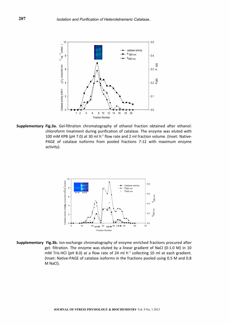
Supplementary Fig.3a. Gel-filtration chromatography of ethanol fraction obtained after ethanol: chloroform treatment during purification of catalase. The enzyme was eluted with 100 mM KPB (pH 7.0) at 30 ml h-1 flow rate and 2 ml fraction volume. (Inset: Native-PAGE of catalase isoforms from pooled fractions 7-12 with maximum enzyme activity).
0
0 10 20 30 40 50 60 70
0
2
4
6
8
10
A2
80 n
m
0.0
0.2
0.4
0.6
0.8Catalase activityA280 nmA405 nm
Cata
lase a
ctiv
ity (
mM
H 2O2
consum
ed m
in-1m
g-1pro
tein
)
A4
05
nm
Fraction Number
0.5 M 0.8 M 1.0 M
0.5 M 0.8 M
Supplementary Fig.3b. Ion-exchange chromatography of enzyme enriched fractions procured after gel- filtration. The enzyme was eluted by a linear gradient of NaCl (0-1.0 M) in 10 mM Tris-HCl (pH 8.0) at a flow rate of 24 ml h -1 collecting 10 ml at each gradient. (Inset: Native-PAGE of catalase isoforms in the fractions pooled using 0.5 M and 0.8 M NaCl).
JOURNAL OF STRESS PHYSIOLOGY & BIOCHEMISTRY Vol. 9 No. 1 2013
207
Page 25
Journal of Stress Physiology & Biochemistry, Vol. 9 No. 1 2013, pp. 184-208 ISSN 1997-0838Original Text Copyright © 2013 by Kapoor, Tripathi and Shrivastava
Isoform Subunits P Tetramer IEF catalase
Sequencing ~320kDa
Supplementary Fig. 4. Schematic representation of heterotetrameric structure of catalase on the basis of internal amino acid sequence analysis.
Amino acids CATA CATB Total % of total amino acids
Tryptophan 10 9 19 1.31
Tyrosine 17 16 33 2.28
Alanine 54 55 109 7.53
Valine 51 50 101 6.98
Isoleucine 27 34 61 4.21
Leucine 56 60 116 8.022
Methionine 11 12 23 1.59
Phenylalanine 42 42 84 5.80
Cysteine 1 1 2 0.138
Supplementary Fig. 5. Hydrophobic amino acids in catalase structure of L.arboricola as revealed from amino acid sequencing
temperature (oC)
pH
Re
lativ
e a
ctiv
ity(%
)
0
20
40
60
80
100
120
pHtemperature
3.5 4.5 5.5 6.5 7.0 7.5 8.0 8.5 9.5 10.5
20 30 40 50 60 70 80 90
Supplementary Fig.6. Effect of pH, temperature on purified catalase from L. arboricola. Catalase activity was assayed at different pH and temperature as described in Materials and methods.
JOURNAL OF STRESS PHYSIOLOGY & BIOCHEMISTRY Vol. 9 No. 1 2013
1
2
3/4
CatB
CatA
CatA
CatB
208