15
Absorption microscop y Kuliah Karakteristik Mikroskopi Basic absorption microscop y
| Date post: | 03-Jun-2018 |
| Category: |
Documents |
| Upload: | fadli-ikhsan |
| View: | 226 times |
| Download: | 0 times |

8/12/2019 Materi 2_ Absorption Microscopy
http://slidepdf.com/reader/full/materi-2-absorption-microscopy 1/20
Absorption microscopy
Kuliah Karakteristik Mikroskopi
8/12/2019 Materi 2_ Absorption Microscopy
http://slidepdf.com/reader/full/materi-2-absorption-microscopy 2/20
Basic absorption microscopy

8/12/2019 Materi 2_ Absorption Microscopy
http://slidepdf.com/reader/full/materi-2-absorption-microscopy 3/20

8/12/2019 Materi 2_ Absorption Microscopy
http://slidepdf.com/reader/full/materi-2-absorption-microscopy 4/20
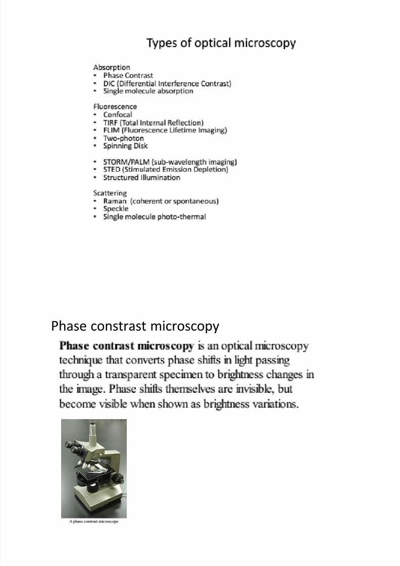
Phase constrast microscopy

8/12/2019 Materi 2_ Absorption Microscopy
http://slidepdf.com/reader/full/materi-2-absorption-microscopy 5/20
When light wave travels through a medium other than vaccum,
interaction with medium causes the wave amplitude and phase to
change in a manner dependent on the properties of medium. Changesin amplitude (brightness) arise from the scattering and absorption of
light, which is often wavelength dependent and may give rise to colors.
Photographic equipment and human eyes are only sensitive to
amplitude variation. Without special arragements, those changes are
therefore invisible. Yet, phase change often, carry important information.(wikipedia)
8/12/2019 Materi 2_ Absorption Microscopy
http://slidepdf.com/reader/full/materi-2-absorption-microscopy 6/20
Working principle
8/12/2019 Materi 2_ Absorption Microscopy
http://slidepdf.com/reader/full/materi-2-absorption-microscopy 7/20
The images constrast is increase in two steps:
1. The background light is phase shifted -90 degree by passing it
through a phase shift ring. This eliminates the phase different
between backgorund light and the scattered light, leading to an
increase intensity between foregound and background.
2. To further increase constrast, the background is dimmed by gray
filter ring.

8/12/2019 Materi 2_ Absorption Microscopy
http://slidepdf.com/reader/full/materi-2-absorption-microscopy 8/20
Differential Interference Constrast Microscopy
Patented by Georges Nomarski at 1952.
The microscope enhances constrast by creating
artificial shadows as if the object is illuminated
from the side. This DIC microscopy uses Polarized light,
making it unsuitable when the object alter
polarization
Replaced by Hoffmann modulation contrast
microscopy. Invented by Robert Hoffmann at1975

8/12/2019 Materi 2_ Absorption Microscopy
http://slidepdf.com/reader/full/materi-2-absorption-microscopy 9/20
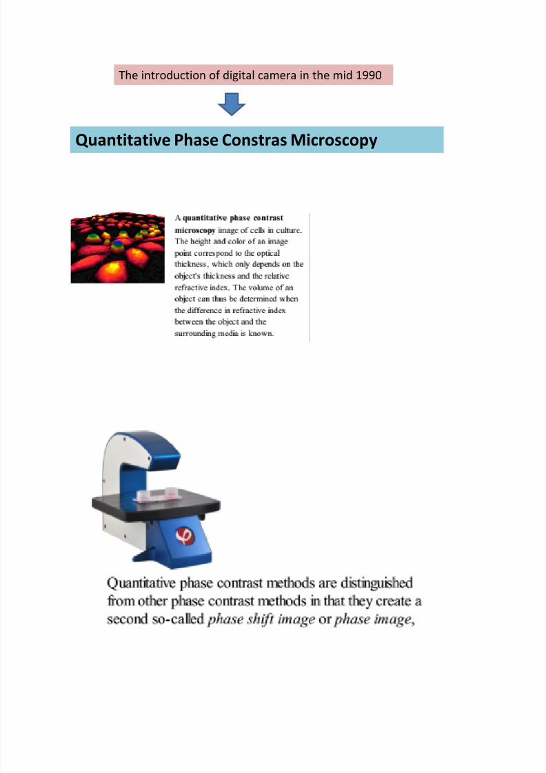
The introduction of digital camera in the mid 1990
Quantitative Phase Constras Microscopy

8/12/2019 Materi 2_ Absorption Microscopy
http://slidepdf.com/reader/full/materi-2-absorption-microscopy 10/20

8/12/2019 Materi 2_ Absorption Microscopy
http://slidepdf.com/reader/full/materi-2-absorption-microscopy 11/20
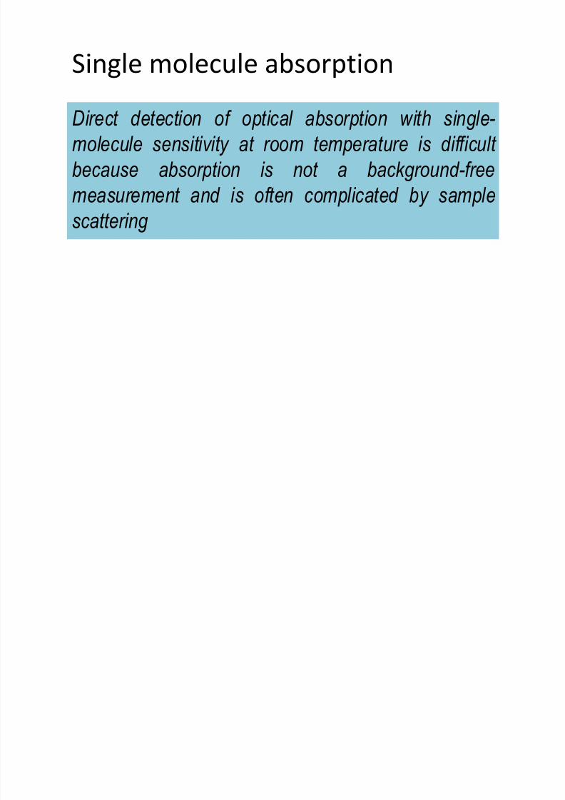
Single molecule absorption
Direct detection of optical absorption with single-
molecule sensitivity at room temperature is difficult
because absorption is not a background-free
measurement and is often complicated by samplescattering
8/12/2019 Materi 2_ Absorption Microscopy
http://slidepdf.com/reader/full/materi-2-absorption-microscopy 12/20
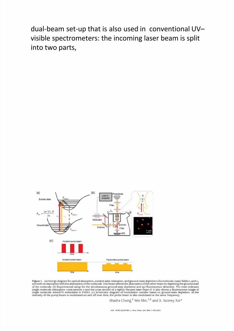
• Celebrano et al. 8 described in detail in Nature
Photonics, allows for the direct imaging ofindividual molecules by monitoring the
attenuation induced in probe light.
The molecular absorption cross-sections, often < than afew square angströms,
a tightly focused light beam, as used in optical
microscopes, reveal that less than one photon in a
million is absorbed.

8/12/2019 Materi 2_ Absorption Microscopy
http://slidepdf.com/reader/full/materi-2-absorption-microscopy 13/20
Direct observation of a single molecule by measuring its
Attenuation (serapan) of a light beam
Three different method that allow detection of
light absorption by single molecule
8/12/2019 Materi 2_ Absorption Microscopy
http://slidepdf.com/reader/full/materi-2-absorption-microscopy 14/20
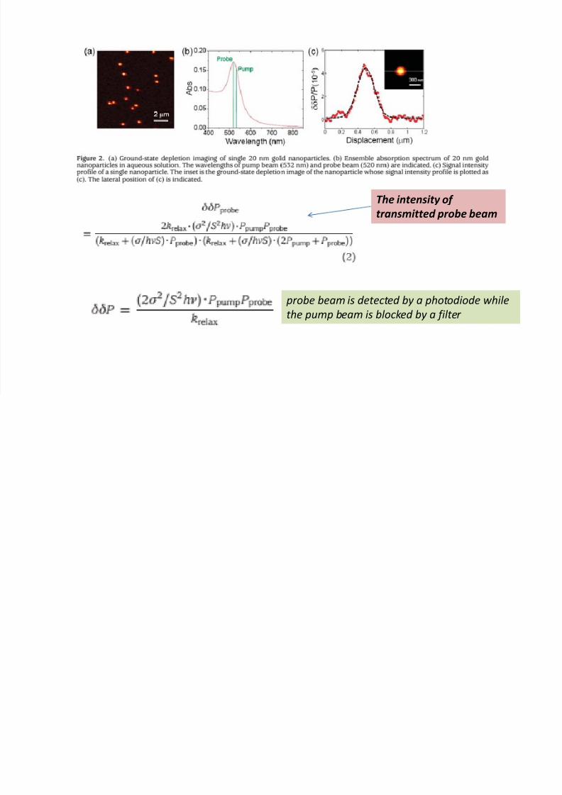
The local heating induced by light absorption results in tiny changes
in the local refractive index, which can be probed by the enhanced
backscattering of a second, probe, beam(red). Fast intensitymodulation of the absorbed blue beam results in measureable
modulations in backscattering intensity with the same frequency.
“Double beam”
8/12/2019 Materi 2_ Absorption Microscopy
http://slidepdf.com/reader/full/materi-2-absorption-microscopy 15/20
Modulation of the blue beam influences the absorption of
the green beam by depleting (menghabiskan) the ground
state.

8/12/2019 Materi 2_ Absorption Microscopy
http://slidepdf.com/reader/full/materi-2-absorption-microscopy 16/20

8/12/2019 Materi 2_ Absorption Microscopy
http://slidepdf.com/reader/full/materi-2-absorption-microscopy 17/20

8/12/2019 Materi 2_ Absorption Microscopy
http://slidepdf.com/reader/full/materi-2-absorption-microscopy 18/20
The attenuated Power (P) of the incident beam (P) by the single
molecule:
hv is the photon energy; is the absorption crosssection for the single chomophore
(single molecule) (~ 10-16 cm2 at room temperature);
S is the beam waist (pinggang) area .

8/12/2019 Materi 2_ Absorption Microscopy
http://slidepdf.com/reader/full/materi-2-absorption-microscopy 19/20
K relax
is k relax2 : the rate constant of the rate-limiting step in the
sequential relaxation process from the vibronic states prepared by theoptical excitation to the lowest vibrational level in the ground electronic
state.

8/12/2019 Materi 2_ Absorption Microscopy
http://slidepdf.com/reader/full/materi-2-absorption-microscopy 20/20


