Seizures: Abnormal electrical discharge in the brain with an associated altered level of consciousness and rhythmic movements followed by a post‐ictal state.•Associated Symptoms: incontinence of bowel and bladder, apnea, cyanosis. May be preceded by an aura.•Common causes: fever, ingestion, electrolyte disturbance, infections or tumor.
•Most important aspect of diagnosis is detailed history of event, type of seizure and resulting disability • Family history of seizures? Travel History or sick contacts?• Labs: CBC, electrolytes (glucose, calcium sodium and magnesium), drug levels and toxicology screen.
• ED: lasts >5 minutes, head injury, high fever, compromised resp or cardiac function.
Bilateral hemispheres are engagedSudden loss of toneOriginates in one hemisphere
ClonicTonic
Rhythmic repetitive movementsSustained extension or flexion of head trunk or extremities
Febrile Occur on rise of fever, usually between ages 6‐60 months, generalized, last less than 15 minutes and do not recur within 24hrs. No testing or neuroimaging necessary unless meningeal signs.
Status Epilepticus A single seizure lasting longer than 30 minutes or two or more consecutive seizures without returning to baseline LOC.
Referral to the ED
First line therapy ‐ABC, then benzodiazepines PR, IM, IV.Secondary therapy with AED’s. Dilantin, Keppra or phenobarbital load.Refractory‐may require drug induced coma‐EEG for burst suppression or surgical resection. Vagal nerve stimulator may be implanted.
• Stabilize‐0‐5 min, ABC’s, IV, critical labs, drug levels• Initial Therapy‐ 5‐20 min, Benzodiazepines‐IM,IV, PR• Secondary Line Therapy 20‐40 min, Keppra, Phosphenytoin, valproate, last choice‐phenobarbital
• Third Line Therapy‐ 40‐60 min, repeat dosing of above, then anesthetic dosing of midazolam, propofol, thiopental or pentobarbital with continuous EEG monitoring(neurology).
‐Seizure associated injury can occur within 30 minutesCourtesy of American Epilepsy Society
• EEG: Non‐invasive, provides seizure location and abnormalities such as spikes, slowing or silence.
‐Records the frequency , amplitude and characteristics of brain waves.Indication: AMS, seizures, subclinical seizures, identify location of seizures or adjunct in brain death.• Type‐short, sleep deprived, 24hour or continuous.
Advantages• Sensitive to presence of blood, bones and lesions.• Can be done quickly•More readily available• Fluid changes seen‐hydrocephalus/cerebral edema.
Disadvantages• Exposure to radiation‐highest with abdomen.• Need to think of cumulative affects.
• Indication: infectious, autoimmune or inflammatory process to include meningeal signs of neck stiffness and + Kernig and/or Brudzinski signs.
• Age Specific‐ infant aged 6 to 12 months has a seizure and fever without immunizations for Haemophilus influenzae type b (Hib) or Streptococcus pneumoniae or if his or her immunization status is unknown. Adolescent: Therapeutic to remove CSF in Pseudotumor cerebri
• LP also should be considered when a child with a seizure and fever has been pre‐treated with antibiotics, because antibiotics can mask the signs and symptoms of meningitis but may not be sufficient to eradicate it.
• Measure opening pressure• Contraindicated with high ICP‐perfom CT first
• Procedure: Lateral decubitus position or upright with procedural sedation prnand 1% lidocaine for local anesthesia
• LP below the level of the conus medullaris which ends at L1‐2• Iliac crest lines up with L3‐4. Most common is completed at L4‐L5. Can be L3‐4 or L5‐S1.
• Perform with sterile field, time‐out and consent• Pre‐assemble manometer and collection tubes numerically for quick access and ensure special labs are ordered.
• Replace stylus before withdrawing needle• Post‐procedure‐watch for CSF leak, headache, lower extremity sensory changes, hematoma at the site or AMS.
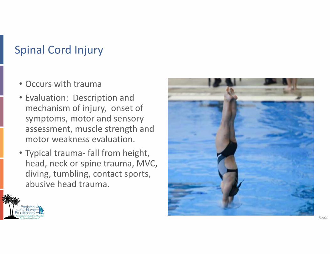
• Occurs with trauma• Evaluation: Description and mechanism of injury, onset of symptoms, motor and sensory assessment, muscle strength and motor weakness evaluation.
• Typical trauma‐ fall from height, head, neck or spine trauma, MVC, diving, tumbling, contact sports, abusive head trauma.
• Age specific considerations: • Infants have poorly developed cervical musculature, head is disproportionately large
• Children less than age 9 have wedge shaped vertebral bodies, angled horizontally.
• Young children have cartilaginous endplates with lax interspinous ligaments, so they are more prone to SCIWORA (spinal cord injury without radiological abnormality).
• Children with Down syndrome are prone to atlanto‐axial subluxation as a result of acute flexion injury
Diagnostics: • Radiographs: head and neck films with lateral view, odontoid views
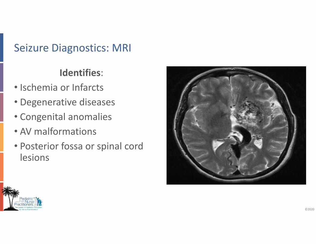
• Neck CT or MRI
Management:• Manage airway• Immobilize C‐spine• High dose IV steroids (30mg/kg)*• Manage neurogenic shock with fluids, alpha‐adrenergic agents, continuous monitoring. Spinal shock can last several days, causing paralysis and loss of tone with hypovolemia and hypotension.
• Traumatic Brain Injury: direct trauma as primary injury or secondary injury from systemic (hypotension, hypoxia, anemia, etc) or intracranial problems such as tumor, cerebral edema, seizures or infection
• CT Scan: To scan or not to scan? Use PECARN or CHALICE Criteria• Acute monitoring: ICP monitor, cerebral perfusion pressure (CPP) monitoring
• Goal of TBI management: Normal ICP, Optimize CPP, oxygenation and ventilation with appropriate cardiac output, B/P, surgical evacuation of masses, CSF or blood.
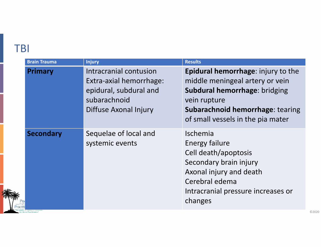
Primary Intracranial contusionExtra‐axial hemorrhage: epidural, subdural and subarachnoidDiffuse Axonal Injury
Epidural hemorrhage: injury to the middle meningeal artery or veinSubdural hemorrhage: bridging vein ruptureSubarachnoid hemorrhage: tearing of small vessels in the pia mater
Secondary Sequelae of local and systemic events
IschemiaEnergy failureCell death/apoptosisSecondary brain injuryAxonal injury and deathCerebral edemaIntracranial pressure increases or changes
• Follow Critical Pathway for Treatment of Established Intracranial Hypertension in Pediatric Trauma (Society of Critical Care Medicine (2012). ( available on line: www.braintrauma.org/pdf/guidelines_pediatric2.pdf)
• Continued evaluation of symptoms is extremely important in children with moderate to severe brain injuries, but children with mild injury should also be monitored for changes in status and sequelae
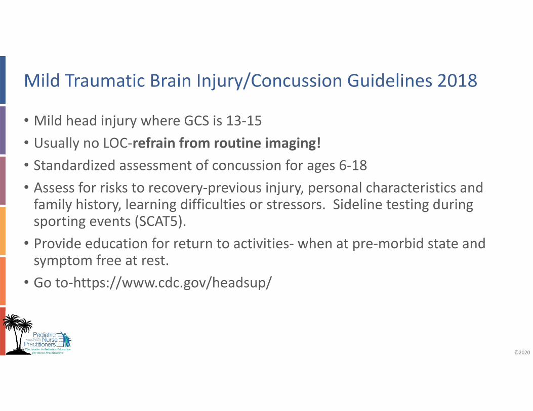
• Mild head injury where GCS is 13‐15• Usually no LOC‐refrain from routine imaging!• Standardized assessment of concussion for ages 6‐18• Assess for risks to recovery‐previous injury, personal characteristics and family history, learning difficulties or stressors. Sideline testing during sporting events (SCAT5).
• Provide education for return to activities‐ when at pre‐morbid state and symptom free at rest.
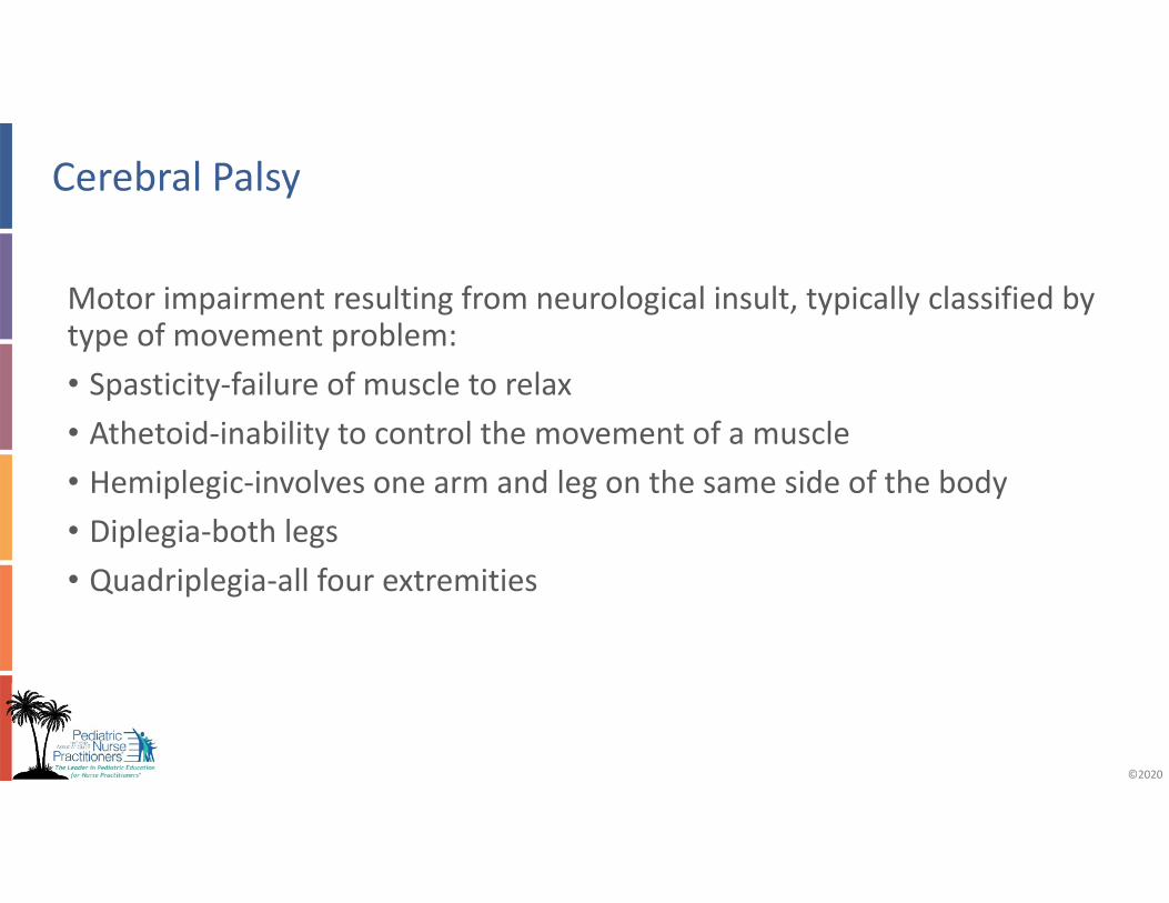
Motor impairment resulting from neurological insult, typically classified by type of movement problem:• Spasticity‐failure of muscle to relax• Athetoid‐inability to control the movement of a muscle• Hemiplegic‐involves one arm and leg on the same side of the body• Diplegia‐both legs• Quadriplegia‐all four extremities
• Management: treatment of known cause and neurology consult with recommended diagnostic work up. EEG abnormal 90% cases. Therapy should be initiated with acyclovir in any young infant or child who is suspected of having viral encephalitis, especially those who appear ill. Cause often never known, may have irreversible brain damage.
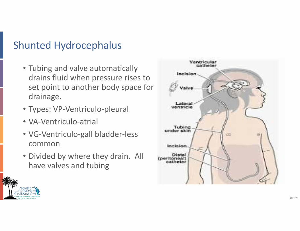
• An excess of CSF in the cranial vault due to either excess production, obstructed flow or defective drainage paths.
• May result in ventriculomegaly, increased ICP, macrocephaly and cognitive impairments.
• Signs and Symptoms: Rapid head growth, full or tense fontanel, irritability, vomiting, strabismus, inability to look up, apnea, dysphagia, hoarse or weak cry
Infection: 5‐10%. Fever, Neck stiffness, Pain, Tenderness, Redness, Drainage from the shunt incisions or tract, AMS or Abdominal pain.Malfunction: Blocked or broken: Seizures, pain, worsening cognitive function, speech impairment or dysphagia, limb or balance problems.Overdraining or underdraining: Can cause hemorrhage, alter brain growth or hydrocephalus persists‐must adjust valve, close follow up.Diagnosis: Shunt series and CT scan, CSF tap and studiesTreatment: EVD and antibiotics with replacement of device when infection has cleared.
• Premature fusion of one or more of the cranial suture lines causing abnormal skull growth deformities and restriction if corrective surgery is not performed.
• Syndromes affected in 40% of cases‐Aperts, Crouzons, Pierre‐ Robin, Turner, Goldenhar, VATER, Dandy‐Walker.
• Associated brain abnormalities‐hydrocephalus, Chiari malformation and increased ICP.
• Other associated abnormalities: Hearing loss, vision problems or limb abnormalities. May have cognitive or behavioral impairments.
• Diagnostics‐skull film, CT, 3‐D CT, MRI• Management: Surgery: Complex vault repair based on defect, bones broken. Endoscopic approaches now used with less complications but may be more costly.
• ‐Post‐op Complications: Pain and Bleeding! May also see: SIADH, bradycardia, fever, seizures and facial edema or transfusion related complications.
• Younger patients have less complications, many have late diagnosis.
Botulism: progressive, descending, symmetrical, neuromuscular weakness as a result of presence of C. botulinum spores in stool. • Infant less than the age of 12 months,may have exposure to contaminated soil, honey or water.
• Diagnosis: identification of C. botulinum spores in feces/clinical features. • Treatment is botulism specific IVIG (BIG‐IV) obtained only through the California Department of Health.
Guillain‐Barre Syndrome: progressive neuromuscular weakness • Presents 4 – 6 weeks after a viral illness or prior infection. • Symptoms are progressive and symmetrical with ascending paralysis. • Pain, numbness, tingling of the extremities and sensory loss with gait disturbances.
• Management: diagnosis is through CSF protein measurement and clinical findings. Treatment IVIG and or plasmapheresis .
Muscular Dystrophy: diagnosed by age 2. The limb girdle muscular dystrophies have progressive, symmetrical proximal weakness with genetic link. • Concern for progressive neuromuscular weakness which will eventually involve respiratory center and require various ventilation support.
Spinal Muscular Atrophy (SMA): neuromuscular disease of childhood. Symptoms include weakness at birth or within the first year of life, feeding, breathing difficulties. • Type I or Werdnig Hoffman, early diagnosis by 6 months. • Type 2 = intermediate, usually can sit, but not stand or ambulate. Fine motor tremor of hands, tongue fasiculations
• Type 3 or Kugelberg‐Welander, presents after age 18 months and walk. Weakness to proximal muscle groups. Weak lower extremities. Could live full life but wheelchair dependent.
• Stroke: Sudden interruption of arterial or venous blood flow to a focal region of the brain.
Ischemic: Disruption of blood flow leads to brain dysfunction from hypoperfusion, thrombus or embolism.Hemorrhagic: A rupture of a vessel or aneurysm that leaks into surrounding tissues and cells.Presentation: AMS, seizures, focal deficits, aphasia, visual deficitsFollow AHA National Stroke guidelines
• Acute Disseminated Encephalomyelitis‐ADEMBrief but widespread inflammation of the brain and spinal cord that damages the myelin(white matter)‐usually follows bacterial or viral illness.Symptoms: Rapid onset of Encephalitis like illness‐fever, fatigue, headache, N/V. Severe‐seizures and coma, vision loss, weakness or paralysis.Treatment: Steroids, IVIG or plasmapheresis, can relapse.
• Posterior Reversible encephalopathy Syndrome‐PRESConstellation of symptoms and radiologic abnormalities resulting from disruption in the blood brain barrier with radiologic evidence of vasogenicedema at occurs most frequently in the posterior brain circulation but can be seen elsewhere.• Most instances is reversible, may occur in other areas of the brain and rarely the spinal cord.
• Most common presenting symptom in pediatrics is: SEIZURES
• Other sxs: Hypertension, headaches, visual disturbances, focal neurological deficits, N/V, altered level of consciousness
• Brain Death• Clinical diagnosis defined as the irreversible loss of all brain functions (including brain stem).
• Coma, apnea, and the absence of brainstem reflexes are essential findings in brain death.
• Potential reversible causes of coma should be excluded• Two examinations performed by different attending physicians must be completed separated by an observation period of 24 hours for neonates (37 weeks gestation to term infants 30 days of age) and 12 hours for infants and children (>30 days to 18 years). State by state variability.
• Pseudotumor Cerebri• Rapid reproduction of ones’ own cerebral spinal fluid. • Visual loss is a potential complication. • Goal of treatment is vision preservation and alleviation of symptoms. • Visual field testing, dilated fundoscopic exam and imaging of optic disks are necessary for proper diagnosis
• Lumbar puncture is diagnostic and is a treatment for PTCS. Hallmark opening pressure would be greater than 280 mm
• Arteriovenous malformation AVM• Congenital intracranial malformation distinguished by a persistently abnormal connection between arteries and veins within the brain without an interposed or developed capillary bed
• associated with neurological deficits such as hemiparesis, seizures and speech may be affected.
• Management includes ABCD, close ICP control • Cerebral angiography is considered the gold standard study to identify the arteries involved
• May need surgical clipping, craniectomy or embolization
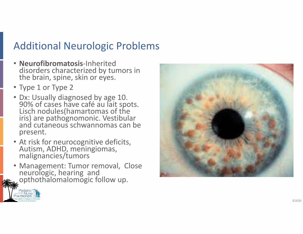
• Neurofibromatosis‐Inherited disorders characterized by tumors in the brain, spine, skin or eyes.
• Type 1 or Type 2• Dx: Usually diagnosed by age 10. 90% of cases have café au lait spots. Lisch nodules(hamartomas of the iris) are pathognomonic. Vestibular and cutaneous schwannomas can be present.
• At risk for neurocognitive deficits, Autism, ADHD, meningiomas, malignancies/tumors
• Management: Tumor removal, Close neurologic, hearing and opthothalomalomogic follow up.
• Tuberous Sclerosis Complex Autosomal dominant disorder, develop benign tumors that develop in the brain, skin, kidneys.
• Features: hypomelanotic fibromas, cortical tubers, subependymal nodules or astrocytoma, facial angiofibromas, fibromas, lymphangioleiomyomatosis, hamartomas, bone or renal cysts
• Diagnosis: Definite: 2major features, 1major and 2 minor.• Mild to severe. Infants with seizures, cardiac rhabdomyomas, ash leaf spots. Seizures can be refractory.
• Management: Symptom dependent. Seizure control. Immunosuppressant therapy may be helpful (not approved). Tumor removal.
• Multi‐system problems!• Complex care/Medically fragile • Rehabilitation and medical home management.• Chronic care of child with TBI or CP includes collaboration with services such as Rehab medicine, physical therapy, speech and language, school services, neurology and other sub‐specialty services.
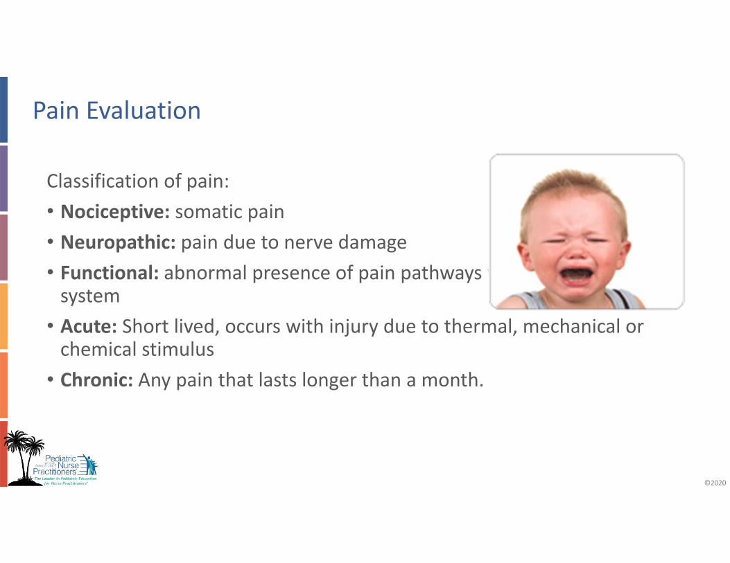
Classification of pain:• Nociceptive: somatic pain• Neuropathic: pain due to nerve damage• Functional: abnormal presence of pain pathways within the nervous system
• Acute: Short lived, occurs with injury due to thermal, mechanical or chemical stimulus
• Chronic: Any pain that lasts longer than a month.
Methadone: physiologically dependent on opioids.Buprenorphine: Used for mild‐to‐moderate opioid withdrawal to reduce risk of precipitated withdrawal. Naltrexone: Recommended for preventing relapse.
manifested by withdrawal syndrome produced by abrupt
cessation of the drug, rapid dose reduction, decreasing blood level of the drug, and/or administration of
an antagonist. (Nierengarten, M. Pediatrics, 2016)
AddictionPrimary, chronic, neurobiological disease with genetic, psychosocial, and environmental factors that influence its development, and is characterized by such behaviors as impaired control over opioid use, compulsive use, continued use despite harm, and craving.
State Behavioral Score‐Respiratory drive, coughing, best response to stimulation, attentiveness to provider, tolerance to care, movement to consoled.Scores from ‐3 to 2 or unresponsive to agitated. Goal is 0.
Richmond Agitation Scoring System• Longer range of scores from dangerous to self to frequent non‐purposeful to goal 0 calm to lightly sedated, moderate and deeply sedated.
• Scores from +4 to ‐5 or from Combative to unarouseable.
Sedation Scores‐Those supported on mechanical ventilation scored on the following:
• Safe to sedate??‐Screen them! ‐AMPLE mnemonicAllergies, Medications, Past Illnesses, Last oral intake, Events leading up to injury• Consent • Age and developmental specific differences• Underlying problems or behavior disorders‐may require alternative medications or have unexpected effects of medications
• Team sport‐need nurse, respiratory support, safe room with immediate access to emergency age appropriate equipment
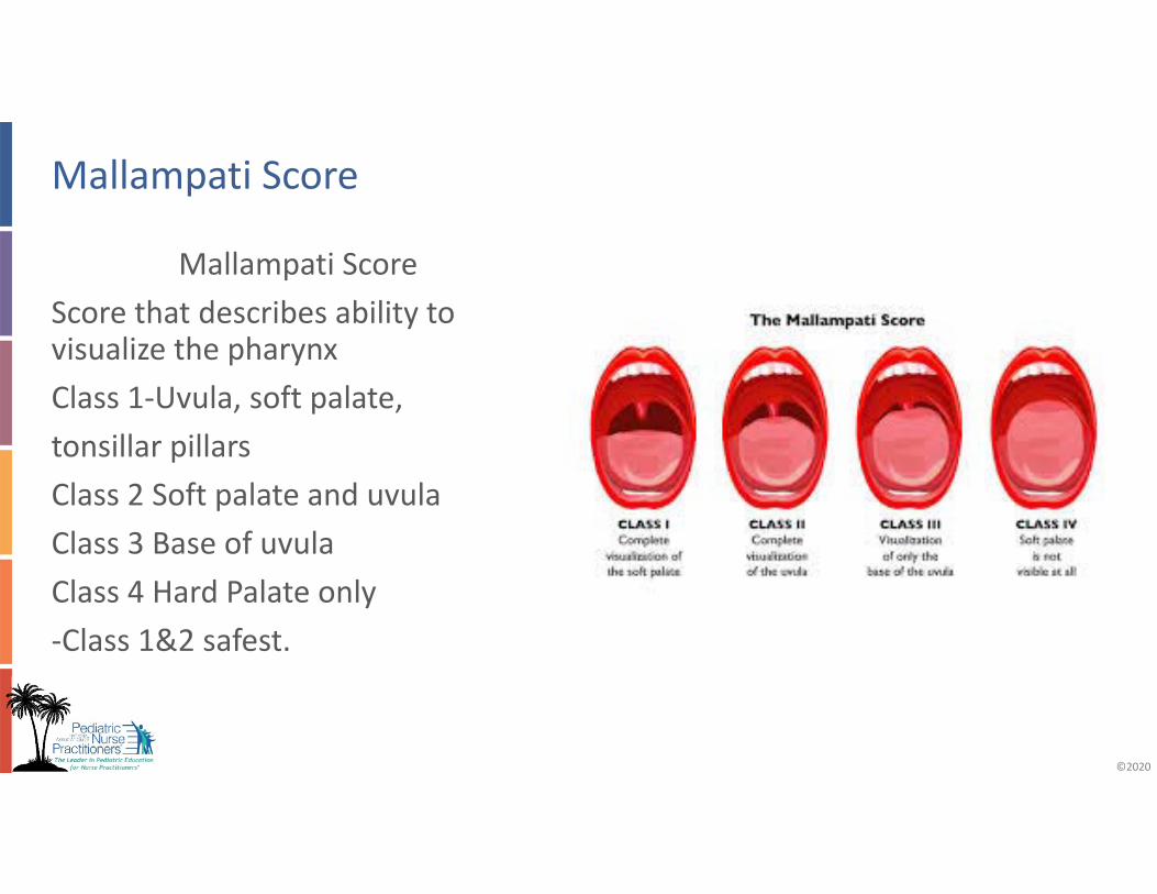
Mallampati ScoreScore that describes ability to visualize the pharynxClass 1‐Uvula, soft palate,tonsillar pillarsClass 2 Soft palate and uvulaClass 3 Base of uvulaClass 4 Hard Palate only‐Class 1&2 safest.
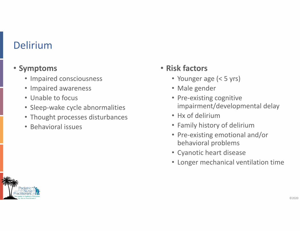
• What is delirium? • “a disturbance of consciousness and cognition that develops acutely with a fluctuating course of inattention and an impaired ability to receive, process, store or recall information”
‐‐Smith et al, 2009
• A frequent and serious complication of critical illness with links to:• Increased mortality • Prolonged hospital stays• Long‐term disability• A complication of hospitalization
• Non‐pharmacologic‐circadian reset, frequent re‐orientation, schedule for the day.
• Antipsychotics‐Haldol most widely used across the lifespan. Prolonged Qtcor dystonic reactions.
• Atypical Antipsychotics‐ Resperidone, Zyprexa or Geodon have lower side effects. Blocks at multiple receptor sites, not just dopaminergic receptor sites as with haldol.
• Obstructive Sleep Apnea‐could affect growth‐need sleep study to formally evaluate
• ENT evaluation for possible T & A or obstruction• Nutrition consult if obese• Screen for other conditions‐night terrors, enuresis, anxiety• Delirium‐Assess for presence, hospital not a place for rest• Medication side effect‐Stimulants, OTC medications, watch for “natural” sleep aids.
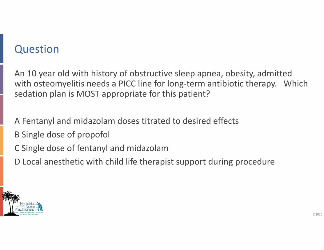
An 10 year old with history of obstructive sleep apnea, obesity, admitted with osteomyelitis needs a PICC line for long‐term antibiotic therapy. Which sedation plan is MOST appropriate for this patient?
A Fentanyl and midazolam doses titrated to desired effectsB Single dose of propofolC Single dose of fentanyl and midazolamD Local anesthetic with child life therapist support during procedure
An 10 year old with history of obstructive sleep apnea, obesity, admitted with osteomyelitis needs a PICC line for long‐term antibiotic therapy. Which sedation plan is MOST appropriate for this patient?
D. Local anesthetic with child life therapist support during procedure
A 1 year old with head injury develops acute irritability with decreased sensorium and bradycardia. The most important urgent management includes intubation and:
A. MRI brain and administration of mannitol B. CT Brain and administration of hypertonic salineC. CT brain and administration of furosemideD. MRI brain and administration of hypertonic saline
A 1 year old with head injury develops acute irritability with decreased sensorium and bradycardia. The most important urgent management includes intubation and:
B. CT Brain and administration of hypertonic saline
An afebrile sedentary adolescent female is seen in the ER for headache and worsening vision disturbances. The family is concerned for meningitis. What is the next course of treatment?
A Head CTB Lumbar punctureC Formal eye examD Nutrition consult
An afebrile sedentary adolescent female is seen in the ER for headache and worsening vision disturbances. The family is concerned for meningitis. What is the next course of treatment?