1
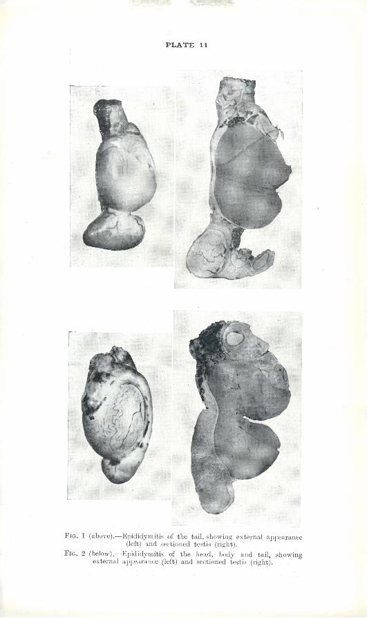
PLATE 11 Fig. 1 (above).—Epididymitis of the tail, showing external appearance (left) and sectioned testis (right). Fig. 2 (below).—Epididymitis of the head, body and tail, showing externa] appearance (left) and sectioned testis (right).