1 Prostate Immunohistochemistry Murali Varma Cardiff, UK [email protected]Sarajevo Nov 2013 IHC Interpretation: General Principles (1) Must be aware of staining pattern of antibody in the relevant tissue Nuclear/cytoplasmic/membranous eg. b-Catenin used for colon cancer vs. prostate cancer • Nuclear positivity only in colon cancer • Membranous/cytoplasmic positivity in up to 88% of prostate cancer IHC Interpretation: General Principles (2) Not just positive or negative • Quantitative aspect has to considered • Immunoreactivity is a continuous variable • Focal positivity must be interpreted with caution • Negativity in limited material not diagnostic IHC must always be interpreted in the context of morphology Literature Interpretation: Caveats Interpret literature data critically due to differences in: Staining techniques • Antibody type, fixation, antigen retrieval Characteristics of tumours studied • Effect of grade o PSA/PSAP expression lower in high-grade prostate carcinoma o Uroplakin expression lower in high-grade TCC Literature interpretation: Caveats (2) Definition of positivity may vary between studies • Cut-offs used to consider CK7/CK20 as positive varies from any positivity to >10% cells positive Microarray techniques may overestimate specificity and underestimate sensitivity especially if staining patchy Interpreting Immunohistochemistry: A Systematic Approach 1. Check controls a) Appropriate tissue for control b) Appropriate staining reaction 2. Exclude spurious positivity (pigment, biotin etc) 3. Pattern of positivity a) Correlate with morphology • Necrosis, entrapped benign tissue etc. b) Nuclear/cytoplasmic/membranous
3. Pattern of positivity a) Correlate with morphology
• Necrosis, entrapped benign tissue etc.
b) Nuclear/cytoplasmic/membranous
2
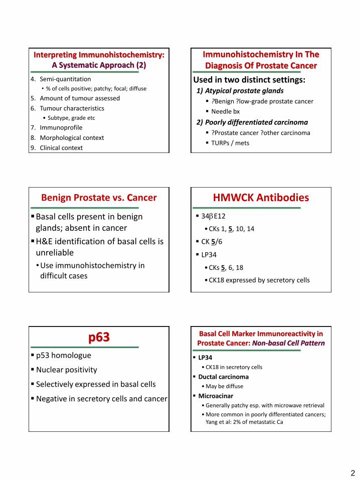
Interpreting Immunohistochemistry: A Systematic Approach (2)
4. Semi-quantitation
• % of cells positive; patchy; focal; diffuse
5. Amount of tumour assessed
6. Tumour characteristics
• Subtype, grade etc
7. Immunoprofile
8. Morphological context
9. Clinical context
Immunohistochemistry In The Diagnosis Of Prostate Cancer
1) Atypical prostate glands
?Benign ?low-grade prostate cancer
Needle bx
2) Poorly differentiated carcinoma
?Prostate cancer ?other carcinoma
TURPs / mets
Used in two distinct settings:
Benign Prostate vs. Cancer
Basal cells present in benign glands; absent in cancer
H&E identification of basal cells is unreliable
•Use immunohistochemistry in difficult cases
34bE12
• CKs 1, 5, 10, 14
CK 5/6
LP34
• CKs 5, 6, 18
• CK18 expressed by secretory cells
HMWCK Antibodies
p63 p53 homologue
Nuclear positivity
Selectively expressed in basal cells
Negative in secretory cells and cancer
Basal Cell Marker Immunoreactivity in Prostate Cancer: Non-basal Cell Pattern
LP34
• CK18 in secretory cells
Ductal carcinoma
• May be diffuse
Microacinar
• Generally patchy esp. with microwave retrieval
• More common in poorly differentiated cancers; Yang et al: 2% of metastatic Ca
3
Basal Cell Marker Immunoreactivity in Prostate Cancer: Basal Cell Pattern
Ductal type prostate cancer
• ?intraductal spread of tumour
Microacinar prostate cancer
• Very rare
• Oliai et al Am J Surg Pathol 2002;26:1151-60.
• 1.1% cases in referral material
• ?entrapped outpouchings of PIN
• ?basal cells retained in early invasive cancer
Comparison Of Basal Cell Markers
CK5/6 more sensitive than 34bE12 • Abrahams et al. Histopathology 2002;41:35-41.
p63 slightly more sensitive than 34bE12 • Shah et al. Am J Surg Pathol 2002;26:1161-68.
• Weinstein et al. Mod Pathol 2002;15:1302-08.
LP34 more sensitive than CK5/6 and CK14 Freeman et al. Histopathology 2002;40:492-4.
Which Is The Best Basal Cell Marker?
The one that works best in your lab!
Every case is a quality control •Check out the background benign glands
Consider HMWCK + p63 combination rather than HMWCK + CK5/6
Limitations Of Basal Cell Markers
Often used as a single marker Cancer diagnosis based on negative
staining reaction Benign glands are occasionally
negative Basal cell layer fragmented in high-
grade PIN and adenosis
Basal Cell Marker Negative Small Glandular Proliferations
Prostate cancer
Outpouchings of high-grade PIN
Adenosis
Alpha-Methylacyl-CoA-Racemase
A revolutionary new positive marker for prostate cancer
Identified by cDNA library subtraction and microarray techniques
• First of many
• Prostein (P501S): a recently described prostatic marker (benign and malignant)
Rabbit monoclonal antibody
• Higher affinity than conventional mouse monoclonals, especially for small epitopes
4
When is AMACR Immunoreactivity Considered Positive?
Only circumferential staining of the luminal cells that can be identified at low (100x) magnification with no more than weak, non-circumferential staining of adjacent benign glands.
Basal cell markers vs. AMACR
Basal cell markers
• Difference between benign glands and cancer is qualitative
• No basal cells in cancer
AMACR
• Difference is quantitative
• AMACR over-expressed in cancer but also expressed in benign glands
• Hence titration of sensitivity is critical
Basal cell markers vs. AMACR Evaluation of immunoreactivity
AMACR
• Positive or negative
Basal cell markers
• Positive or negative
• Pattern of immunoreactivity
• “basal cell pattern”
Limitations of AMACR
AMACR immunoreactivity in focus of cancer often heterogeneous
• Initial studies: 97-100% sensitivity
• More recent: up to 20% of limited cancers AMACR negative
Commonly positive in high-grade PIN and its outpouchings
AMACR immunoreactivity weaker in prostate cancer variants (pseudo-hyperplastic, foamy gland)
AMACR: Notes of caution
Always use in conjunction with basal cell marker(s)
Do not downgrade (cancer to suspicious or suspicious to benign) based on negative AMACR staining
Do not upgrade from “suspicious ?outpouchings of HG-PIN” to cancer based on positive AMACR staining
Limitations of AMACR
Positive in
• Adenosis (15%)
• Nephrogenic adenoma (50%)
• Variety of other benign mimickers
• Atrophy, PAH etc
• Occasional benign glands may show generally weak positivity
5
Suspicious to Cancer “Based” on Racemase Positivity
1. Architecturally atypical foci with insufficient cytologic atypia
2. Foci with crush artefact
3. Too few atypical glands
differential diagnosis NOT outpouchings of high-grade PIN or nephrogenic adenoma and beware of adenosis!
AND
Sections for Immunohistochemistry
Intervening levels must be retained on treated slides for potential immunostaining
•Small atypical foci may not be present in deeper levels
Tissue ribbons
Level 1 Level 2 Level 3
Glass slides
H&E
Immuno
Spares
Cardiff Protocol for Processing Prostate Biopsies Advantages of Cardiff Protocol
Immunostained section corresponds more closely with H&E section
All 3 levels immunostained
•Basal cell marker positivity may not be present in all levels
Antibody Cocktails
Advantages
• Multiple markers can be performed on limited number of spares available.
• Cost and time savings
Disadvantages
• Antibody dilutions cannot be optimised separately for local conditions.
• Single retrieval method for both markers.
• One marker may mask other if single colour detection used (AMACR + p63)
Interpreting Prostate IHC (1)
First evaluate glands away from suspect focus
• Quality assurance
• Some patients have very fragmented pattern in benign glands
• May be microscopic focus missed on H&E
Evaluate immunostaining in focus as a whole rather than in individual glands
• HMWCK (-) glands in adenosis morphologically identical to HMWCK (+) glands within the focus
6
Factors to consider: Morphological differential diagnosis ?PIN ?Ca: AMACR not useful ?Nephrogenic adenoma: PSA/PSAP rather than HMWCK/AMACR
Size of suspect focus • Small HMWCK(-): Suspicious • Larger HMWCK(-): Cancer
Degree of atypia • Architectural • Cytological
Pattern in adjacent benign glands
Interpreting Prostate IHC (2) Prostate Cancer vs. TCC
Prostatic markers
•PSA, PSAP, PSMA, Prostein (P501S), Leu7 (CD57)
Urothelial markers
•HMWCK, p63, CK7, CK20, uroplakin III
PSA: Sensitivity Very sensitive in low-grade prostate cancer
PSA expression lower in high-grade prostate cancer
Goldstein (Am J Clin Path 2002;117:471-7)
• 225 prostate cancers various of grades
• All Gleason 6 tumours >50% cells PSA+
• 26% Gleason 10 tumours <5% cells PSA+
• PSA negativity in high-grade cancer does not exclude prostatic origin
PSA: Sensitivity Varma et al. (Am J Clin Path 2002;118:202-7)
20 Gleason score 10 prostate cancers
Monoclonal anti-PSA: 35% <10% cells positive
Polyclonal PSA: 95% >50% cells +
Polyclonal anti-PSA significantly more sensitive than monoclonal anti-PSA in poorly differentiated prostate cancer
PSA: Specificity
PSA immunoreactivity reported in a variety of non-prostatic tumours
• Almost always weak and focal
• Reflection of large number of cases studied
PSA: Quality Control Issues
PSA less expressed in high-grade prostate cancer compared to benign prostate /low-grade prostate cancer
Polyclonal anti-PSA significantly more sensitive than monoclonal anti-PSA in high-grade prostate cancer
? Use high-grade tumour for positive control and optimising dilutions
7
Choice of Prostatic Marker
A survey of UK laboratories found that all used PSA but only about half used PSAP.
PSA negative poorly differentiated prostate cancers may diffusely PSAP (+)
Use of both PSA and PSAP is recommended when evaluating poorly differentiated tumours