Purification of Recombinant Helicobacterpylori UreaseApoenzyme Encoded by ureA and ureB
LI-TAI HU, PAUL A. FOXALL, ROBERT RUSSELL, AND HARRY L. T. MOBLEY*
Division of Infectious Diseases, Department of Medicine, University of MarylandSchool of Medicine, 10 South Pine Street, Baltimore, Maryland 21201
Received 20 November 1991/Accepted 16 April 1992
Helicobacter pyloni, a gram-negative, microaerophilic, spiral-shaped bacterium, is an etiologic agent ofhuman gastritis and peptic ulceration and is highly restricted to the gastric mucosa of humans. Urease,synthesized at up to 6% of the soluble cell protein, hydrolyzes urea, thereby releasing ammonia, which may
neutralize acid, allowing survival of the bacterium and initial colonization of the gastric mucosa. The urease
protein is encoded by two subunit genes, ureA and ureB; however, accessory genes are necessary for enzymeactivity. H. pylori urease genes were isolated from a cosmid gene bank and subcloned on a 5.8-kb Sau3A partialfragment carrying ureCDAB, corresponding to four open reading frames described by A. Labigne, V. Cussac,and P. Courcoux (J. Bacteriol. 173:1920-1931, 1991). Clones were confirmed as urease gene sequences bypolymerase chain reaction amplification. The recombinant enzyme was purified from the soluble protein ofFrench press lysates of Escherichia coli DH5ox(pHP402) by chromatography on DEAE-Sepharose, Phenyl-Sepharose, Mono-Q, and Superose 6 resins. Fractions containing a catalytically inactive apoenzyme were
identified by an enzyme-linked immunosorbent assay (ELISA) by using antisera to native UreA (29.5 kDa) andUreB (66 kDa). Purified recombinant urease was indistinguishable from native enzyme on a Superose 6 columnand on Coomassie blue-stained sodium dodecyl sulfate-polyacrylamide gels. The protein reacted specifically onWestern blots (immunoblots) with anti-UreA and anti-UreB antibodies and was recognized with an intensityequal to that of the native enzyme in an ELISA using human sera. Clones containing only ureA and ureB alsoproduced an assembled but inactive enzyme. Enzyme activity was not restored by in trans complementationwith cloned urease accessory gene sequences from Proteus mirabilis or Morganella morganii. H. pylori urease
genes (ureCDAB) subcloned into pACYC184 were also not complemented with any of 1,000 cosmid clonescontaining H. pyloni chromosomal sequences. However, larger clones containing 4.5 kb of DNA downstream ofureB synthesized catalytically active urease when grown in minimal medium. These data indicate that the ureAand ureB genes encoding H. pyloni urease are transcribed and translated in E. coli and that these genes aloneare sufficient for the synthesis and assembly of the native size enzyme. Genes downstream of ureB, however,are necessary for production of a catalytically active urease.
Helicobacter pylon, a gram-negative, microaerophilic,spiral-shaped bacterium, is an etiologic agent of humangastritis and peptic ulceration. This species, whose niche ishighly restricted to the gastric mucosa of humans, hasadopted a strategy of survival that includes the production ofbacterial urease, which is synthesized at up to 6% of thesoluble cell protein (25). Urease appears to be critical for H.pylon colonization of the gastric mucosa (12). In vitro, thebacterium is quite sensitive to the effects of a low pH (22)unless urea is present (36). It is postulated that the organismhydrolyzes urea, thereby releasing ammonia, which neutral-izes acid, allowing survival of the bacterium and initialcolonization. Urease-negative mutants of H. pylon, gener-ated by nitrosoguanidine mutagenesis (12) or by the selectionof naturally occurring mutants (5, 8), are unable to colonizewhen the gnotobiotic piglet model of infection is used (12). Inaddition to the survival benefit of urease, there is evidencethat ammonium hydroxide, generated by urea hydrolysis,contributes to histological damage (46). Urease is a keyprotein used for detection of the organism by measuringserum antibody to the protein (10, 23, 43, 44), by detectingenzyme activity directly in a gastric biopsy (37), or bydetecting the product of hydrolysis by using urea breath tests(3, 17).
* Corresponding author.
The H. pylon urease is a high-molecular-mass (550-kDa)multimeric enzyme comprising two distinct subunits of 66and 29.5 kDa (11, 13, 25, 38). The ratio of subunits, deter-mined by densitometric scanning of stained polyacrylamidegels of purified preparations, is approximately 1:1, suggest-ing a stoichiometry of (29.5 kDa-66 kDa)6 for the nativeenzyme (25). Like all other ureases (20, 39), nickel ions are
present in the native, catalytically active enzyme structure(21), probably as a component of the enzyme active site.
Purified H. pylon urease has a higher affinity for urea thanother ureases studied to date. With a Km for urea of 0.2 to 0.3mM (11, 25, 38), this enzyme is well suited for urea concen-
trations in the stomach (4 mM) or blood (1.7 to 3.4 mM) (38).The ureases of uropathogenic species have significantlyhigher Kms and function well below their Vmax at these lowsubstrate concentrations (27).The urease protein is encoded by two subunit genes, ureA
and ureB; however, seven other genes are either necessaryfor activity or associated with these gene sequences (9, 30).To determine which genes are essential for enzyme subunitexpression, enzyme assembly, and production of a catalyti-cally active urease, we have isolated H. pylon urease genesequences and purified the recombinant urease apoenzyme.Our data suggest that only ureA and ureB are necessary forthe expression and assembly of the H. pylori apoenzyme.Additional genes downstream of ureB are necessary for a
modification that results in a catalytically active enzyme. To
our knowledge, this is the first report of the purification of arecombinant H. pylon protein from E. coli. A preliminaryaccount of this work has appeared previously (24).
MATERIALS AND METHODS
Bacterial strains and culture conditions. H. pyloriUMAB41 was obtained by biopsy from a patient withsuspected gastritis at the time of endoscopic examination atthe University of Maryland Hospital in Baltimore. Biopsiedtissue was used to inoculate brucella agar plates (DifcoLaboratories, Detroit, Mich.) containing 5% sheep bloodand Skirrow's supplement (45) and was cultured for 4 days at37°C under microaerobic conditions (CampyPak; BBL Mi-crobiology Systems, Cockeysville, Md.). A gram-negativespiral bacterium that tested positive for catalase, oxidase,and urease was identified as H. pylon. Pure cultures werestored in brucella broth supplemented with 15% (vol/vol)glycerol at -70°C.
E. coli HB101 [supE44 hsdS20(r-B m) recA13 ara-14proA2 lacYl galK2 rpsL20 xyl-5 mtl-1] and E. coli DH5ao(supE44 AlacU169/480 lacZ AMJ5IhsdRl7 recAl endAIgyrA96 thi-J relA4) were used in cloning experiments (1). E.coli SE5000 [F- araD193 A(argF lac) U169 rpsllS0 relAlflbB5301 deoClptsF25 rbsR recA56] containing recombinantplasmids was used for maxicell analysis (16).To assess the urease activity of recombinant clones, a
nitrogen-limited minimal salts agar (a concept introduced inreference 9) was used (M9 medium [34] without NH4Cl andCasamino Acids but supplemented with 10 mM L-arginine,0.6% [wt/vol] urea, 0.0035% phenol red, and 2% agar).
Polymerase chain reaction (PCR) amplification. Pairs ofoligonucleotide primers were used to detect the presence ofurease structural genes, ureA-ureB, in H. pylon UMAB41and respective cosmid clones (15). To amplify the 2,409-bpfragment containing the complete ureA and ureB genes, twooligonucleotides with the recognition sequences 5'AGGAGAATGAGATGA3' (bp 53 to 67 [6]) and 5'ACTFITATTGGCTGGT3' (bp 2462 to 2448 [6, 15]) were used. For amplifi-cation of a 360-bp fragment covering the ureA-ureB junction,two oligonucleotides with the recognition sequences 5'GATGATTAGATCCAGTTCTTCT3' (bp 993 to 972 [6]) and5'TGGATTl'TAACGCGTTGGTTGA3' (bp 651 to 671 [6])were used.PCR primers were designed for cloning and expression of
the ureA and ureB genes from their H. pylon ribosomebinding site and promoter. One oligonucleotide primer rec-ognized the sequence 5'GTCATGATAGATGTGGGTGT3'(bp 2233 to 2251 [30]) and had an EcoRI restriction site addedto the 5' end of the oligonucleotide. This was used in con-junction with a primer with the recognition sequence 5'CTAGAAAATGCTAAAGAGTTGC3' (bp 5088 to 5067 [30]),which had a KpnI restriction site added to the 5' end. For allPCR amplifications, conditions were 30 cycles at an anneal-ing temperature of 50°C for 2 min, an extension period at72°C for 2 min, and denaturation at 94°C for 1 min.
Oligonucleotides. Oligonucleotides were synthesized bythe phosphoramidite method on an Applied Biosystemsautomated DNA synthesizer (model 380B) and purified on aPharmacia fast protein liquid chromatography system with aMono-Q column eluted with a 500 to 900 mM sodiumchloride gradient.Cosmid clones were screened with three oligonucleotides
based on urease gene and protein sequences. Probe A, theoligonucleotide 5'ATGAAACTCACCCCAAAAGAG3', wasbased on the H. pyloni urease N-terminal amino acid se-
AK 1 2
B C1 2 L 1 2 K
FIG. 1. PCR amplification of urease sequences and HaeIII re-striction digest patterns of PCR products. DNA was amplified fromH. pylori and a cosmid gene bank clone thought to encode H. pylonurease. Samples were electrophoresed on 0.7% agarose gels. (A)Amplification of a 360-bp fragment containing the ureA-ureB junc-tion; (B) amplification of the 2,409-bp complete urease structuralgenes; (C) HaeIII digestion of amplified urease structural genes.Lanes: K, 1-kb ladder; L, lambda HindIII (23.2-, 9.4-, 6.6-, 4.4-,2.3-, and 2.0-kb sequences were observed); 1, H. pylori UMAB41;2, cosmid clone pHP9D11.
quence (25) and the published DNA sequence of H. pyloniureA (6); probe B, the oligonucleotide 5"TTTGGCCCAACAACAGGCGAT3', corresponded to amino acids 283 to 289 ofjack bean urease (33), amino acids 12 to 18 of Klebsiellaaerogenes UreC (42), and the nucleotide sequence from P.mirabilis ureC (amino acids 13 to 19) (29); and probe D, theoligonucleotide 5'GCGTTlfllCCTTGCTCAGT3', was basedon nucleotides 1994 to 2111 of ureD (30).Chromosomal DNA isolation. Chromosomal DNA was
isolated from bacterial cells lysed with sodium dodecylsulfate (SDS); treated with proteinase K; and extracted withphenol, chloroform, and ether by the method of Marmur(35).Gene bank construction and cloning. A gene bank of H.
pylon UMAB41 chromosomal DNA was constructed in E.coli HB101 (41). Briefly, isolated chromosomal DNA waspartially digested with Sau3A, ligated into the BamHI site ofcosmid vector pHC79 (34), and packaged in vitro by usingthe Gigapack lambda packaging kit (Stratagen Cloning Sys-tems, San Diego, Calif.). E. coli HB101, grown in Luriabroth supplemented with 0.2% (wt/vol) maltose, was trans-fected, and cosmid-containing clones were selected on Luriaagar containing ampicillin (200 ,ug/ml).
Plasmid DNA isolation. Plasmid DNA was isolated byalkaline SDS extraction (4) from cultures (200 ml) of E. coliHB101 or DH5cx. DNA was purified by centrifugation toequilibrium in cesium chloride-ethidium bromide densitygradients (34).
Colony blot hybridization. Bacterial colonies grown onnitrocellulose filters were hybridized with DNA probes asdescribed by Maniatis et al. (34). Oligonucleotides were endlabeled with [32P]ATP (1).
SPH IFIG. 2. Restriction map of plasmids pHP402 and pHP802, encoding H. pylon urease. Plasmids represent subclones of cosmid clone
pHP9D11. Inserts (Sau3A partial digests) were cloned in opposite orientations into BamHI-digested pACYC184. The positions of ureC, ureD,ureA, and ureB were based on PCR amplification, maxicell analysis, Western blots, oligonucleotide hybridization, and restriction analysis andcorrespond to open reading frames described by Labigne et al. (30). CM, chloramphenicol; ORI, origin of replication. Sizes (in kilobases)represent total plasmid sizes.
Southern blot analysis. Chromosomal DNA was digestedwith restriction enzymes, electrophoresed on a 0.7% agarosegel, and transferred to nitrocellulose. Blots were hybridizedwith oligonucleotide probes as described by Maniatis et al.(34), washed, dried, and autoradiographed. Data were usedfor the mapping of recombinant urease gene sequences.Bal 31 deletion mutagenesis. Plasmid DNA (10 ptg) was
linearized with restriction enzymes that cut uniquely at thedesired position and then digested with 1.13 or 2.25 U of Bal31 for a period suitable to give the desired deletion (deter-mined by pilot experiment). DNA was treated with theKlenow fragment of DNA polymerase and T4 DNA poly-merase, blunt-end ligated, and transformed into E. coliDH5at (34).Antiserum production. Polyclonal antiserum to each of the
purified H. pylon urease subunits, UreA and UreB, wasraised in New Zealand White rabbits as previously described(25).Enzyme-linked immunosorbent assay (ELISA). Serum
immunoglobulins to H. pylon antigens was measured by themethod of Dent et al. (10). Antigens were purified asdescribed in this report and in reference 25. Antigen concen-trations of 0.5 ,ug per well were used in all assays. Test serahave been described by Hopkins et al. (23). Additionally,column fractions were monitored by similar methods usingrabbit anti-UreA and anti-UreB antibodies.PAGE. SDS-polyacrylamide gel electrophoresis (PAGE)
was carried out by the method of Laemmli (31) or forgradient gels by the method of Hames (18). Nondenaturing (4to 15% polyacrylamide gradient) PAGE was done as de-scribed previously (27).
Urease preparations. E. coli HB101 or DH5ot containingplasmid clones or vector alone was grown in 100 ml of Luriabroth at 37°C for 18 h with aeration. Cells were harvested bycentrifugation (8,000 x g, 4°C, 10 min); washed in 20 mMsodium phosphate (pH 6.8); resuspended in 3 ml of 20 mMsodium phosphate with 10 mM P-mercaptoethanol, 1 mMphenylmethylsulfonyl fluoride, and 1 mM EDTA; and pas-saged through a French pressure cell at 20,000 lb/in2. Insol-uble material was removed by centrifugation (10,000 x g, 10min, 4°C). Soluble protein was used for Western blotting(immunoblotting).Western blot. Protein was electrophoresed on 10 to 17%
polyacrylamide gradient gels and blotted onto nitrocellulose.Western blots were developed as described by Towbin et al.(47) with the modifications of Batteiger et al. (2). All incu-bations were carried out at room temperature. Rabbit anti-serum, raised against each of the purified H. pylon UreA andUreB urease subunit polypeptides, was diluted in 0.05%Tween 20 in 137 mM sodium chloride-2.7 mM potassiumchloride-8.1 mM disodium hydrogen phosphate-1.5 mMpotassium dihydrogen phosphate, pH 7.3 (PBS; Oxoid USA,Columbia, Md.). Nitrocellulose filters were incubated inantisera for 1 h. After being washed in 0.05% Tween 20 in
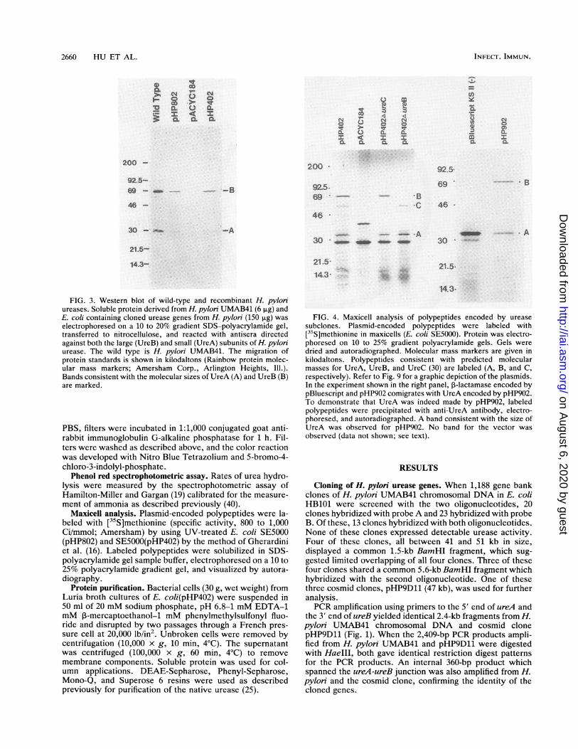
FIG. 3. Western blot of wild-type and recombinant H. pylonureases. Soluble protein derived from H. pylon UMAB41 (6 p.g) andE. coli containing cloned urease genes from H. pylon (150 ,ug) waselectrophoresed on a 10 to 20% gradient SDS-polyacrylamide gel,transferred to nitrocellulose, and reacted with antisera directedagainst both the large (UreB) and small (UreA) subunits of H. pyloniurease. The wild type is H. pylon UMAB41. The migration ofprotein standards is shown in kilodaltons (Rainbow protein molec-ular mass markers; Amersham Corp., Arlington Heights, Ill.).Bands consistent with the molecular sizes of UreA (A) and UreB (B)are marked.
PBS, filters were incubated in 1:1,000 conjugated goat anti-rabbit immunoglobulin G-alkaline phosphatase for 1 h. Fil-ters were washed as described above, and the color reactionwas developed with Nitro Blue Tetrazolium and 5-bromo-4-chloro-3-indolyl-phosphate.
Phenol red spectrophotometric assay. Rates of urea hydro-lysis were measured by the spectrophotometric assay ofHamilton-Miller and Gargan (19) calibrated for the measure-ment of ammonia as described previously (40).
Maxicell analysis. Plasmid-encoded polypeptides were la-beled with [35S]methionine (specific activity, 800 to 1,000Ci/mmol; Amersham) by using UV-treated E. coli SE5000(pHP802) and SE5000(pHP402) by the method of Gherardiniet al. (16). Labeled polypeptides were solubilized in SDS-polyacrylamide gel sample buffer, electrophoresed on a 10 to25% polyacrylamide gradient gel, and visualized by autora-diography.
Protein purification. Bacterial cells (30 g, wet weight) fromLuria broth cultures of E. coli(pHP402) were suspended in50 ml of 20 mM sodium phosphate, pH 6.8-1 mM EDTA-1mM I-mercaptoethanol-1 mM phenylmethylsulfonyl fluo-ride and disrupted by two passages through a French pres-sure cell at 20,000 lb/in2. Unbroken cells were removed bycentrifugation (10,000 x g, 10 min, 4°C). The supernatantwas centrifuged (100,000 x g, 60 min, 4°C) to removemembrane components. Soluble protein was used for col-umn applications. DEAE-Sepharose, Phenyl-Sepharose,Mono-Q, and Superose 6 resins were used as describedpreviously for purification of the native urease (25).
FIG. 4. Maxicell analysis of polypeptides encoded by urease
subclones. Plasmid-encoded polypeptides were labeled with[35S]methionine in maxicells (E. coli SE5000). Protein was electro-phoresed on 10 to 25% gradient polyacrylamide gels. Gels were
dried and autoradiographed. Molecular mass markers are given inkilodaltons. Polypeptides consistent with predicted molecularmasses for UreA, UreB, and UreC (30) are labeled (A, B, and C,respectively). Refer to Fig. 9 for a graphic depiction of the plasmids.In the experiment shown in the right panel, I-lactamase encoded bypBluescript and pHP902 comigrates with UreA encoded by pHP902.To demonstrate that UreA was indeed made by pHP902, labeledpolypeptides were precipitated with anti-UreA antibody, electro-phoresed, and autoradiographed. A band consistent with the size ofUreA was observed for pHP902. No band for the vector was
observed (data not shown; see text).
RESULTS
Cloning of H. pylori urease genes. When 1,188 gene bankclones of H. pyloni UMAB41 chromosomal DNA in E. coliHB101 were screened with the two oligonucleotides, 20clones hybridized with probe A and 23 hybridized with probeB. Of these, 13 clones hybridized with both oligonucleotides.None of these clones expressed detectable urease activity.Four of these clones, all between 41 and 51 kb in size,displayed a common 1.5-kb BamHI fragment, which sug-gested limited overlapping of all four clones. Three of thesefour clones shared a common 5.6-kb BamHI fragment whichhybridized with the second oligonucleotide. One of thesethree cosmid clones, pHP9D11 (47 kb), was used for furtheranalysis.PCR amplification using primers to the 5' end of ureA and
the 3' end of ureB yielded identical 2.4-kb fragments from H.pylori UMAB41 chromosomal DNA and cosmid clonepHP9D11 (Fig. 1). When the 2,409-bp PCR products ampli-fied from H. pylori UMAB41 and pHP9D11 were digestedwith HaeIII, both gave identical restriction digest patternsfor the PCR products. An internal 360-bp product whichspanned the ureA-ureB junction was also amplified from H.pylon and the cosmid clone, confirming the identity of thecloned genes.
FIG. 5. Western blot of a nondenaturing polyacrylamide gel ofurease from H. pylon and E. coli containing recombinant plasmids.Purified H. pylon urease and soluble protein from H. pylon UMAB41and E. coli DH5a containing recombinant plasmids were electro-phoresed on a nondenaturing 4 to 15% polyacrylamide gel, transferredto nitrocellulose, and reacted with pooled anti-UreA and anti-UreBantibodies. Western blots were developed by the standard technique ofBatteiger et al. (2) (A) or by the enhanced chemiluminescence method(B) as directed by the manufacturer (Amersham). Arrows indicate theelectrophoretic mobility of active H. pylon urease.
For subcloning, cosmid DNA of pHP9D11 was partiallydigested with Sau3A, yielding fragments ranging from 5 to 6or 7 to 10 kb. Alkaline phosphatase-treated BamHI-digestedplasmid vector pACYC184 was ligated with each range ofSau3A fragments and transformed into E. coli DH5oa. Forty-nine of 131 chloramphenicol-resistant, tetracycline-sensitiveclones hybridized with probe A. Three of these subclones,the 10.0-kb pHP402, the 12.1-kb pHP802 (Fig. 2), and the15.2-kb pHP808 (see below), were used for further analysis.An additional clone, pHP902, was constructed by PCRamplification of a 2.8-kb fragment carrying only the ureA andureB gene sequences. This fragment, amplified to includeEcoRI and KpnI restriction sites at the 5' and 3' ends,respectively, was cloned into the EcoRI-IpnI site of pBlue-script KSII(-).
Expression of urease subunits and accessory polypeptides.To demonstrate that cloned sequences encoded urease poly-peptides, Western blots were prepared with cell lysates fromthe subclones pHP402 and pHP802, vector pACYC184, andwild-type H. pylon UMAB41. Subclones pHP402 andpHP802 synthesized polypeptides of 66 and 29.5 kDa thatwere recognized by rabbit antisera raised against both theUreA small subunit and UreB large subunit (Fig. 3). Thesepolypeptides were also recognized in cell lysates of wild-type H. pylon UMAB41 and corresponded to the expectedsizes of the UreA and UreB subunit polypeptides of H.pylon urease.
Maxicell analysis revealed three polypeptides of 66, 47,and 29.5 kDa (apparent molecular masses), which corre-
Recombinant H. pylorl Urease
Superose 6
2.00
1.50
S
c
0
a.
0
1.00
0.50
0.00
0 10 20 30
Elution Volume (ml)FIG. 6. Superose 6 chromatography of recombinant urease. Urease-containing fractions from a Mono-Q column were pooled and chromato-
graphed on Superose 6. Protein was monitored at 280 nm. Urease for each fraction was measured by an ELISA, which was read at 405 nm.
sponded to the predicted molecular sizes from the ureaseopen reading frames (ureB, ureC, and ureA), which, alongwith ureD, are essential for urease activity when expressedin Campylobacterjejuni (30) (Fig. 4, left panel). Although apolypeptide of 14 kDa corresponding to UreD was notobserved, an oligonucleotide (probe D) specific for this openreading frame hybridized with both pHP402 and pHP802.These data suggested that these clones carried ureCDAB.Plasmid pHP902 carrying only ureA and ureB sequencessynthesized polypeptides of 66 and 29.5 kDa (apparentmolecular masses) (Fig. 4, right panel). The radiolabeled29.5-kDa polypeptide could be immunoprecipitated withanti-UreA rabbit antiserum and visualized (clearly butweakly) on an autoradiograph of an SDS-polyacrylamide gel(data not shown). Immunoprecipitation was necessary todemonstrate the presence of UreA, since ,B-lactamase, en-coded by the vector, comigrates with the smaller ureasesubunit. Anti-UreA antiserum precipitated a total of 7,280cpm from the pHP902 preparation and only 159 cpm from thepBluescript KSII(-) vector maxicell preparation.
Deletion mutants of ureC and ureB. A Bal 31 deletionmutation within the ureC open reading frame was con-structed by linearizing pHP402 with NsiI, which cutuniquely within ureC. One derivative with a 200-bp deletionwhich was designated pHP402AureC was selected. Thisdeletion corresponded to the loss of a 47-kDa polypeptideobserved on the gel of maxicell-labeled polypeptides (Fig. 4).A similar procedure was used to construct a ureB mutant inwhich plasmid pHP402 was linearized with BamHI. Thisresulted in two clones, one with a 300-bp deletion and onewith a 600-bp deletion within ureB, the former of which wasdesignated pHP402AureB. Likewise, this deletion corre-sponded to the loss of a 66-kDa polypeptide on the gel ofmaxicell-labeled polypeptides (Fig. 4).The electrophoresis of soluble proteins from these mu-
tants and the detection of urease gene products on nonde-naturing polyacrylamide gels were used as an index ofenzyme assembly. A Western blot was prepared from thesegels by using pooled anti-UreA and anti-UreB antibodies(Fig. 5). E. coli DH5at containing pHP402 and pHP402AureCproduced urease proteins with the same electrophoreticmobility as purified native enzyme (25) or enzyme from celllysates of the parent strain H. pyloni UMAB41. E. colicontaining vector alone or pHP402AureB (300-bp deletion)did not produce an assembled protein that corresponded tothe wild-type urease enzyme. E. coli (pHP902), encodingonly ureA and ureB, however, did synthesize an assembledbut inactive urease.
Purification of recombinant urease. Recombinant ureaseexpressed by E. coli(pHP402) was purified to homogeneityby chromatography on four resins: DEAE-Sepharose, Phe-nyl-Sepharose, Mono-Q, and Superose 6 (Fig. 6). Fractionsin which urease was detected by Western blot or ELISAwere pooled and applied to the next column resin in thepurification scheme. For each column, recombinant ureaseeluted under the same conditions as those observed previ-ously for the native enzyme (25), suggesting that the recom-binant enzyme was indeed assembled in E. coli. This alsoindicated that the surface charge, relative hydrophobicity,stoichiometry of subunits, quaternary structure, and molec-ular weight were identical for native and recombinant ure-ase.
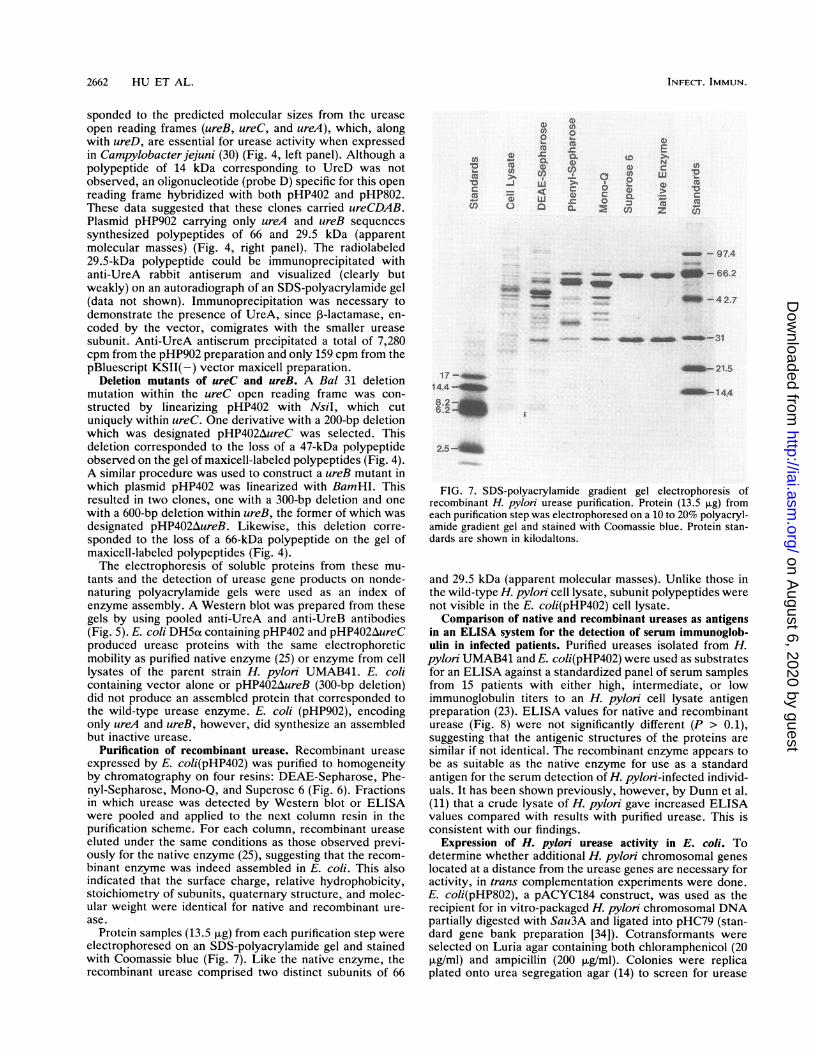
Protein samples (13.5 ,ug) from each purification step wereelectrophoresed on an SDS-polyacrylamide gel and stainedwith Coomassie blue (Fig. 7). Like the native enzyme, therecombinant urease comprised two distinct subunits of 66
o
E
c~ ~0 X N U)
u 0 L. 4)Q0_-7 .>
Cf, L~) 0CLc l
-~ 97.4
- MM- - 66.2
- m-42.7
- - _ _4_-31
Am-21.517-__
14.4-
8.2-6.2_
4i -144
FIG. 7. SDS-polyacrylamide gradient gel electrophoresis ofrecombinant H. pyloni urease purification. Protein (13.5 ,ug) fromeach purification step was electrophoresed on a 10 to 20% polyacryl-amide gradient gel and stained with Coomassie blue. Protein stan-dards are shown in kilodaltons.
and 29.5 kDa (apparent molecular masses). Unlike those inthe wild-type H. pyloni cell lysate, subunit polypeptides werenot visible in the E. coli(pHP402) cell lysate.Comparison of native and recombinant ureases as antigens
in an ELISA system for the detection of serum immunoglob-ulin in infected patients. Purified ureases isolated from H.pylon UMAB41 and E. coli(pHP402) were used as substratesfor an ELISA against a standardized panel of serum samplesfrom 15 patients with either high, intermediate, or lowimmunoglobulin titers to an H. pylon cell lysate antigenpreparation (23). ELISA values for native and recombinanturease (Fig. 8) were not significantly different (P > 0.1),suggesting that the antigenic structures of the proteins aresimilar if not identical. The recombinant enzyme appears tobe as suitable as the native enzyme for use as a standardantigen for the serum detection of H. pylon-infected individ-uals. It has been shown previously, however, by Dunn et al.(11) that a crude lysate of H. pylon gave increased ELISAvalues compared with results with purified urease. This isconsistent with our findings.
Expression of H. pylori urease activity in E. coli. Todetermine whether additional H. pylon chromosomal geneslocated at a distance from the urease genes are necessary foractivity, in trans complementation experiments were done.E. coli(pHP802), a pACYC184 construct, was used as therecipient for in vitro-packaged H. pylon chromosomal DNApartially digested with Sau3A and ligated into pHC79 (stan-dard gene bank preparation [34]). Cotransformants wereselected on Luria agar containing both chloramphenicol (20jig/ml) and ampicillin (200 jig/ml). Colonies were replicaplated onto urea segregation agar (14) to screen for urease
FIG. 8. Comparison of native and recombinant ureases as antigens in an ELISA for the detection of serum immunoglobulin to H. pylon.Purified native urease (Superose 6 fraction) (25) and purified recombinant H. pylon ureases were used as antigens in an ELISA against astandardized panel of sera from individuals with either high, intermediate, or low immunoglobulin titers to an H. pylon cell lysate antigenpreparation. For comparison, antigen preparations obtained during purification of native enzyme (crude lysate, cytosol, DEAE-Sepharose,Phenyl-Sepharose, and Mono-Q [25]) were also included. Unique symbols represent the serum samples from 15 different individuals. Linesare used for the comparison of native and recombinant enzymes with the other antigen preparations for a given serum sample.
production. None of approximately 1,000 colonies screenedwas positive for urease activity.The possibility existed that H. pylon genes necessary for
an active urease were not synthesized or not functional in E.coli. To determine whether urease accessory genes fromenteric species could complement activity, E. coli DH5a(pHP802) was cotransformed with transposon mutants ofcloned urease sequences from M. morganii and P. mirabilis.For M. morganii, plasmids pMOM203::TnS-7, -8, -22, -31,and -33 represented transposon insertions that spanned 4 kbof these urease gene sequences (26). For P. mirabilis,plasmid pMID1417, a TnS insertion that abolished structuralsubunit production, was used (28). None of the six cotrans-formants synthesized an active urease.Cussac et al. (9) reported recently that nitrogen-limiting
conditions were necessary for the expression of a catalyti-cally active recombinant enzyme. To test whether nitrogenlimitation would yield a catalytically active urease, ourclones containing ureCDAB (pHP402 or pHP802) or a plas-mid bearing these genes plus DNA downstream of thisregion (plasmid pHP808) were cultured on minimal saltsagar, with 10 mM arginine replacing NH4Cl, conditionsdescribed as optimal (Fig. 9). Under these conditions, E. coliDH5a(pHP808) was qualitatively positive for urease activ-ity. E. coli(pHP402) and E. coli(pHP802) were negative forurease activity under these growth conditions.
DISCUSSION
The urease subunit genes of H. pyloni, ureA and ureB, areexpressed in E. coli, and these genes alone are sufficient toencode a fully assembled but catalytically inactive apoen-zyme. The subunit genes are apparently transcribed andtranslated in E. coli by using the H. pylon promoter sincegenes are equally well expressed when cloned in eitherorientation (pHP402 or pHP802) into pACYC184 (Fig. 3).Deletion mutations in ureC (Fig. 9), an accessory geneupstream from ureA and ureB, do not significantly affect thesynthesis and assembly of the apoenzyme. UreC mutantssynthesize a protein, recognized by anti-UreA and anti-UreB antibodies, which has the same electrophoretic mobil-ity on nondenaturing polyacrylamide gels as do purifiednative urease and recombinant urease synthesized frompHP402, suggesting that enzyme assembly is not affected bya lack of the UreC polypeptide (Fig. 5). In addition, pHP902,a clone containing only ureA and ureB, also synthesized afully assembled but inactive apoenzyme.
Purification profiles of recombinant urease synthesized inE. coli indicated that the enzyme was fully assembled intothe intact apoenzyme, yet the protein was catalyticallyinactive. For all steps in the purification, the recombinanturease behaved identically to the native enzyme. This in-cluded a molecular mass estimation of 550 kDa on Superose
FIG. 9. Restriction maps of recombinant plasmids encoding H. pylon urease. Thin lines represent H. pylon sequences. Thick linesrepresent portions of pACYC184 except for pHP902, where the vector is pBluescript KSII(-). RV, EcoRV; B, BamHI; RI, EcoRI; Ns, NsiI.The alignment of ureCDAB gene products (bottom) is based on data from Western blot, maxicell, PCR amplification, and Southern blotanalyses. The alignment of ureIEFGH is based on a comparison of our restriction map with previously published data (9). Cross-hatchedboxes represent sequences deleted by Bal 31 digestion. Positive (+) and negative (-) urease activities were measured qualitatively onnitrogen-limited minimal salts agar. Apoenzyme assembly was assessed on Western blots of nondenaturing gels as follows: +, proteinobserved with an electrophoretic mobility identical to that of the purified native urease; -, no protein observed corresponding to that of thenative urease.
6 (Fig. 6), the value we previously reported for the nativeenzyme (25).The modification of the urease apoenzyme that is neces-
sary for a catalytically active enzyme must be relativelyminor. When active native enzyme purified from H. pyloniUMAB41 was compared with purified recombinant enzyme,no difference could be noted in the charge (elution profilesfrom DEAE-Sepharose and Mono-Q columns), hydropho-bicity (elution profile from Phenyl-Sepharose), apparentmolecular weight and quarternary structure (Superose 6 andnondenaturing polyacrylamide gel profiles), subunit sizes(migration of UreA and UreB on SDS-polyacrylamide gels),or antigenicity (equal recognition by anti-UreA or anti-UreBantibody on Western blots). We speculate that inactivity ofthe recombinant urease is not due to a lack of enzymeassembly but rather to a failure to incorporate nickel (Ni2+)ions into the active site.Evidence from work with enteric urease genes suggests
that certain accessory genes play a role in the incorporationof nickel ions into the active site. Processing may includetransport of the ion into the cell or insertion of the ion intoprotein during polypeptide folding and assembly. The ure-EFG genes of K aerogenes were shown to be necessary forthe production of an enzymatically active recombinant en-zyme (32, 42). When these genes, downstream of the struc-tural subunit genes, were absent, the apoenzyme was pro-duced but no enzyme activity could be detected. Thisobservation is consistent with our data for H. pyloni. PlasmidpHP808, which contains 4.5 kb of DNA downstream ofureCDAB, is capable of synthesizing a weakly active recom-binant urease on nitrogen-limiting minimal salts agar. Plas-mid pHP402, however, which lacks these downstream se-quences, demonstrated no detectable enzyme activity onthis medium.H. pylon urease serves as an excellent antigen for the
sensitive and specific detection of H. pyloni-specific serumimmunoglobulin by ELISA. An immune response to ureasecorrelates with H. pylori infection and the development ofacute gastritis (10, 43, 44). Indeed, Clayton et al. (7) usedhuman serum from an infected patient to screen an H. pylonigene bank in E. coli to identify urease-producing clones. Alarge-scale culture of H. pylori, however, is inherently moredifficult than one for E. coli because of this pathogen'sfastidious growth requirements. Therefore, recombinanturease purified from E. coli can be used for the production ofa standardized ELISA system. Our data indicate that thecatalytically inactive recombinant enzyme is antigenicallyequivalent to the native enzyme and would be suitable forthe production of antigen for standardized tests. It has beenshown previously, however, by Dunn et al. (11), that a crudelysate of H. pylori cells gives increased ELISA valuescompared with those for purified urease. This is consistentwith our findings (Fig. 8).Although nine open reading frames have been shown to be
either essential for enzyme activity or associated with ureasegene sequences (9, 30), only ureA and ureB are necessary forthe synthesis and assembly of the 550-kDa apoenzyme.Additional modification, probably associated with Ni" in-sertion, is carried out by accessory genes downstream ofureB.
ACKNOWLEDGMENTS
This work was supported in part by Public Health Service grantsA125567 and A123328 from the National Institutes of Health.We thank Frank Gherardini and the laboratory of Phillip Bass-
ford, Jr., for their advice on maxicell analysis and Glenn Morris,Robert Hopkins, and Michael O'Donnoghue for assistance with theELISA.
REFERENCES1. Ausubel, F. M., R. Brent, R. E. Kingston, D. D. Moore, J. A.
Smith, J. G. Seidman, and K. Struhl. 1987. Current protocols inmolecular biology. Greene Publishing Associates and WileyInterscience, New York.
2. Batteiger, B., W. J. Newhall, and R. B. Jones. 1982. The use ofTween-20 as a blocking agent in the immunological detection ofproteins transferred to nitrocellulose membranes. J. Immunol.Methods 55:297-307.
3. Bell, G. D., J. Weil, G. Harrison, A. Morden, P. H. Jones, P. W.Gant, J. E. Trowell, A. K. Yoong, T. K. Daneshmend, andR. F. A. Logan. 1987. '4C-urea breath analysis, a non-invasivetest for Campylobacter pyloni in the stomach. Lancet i:1367-1368.
4. Birnboim, H. C., and J. Doly. 1979. A rapid alkaline extractionprocedure for screening recombinant plasmid DNA. NucleicAcids Res. 7:1513-1523.
5. Blaser, M. J., T. L. Cover, R. Steele, K. Eaton, G. Perez-Perez,and A. Labigne. 1990. Characteristics of a urease-negative H.pylori mutant strain. Rev. Esp. Enferm. Dig. 78(Suppl. 1):26.(Abstr.)
6. Clayton, C. L., M. J. Pallen, H. Kleanthous, B. W. Wren, and S.Tabaqchali. 1990. Nucleotide sequence of two genes fromHelicobacterpylori encoding for urease subunits. Nucleic AcidsRes. 18:362.
7. Clayton, C. L., B. W. Wren, P. Mullany, A. Topping, and S.Tabaqchali. 1989. Molecular cloning and expression of Cam-pylobacter pylori species-specific antigens in Eschenchia coliK-12. Infect. Immun. 57:623-629.
8. Cox, D. M., A. McLaren, and M. A. Snowden. 1990. Theisolation and characteristics of urease-negative variants of He-licobacter pylori. Rev. Esp. Enferm. Dig. 78:(Suppl. 1):29.(Abstr.)
9. Cussac, V., R. L. Ferrero, and A. Labigne. 1992. Expression ofHelicobacter pylori urease genes in Escherichia coli grownunder nitrogen-limiting conditions. J. Bacteriol. 174:2466-2473.
10. Dent, J. C., C. A. M. McNulty, J. S. Uff, M. W. L. Gear, andS. P. Wilkinson. 1988. Campylobacter pylori urease: a newserological test. Lancet i:1002.
11. Dunn, B. E., G. P. Campbell, G. I. Perez-Perez, and M. J.Blaser. 1990. Purification and characterization of urease fromHelicobacter pylori. J. Biol. Chem. 265:9464-9469.
12. Eaton, K. A., C. L. Brooks, D. R. Morgan, and S. Krakowka.1991. Essential role of urease in pathogenesis of gastritis in-duced by Helicobacter pylori in gnotobiotic piglets. Infect.Immun. 59:2470-2475.
13. Evans, D. J., Jr., D. G. Evans, S. S. Kirkpatrick, and D. Y.Graham. 1991. Characterization of the Helicobacter pylonurease and purification of its subunits. Microb. Pathol. 10:15-26.
14. Farmer, J. J., III, F. W. Hickman, D. J. Brenner, M. Schreiber,and D. G. Rickenbach. 1977. Unusual Enterobactenaceae:"Proteus rettgen" that "change" to "Providencia stuartii." J.Clin. Microbiol. 6:373-378.
15. Foxall, P. A., L.-T. Hu, and H. L. T. Mobley. 1992. Use ofpolymerase chain reaction-amplified Helicobacten pylon ureasestructural genes for differentiation between isolates. J. Clin.Microbiol. 30:739-741.
16. Gherardini, F. C., M. M. Hobbs, L. V. Stamm, and P. J.Bassford, Jr. 1990. Complementation of an Eschenchia coliproC mutation by a gene cloned from Treponema pallidum. J.Bacteriol. 172:2996-3002.
17. Graham, D. Y., P. D. Klein, D. J. Evans, L. C. Alpert, A. R.Opekun, and T. W. Boutton. 1987. Campylobacter pylondisdetected by the "C-urea test. Lancet i:1174-1177.
18. Hames, B. D. 1985. An introduction to polyacrylamide gelelectrophoresis, p. 1-86. In B. D. Hames and D. Rickwood(ed.), Gel electrophoresis of proteins-a practical approach.IRL Press, Washington, D.C.
19. Hamilton-Miller, J. M. T., and R. A. Gargan. 1979. Rapidscreening for urease inhibitors. Invest. Urol. 16:327-328.
20. Hausinger, R. P. 1987. Nickel utilization by microorganisms.Microbiol. Rev. 51:22-42.
21. Hawtin, P. R., H. T. Delves, and D. G. Newell. 1991. The
demonstration of nickel in the urease of Helicobacterpylon byatomic absorption spectroscopy. FEMS Microbiol. Lett. 77:51-54.
22. Hazell, S. L., and A. Lee. 1985. The adaptation of motile strainsof Campylobacterpylondis to gastric mucus and their associa-tion with gastric epithelial intercellular spaces, p. 189-191. InA. D. Pearson, M. B. Skirrow, H. Lion, and B. Rowe (ed.),Campylobacter III. Proceedings of the Third InternationalWorkshop on Campylobacter Infections. Public Health Labora-tory Service, London.
23. Hopkins, R. J., R. G. Russell, J. M. O'Donnoghue, S. S.Wasserman, A. Lefkowitz, and J. G. Morris, Jr. 1990. Seroprev-alence of Helicobacter pylori in Seventh-Day Adventists andother groups in Maryland. Lack of association with diet. Arch.Intern. Med. 150:2347-2348.
24. Hu, L. T., P. A. Foxall, and H. L. T. Mobley. 1991. RecombinantH. pylon urease is expressed and assembled in E. coli but is notenzymatically active. Microb. Ecol. Health Dis. 4(Suppl):S138.(Abstr.)
25. Hu, L.-T., and H. L. T. Mobley. 1990. Purification and N-ter-minal analysis of urease from Helicobacter pylon. Infect. Im-mun. 58:992-998.
26. Hu, L.-T., E. B. Nicholson, B. D. Jones, M. J. Lynch, andH. L. T. Mobley. 1990. Morganella morganii urease: purifica-tion, characterization, and isolation of gene sequences. J. Bac-teriol. 172:3073-3080.
27. Jones, B. D., and H. L. T. Mobley. 1987. Genetic and biochem-ical diversity of ureases of Proteus, Providencia, and Mor-ganella species isolated from urinary tract infection. Infect.Immun. 55:2198-2203.
28. Jones, B. D., and H. L. T. Mobley. 1988. Proteus mirabilisurease: genetic organization, regulation, and expression ofstructural genes. J. Bacteriol. 170:3342-3349.
29. Jones, B. D., and H. L. T. Mobley. 1989. Proteus mirabilisurease: nucleotide sequence determination and comparison withjack bean urease. J. Bacteriol. 171:6414-6422.
30. Labigne, A., V. Cussac, and P. Courcoux. 1991. Shuttle cloningand nucleotide sequence of Helicobacterpylon genes responsi-ble for urease activity. J. Bacteriol. 173:1920-1931.
31. Laemmli, U. K. 1970. Cleavage of structural proteins during theassembly of the head of bacteriophage T4. Nature (London)227:680-685.
32. Lee, M. H., S. B. Mulrooney, and R. P. Hausinger. 1990.Purification, characterization, and in vivo reconstitution ofKlebsiella aerogenes urease apoenzyme. J. Bacteriol. 172:4427-4431.
33. Mamiya, G., K. Takishima, M. Masakuni, T. Kayumi, K.Ogawa, and T. Sekita. 1985. Complete amino acid sequence ofjack bean urease. Proc. Jpn. Acad. 61:395-398.
34. Maniatis, T., E. F. Fritsch, and J. Sambrook. 1982. Molecularcloning: a laboratory manual. Cold Spring Harbor Laboratory,Cold Spring Harbor, N.Y.
35. Marmur, J. 1961. A procedure for the isolation of deoxyribo-nucleic acid from microorganisms. J. Mol. Biol. 3:208-218.
36. Marshall, B. J., L. J. Barrett, C. Prakesh, R. W. McCallum, andR. L. Guerrant. 1988. Protection of Campylobacterpyloridis butnot Campylobacterjejuni against acid susceptibility by urea, p.402-403. In B. Kaijser and E. Falsen (ed.), Campylobacter IV.University of Goteborg, Goteborg, Sweden.
37. McNulty, C. A. M. 1989. Detection of Campylobacterpylori bythe biopsy urease test, p. 69-73. In B. J. Rathbone and R. V.Heatley (ed.), Campylobacter pylon and gastroduodenal dis-ease. Blackwell Scientific Publications, Oxford.
38. Mobley, H. L. T., M. J. Cortesia, L. E. Rosenthal, and B. D.Jones. 1988. Characterization of urease from Campylobacterpylon. J. Clin. Microbiol. 26:831-836.
39. Mobley, H. L. T., and R. P. Hausinger. 1989. Microbial urease:
significance, regulation, and molecular characterization. Micro-biol. Rev. 53:85-108.
40. Mobley, H. L. T., B. D. Jones, and A. E. Jerse. 1986. Cloning ofurease gene sequences from Providencia stuartii. Infect. Im-mun. 54:161-169.
41. Mobley, H. L. T., L. E. Rosenthal, A. F. Trofa, and B. D. Jones.
1989. Optimization of detection of Campylobacter pylon byurease and DNA hybridization, p. 127-131. In F. Megraud andH. Lamouliatte (ed.), Gastroduodenal pathology and Campylo-bacterpylori. Elsevier Science Publishers, Amsterdam.
42. Mulrooney, S. B., and R. P. Hausinger. 1990. Sequence of theKlebsiella aerogenes urease genes and evidence for accessoryproteins facilitating nickel incorporation. J. Bacteriol. 172:5837-5843.
43. Newell, D. G., and A. Stacey. 1989. Antigens for the serodiag-nosis of Campylobacter pylon. Gastroenterol. Clin. Biol. 13:37B-41B.
44. Perez-Perez, G. I., and M. J. Blaser. 1987. Conservation and
diversity of Campylobacter pyloridis major antigens. Infect.Immun. 55:1256-1263.
45. Skirrow, M. B. 1977. Campylobacter enteritis: a "new" disease.Br. Med. J. 2:9-11.
46. Smoot, D. T., H. L. T. Mobley, G. R. Chippendale, J. F.Lewison, and J. H. Resau. 1990. Helicobacter pylon ureaseactivity is toxic to human gastric epithelial cells. Infect. Immun.58:1992-1994.
47. Towbin, H., T. Staehelin, and J. Gordon. 1979. Electrophoretictransfer of proteins from polyacrylamide gels to nitrocellulosesheets: procedure and some applications. Proc. Natl. Acad. Sci.USA 76:4350-4354.


![Helicobacterpylori inParkinson sDisease ......drugs[4]. H.pylorihas beenassociatedwith avarietyofautoimmunedisorders.AlthoughH. pylori colonizationtakes placemainlyinthe antrum,H.pylori-driven](https://static.documents.pub/doc/80x56/5fbc5630034fd614550b9327/helicobacterpylori-inparkinson-sdisease-drugs4-hpylorihas-beenassociatedwith.jpg)


