367 Revista Facultad de Odontología Universidad de Antioquia - Vol. 27 N. o 2 - Primer semestre, 2016 Abstract. Introduction: orthodontic tooth movement with open apices which have not completed root formation has not been sufficiently studied. There is controversy about the risks associated to this movement, such as root resorption and decreased root length. The goal of this systematic review is to determine the possible effects of lengthening, shortening, or root resorption possibly occurring during orthodontic movement in teeth that have not completed root formation. Methods: electronic search (PubMed, Cochrane, Dentistry and Oral Sciences Source, Science Direct, Google Scholar, IdeA, ProQuest, Embase, Medline, Lilacs, TRIP) and manual search at Universidad El Bosque Juan Roa Vázquez Library since 1990 to 2014. Articles meeting the inclusion criteria, such as randomized clinical trials, prospective and retrospective studies, and studies in early mixed dentition with 2 x 4 system, were evaluated and methodologically qualified by four researchers. Results: this study involved a custom methodological rating taken from Lagravere et al (2005). Four articles were finally selected, three of which were retrospective: Amlani et al (2007), with 26 patients, found root resorption in 8% of the sample, with no statistical significance. Mavragani et al (2002), with a sample of 146 patients, found longer roots in younger teeth, and Kim & Park (2004), with 59 patients, found higher resorption in maxillary lateral incisors. Da Silva et al (2005), with 46 patients, reported a prevalence of 4.4% in root resorption in central incisors. Conclusions: this systematic review must be taken cautiously due to the low and moderate level of evidence found. In general terms, there were no alterations in terms of root length or shape when teeth with open apices were subjected to fixed orthodontic forces. The risk of apical resorption was more related to treatment duration in teeth with both open and closed apexes. Key words: orthodontics, tooth apex, root resorption, systematic review. Wasserman-Milhem I, Bravo-Casanova ML, Caraballo-Moreno FA, Granados-Pelayo DA, Restrepo-Bolívar CP. Orthodontic tooth movement in immature apices. A systematic review. Rev Fac Odontol Univ Antioq 2016; 27 (2): 367-388. DOI: http://dx.doi. org/10.17533/udea.rfo.v27n2a7 RECIBIDO: NOVIEMBRE 11/14 - ACEPTADO: SEPTIEMBRE 29/15 1 Odontólogo ortodoncista, Director de la Clínica de Tratamiento Temprano, posgrado de Ortodoncia, Universidad El Bosque, Bogotá, Colombia. 2 Odontólogo, residente del posgrado de Ortodoncia, Universidad El Bosque, Bogotá, Colombia. EL MOVIMIENTO DENTAL ORTODÓNCICO EN ÁPICES INMADUROS. REVISIÓN SISTEMÁTICA ORTHODONTIC TOOTH MOVEMENT IN IMMATURE APICES. A SYSTEMATIC REVIEW ISAAC WASSERMAN MILHEM 1 , MAYRA LIZBETH BRAVO CASANOVA 2 , FÉLIX ALEXANDER CARABALLO MORENO 2 , DIANA ANTONIA GRANADOS PELAYO 2 , CLAUDIA PATRICIA RESTREPO BOLÍVAR 2 RESUMEN. Introducción: el movimiento dental ortodóncico con ápices abiertos que no han terminado su formación radicular completa no ha sido estudiado suficientemente. Existe controversia sobre los riesgos que se pueden generar por dicho movimiento, como reabsorción radicular o disminución de la longitud radicular. El objetivo de esta revisión sistemática es determinar los posibles efectos de alargamiento, acortamiento o reabsorción radicular que se pudieran presentar durante el movimiento dental ortodóncico en dientes que no han terminado su formación radicular. Métodos: se hizo una búsqueda electrónica (PubMed, Cochrane, Dentistry and Oral Sciences Source, Science Direct, Google Scholar, IdeA, ProQuest, Embase, Medline, Lilacs, TRIP) y una búsqueda manual en la biblioteca Juan Roa Vázquez, de la Universidad El Bosque, desde 1990 a 2014. Los artículos que cumplieron con los criterios de inclusión, como ensayos clínicos aleatorizados, prospectivos, retrospectivos y de dentición mixta temprana con sistema 2 x 4, fueron evaluados por cuatro investigadores y calificados metodológicamente. Resultados: se realizó una calificación metodológica personalizada tomada de Lagravere y colaboradores (2005). Cuatro artículos fueron finalmente seleccionados, de los cuales tres fueron de modalidad retrospectiva: Amlani y colaboradores (2007), con 26 pacientes, encontraron reabsorción radicular en el 8% de la muestra, sin significancia estadística. Mavragani y colaboradores (2002), con una muestra de 146 pacientes, encontraron raíces más largas en dientes más jóvenes, y Kim y Park (2004), con 59 pacientes, encontraron mayor reabsorción en incisivos laterales maxilares. Da Silva y colaboradores (2005), con 46 pacientes, reportaron una prevalencia de 4.4% de reabsorción radicular en incisivos centrales. Conclusiones: esta revisión sistemática debe ser tomada con cautela por el bajo y moderado nivel de evidencia encontrado. En términos generales, no se encontró alteración en la forma ni en la longitud radicular cuando los dientes con ápices abiertos fueron sometidos a fuerzas ortodóncicas fijas. El riesgo de reabsorción apical estuvo más relacionado con la duración del tratamiento, en dientes con ápices tanto abiertos como cerrados. Palabras clave: ortodoncia, ápice del diente, reabsorción radicular, revisión sistemática. Wasserman-Milhem I, Bravo-Casanova ML, Caraballo-Moreno FA, Granados-Pelayo DA, Restrepo-Bolívar CP. El movimiento dental ortodóncico en ápices inmaduros. Revisión sistemática. Rev Fac Odontol Univ Antioq 2016; 27(2): 367-388. DOI: http:// dx.doi.org/10.17533/udea.rfo.v27n2a7 1 Orthodontist, Head of Clínica de Tratamiento Temprano, Graduate Program in Orthodontics, Universidad El Bosque, Bogotá, Colombia. 2 DMD, Intern in the Graduate Program in Orthodontics, Universidad El Bosque, Bogotá, Colombia. SUBMITTED: NOVEMBER 11/14 - ACCEPTED: SEPTEMBER 29/15 REVISIÓN DE LITERATURA LITERATURE REVIEW
Transcript
367Revista Facultad de Odontología Universidad de Antioquia - Vol. 27 N.o 2 - Primer semestre, 2016
Abstract. Introduction: orthodontic tooth movement with open apices which have not completed root formation has not been sufficiently studied. There is controversy about the risks associated to this movement, such as root resorption and decreased root length. The goal of this systematic review is to determine the possible effects of lengthening, shortening, or root resorption possibly occurring during orthodontic movement in teeth that have not completed root formation. Methods: electronic search (PubMed, Cochrane, Dentistry and Oral Sciences Source, Science Direct, Google Scholar, IdeA, ProQuest, Embase, Medline, Lilacs, TRIP) and manual search at Universidad El Bosque Juan Roa Vázquez Library since 1990 to 2014. Articles meeting the inclusion criteria, such as randomized clinical trials, prospective and retrospective studies, and studies in early mixed dentition with 2 x 4 system, were evaluated and methodologically qualified by four researchers. Results: this study involved a custom methodological rating taken from Lagravere et al (2005). Four articles were finally selected, three of which were retrospective: Amlani et al (2007), with 26 patients, found root resorption in 8% of the sample, with no statistical significance. Mavragani et al (2002), with a sample of 146 patients, found longer roots in younger teeth, and Kim & Park (2004), with 59 patients, found higher resorption in maxillary lateral incisors. Da Silva et al (2005), with 46 patients, reported a prevalence of 4.4% in root resorption in central incisors. Conclusions: this systematic review must be taken cautiously due to the low and moderate level of evidence found. In general terms, there were no alterations in terms of root length or shape when teeth with open apices were subjected to fixed orthodontic forces. The risk of apical resorption was more related to treatment duration in teeth with both open and closed apexes.
Wasserman-Milhem I, Bravo-Casanova ML, Caraballo-Moreno FA, Granados-Pelayo DA, Restrepo-Bolívar CP. Orthodontic tooth movement in immature apices. A systematic review. Rev Fac Odontol Univ Antioq 2016; 27 (2): 367-388. DOI: http://dx.doi.org/10.17533/udea.rfo.v27n2a7
RECIBIDO: NOVIEMBRE 11/14 - ACEPTADO: SEPTIEMBRE 29/15
1 Odontólogo ortodoncista, Director de la Clínica de Tratamiento Temprano, posgrado de Ortodoncia, Universidad El Bosque, Bogotá, Colombia.
2 Odontólogo, residente del posgrado de Ortodoncia, Universidad El Bosque, Bogotá, Colombia.
EL MOVIMIENTO DENTAL ORTODÓNCICO EN ÁPICES INMADUROS. REVISIÓN SISTEMÁTICA
ORTHODONTIC TOOTH MOVEMENT IN IMMATURE APICES. A SYSTEMATIC REVIEW
ISAAC WASSERMAN MILHEM1, MAYRA LIZBETH BRAVO CASANOVA2, FÉLIX ALEXANDER CARABALLO MORENO2,
RESUMEN. Introducción: el movimiento dental ortodóncico con ápices abiertos que no han terminado su formación radicular completa no ha sido estudiado suficientemente. Existe controversia sobre los riesgos que se pueden generar por dicho movimiento, como reabsorción radicular o disminución de la longitud radicular. El objetivo de esta revisión sistemática es determinar los posibles efectos de alargamiento, acortamiento o reabsorción radicular que se pudieran presentar durante el movimiento dental ortodóncico en dientes que no han terminado su formación radicular. Métodos: se hizo una búsqueda electrónica (PubMed, Cochrane, Dentistry and Oral Sciences Source, Science Direct, Google Scholar, IdeA, ProQuest, Embase, Medline, Lilacs, TRIP) y una búsqueda manual en la biblioteca Juan Roa Vázquez, de la Universidad El Bosque, desde 1990 a 2014. Los artículos que cumplieron con los criterios de inclusión, como ensayos clínicos aleatorizados, prospectivos, retrospectivos y de dentición mixta temprana con sistema 2 x 4, fueron evaluados por cuatro investigadores y calificados metodológicamente. Resultados: se realizó una calificación metodológica personalizada tomada de Lagravere y colaboradores (2005). Cuatro artículos fueron finalmente seleccionados, de los cuales tres fueron de modalidad retrospectiva: Amlani y colaboradores (2007), con 26 pacientes, encontraron reabsorción radicular en el 8% de la muestra, sin significancia estadística. Mavragani y colaboradores (2002), con una muestra de 146 pacientes, encontraron raíces más largas en dientes más jóvenes, y Kim y Park (2004), con 59 pacientes, encontraron mayor reabsorción en incisivos laterales maxilares. Da Silva y colaboradores (2005), con 46 pacientes, reportaron una prevalencia de 4.4% de reabsorción radicular en incisivos centrales. Conclusiones: esta revisión sistemática debe ser tomada con cautela por el bajo y moderado nivel de evidencia encontrado. En términos generales, no se encontró alteración en la forma ni en la longitud radicular cuando los dientes con ápices abiertos fueron sometidos a fuerzas ortodóncicas fijas. El riesgo de reabsorción apical estuvo más relacionado con la duración del tratamiento, en dientes con ápices tanto abiertos como cerrados.
Palabras clave: ortodoncia, ápice del diente, reabsorción radicular, revisión sistemática.
Wasserman-Milhem I, Bravo-Casanova ML, Caraballo-Moreno FA, Granados-Pelayo DA, Restrepo-Bolívar CP. El movimiento dental ortodóncico en ápices inmaduros. Revisión sistemática. Rev Fac Odontol Univ Antioq 2016; 27(2): 367-388. DOI: http://dx.doi.org/10.17533/udea.rfo.v27n2a7
1 Orthodontist, Head of Clínica de Tratamiento Temprano, Graduate Program in Orthodontics, Universidad El Bosque, Bogotá, Colombia.
2 DMD, Intern in the Graduate Program in Orthodontics, Universidad El Bosque, Bogotá, Colombia.
SUBMITTED: NOVEMBER 11/14 - ACCEPTED: SEPTEMBER 29/15
REVISIÓN DE LITERATURALITERATURE REVIEW
368
EL MOVIMIENTO DENTAL ORTODÓNCICO EN ÁPICES INMADUROS. REVISIÓN SISTEMÁTICA
Revista Facultad de Odontología Universidad de Antioquia - Vol. 27 N.o 2 - Primer semestre, 2016
INTRODUCCIÓN
Diversos estudios en ápices totalmente cerrados re-portan la respuesta dental y de los tejidos de soporte frente al movimiento ortodóncico.1, 2 El movimiento dental con aparatos fijos genera una fuerza constante y continua que permite un remodelado óseo sin aparentes efectos deletéreos,3 según la teoría de presión-tensión,4 según la cual se produce reabsorción en la zona de presión y aposición en la zona de tensión.3 Las células responsables y reguladoras de este mecanismo son los osteoclastos, osteoblastos, osteocitos y sus mediado-res químicos.5, 6 Cuando estas fuerzas son excesivas, ocasionan mayor área de hialinización,7, 8 necrosis y re-absorción ósea, lo cual genera daños en el periodonto, en la pulpa y en las raíces de los dientes.3, 9 De estos efectos, el más atribuido al tratamiento ortodóncico es la reabsorción radicular externa.10, 11 Esta entidad ha sido ampliamente estudiada y se define como la pérdida del componente orgánico e inorgánico de los tejidos duros radiculares, como la dentina y el cemento,12, 13 asociada a otros factores como edad,13, 14 sexo,15 trauma,11 ma-loclusión,16, 17 anatomía radicular,18, 19 tipo de aparato,20 mecánica utilizada,16 características del movimiento dental,21, 22 tipo de fuerza23, 24 y tiempo de tratamiento.25 Su causa, en términos generales, podemos decir que es multifactorial.10
El movimiento dental con ápices abiertos que no han terminado su formación radicular completa no ha sido estudiado suficientemente. Algunos autores26, 27 sugieren que el movimiento dental en ápices inmaduros podría ser un riesgo en la aparición de reabsorciones radiculares o en la disminución de la longitud radicular por cierre apical prematuro, como lo describieron Oppenheimen en 1942 y Phillips en 1955, quienes lo atribuyen a la defor-mación de la Vaina de Hertwig, que altera la calcificación del ápice y por consiguiente el desarrollo radicular, impi-diendo que llegue a su longitud máxima.26, 27 En el 2001, Consolaro y colaboradores señalaron que el movimiento de los dientes con formación radicular incompleta gene-ra una disminución en la longitud radicular por el cierre temprano del ápice —el cual depende de la maduración
INTRODUCTION
Several studies in fully close apices report the response of teeth and supporting tissues to orthodontic movement.1, 2 Tooth movement with fixed appliances creates constant and continuous forces allowing bone remodeling with no apparent deleterious effects,3 according to the theory of presion-tension,4 which states that reabsorption occurs in the area of pressure and apposition in the tension zone.3 The cells responsible for and regulators of this mechanism are osteoclasts, osteoblasts, osteocytes, and their chemical mediators.5, 6 When these forces are excessive, they cause a larger area of hyalinization,7, 8
necrosis, and bone resorption, producing damage in periodontium, pulp, and teeth roots.3, 9 Of these effects, the most commonly attributed to orthodontic treatment is external root resorption.10, 11
This condition has been widely studied and is defined as loss of organic and inorganic component of hard radicular tissue, such as dentin and cement,12, 13 associated with other factors such as age,13, 14 gender,15 trauma,11 malocclusion,16, 17 root anatomy,18, 19 device type,20 used mechanism,16
characteristics of tooth movement,21, 22 force type,23, 24
and treatment time.25 We can say that its cause is in general multifactorial.10
Tooth movement with open apices which have not completed root formation has not been sufficiently studied. Some authors26, 27 suggest that tooth movement in immature apices could be a risk factor for root resorption or reduced root length by premature apical closure, as described by Oppenheimen in 1942 and Phillips in 1955, who attributed it to the deformation of Hertwig sheath, which alters the calcification of apex and therefore root development preventing it from reaching maximum length.26, 27 In 2001, Consolaro et al noted that the movement of teeth with incomplete root formation creates a decrease in root length by early apex closing—which depends on embryo maturation
369
ORTHODONTIC TOOTH MOVEMENT IN IMMATURE APICES. A SYSTEMATIC REVIEW
Revista Facultad de Odontología Universidad de Antioquia - Vol. 27 N.o 2 - Primer semestre, 2016
embrionaria del tejido papilar y pericoronario del folículo dental— y no por reabsorción radicular.28 Por otra parte, algunos autores25, 29 reportan que el movimiento dental no produce efectos adversos en dientes con ápices in-maduros, como lo describen Sameshima y Sinclair en 2001, quienes observaron mayor resistencia a la reab-sorción radicular.25
El objetivo de esta revisión es determinar los efectos a largo plazo del movimiento dental ortodóncico en dien-tes que no han terminado su formación radicular.
MATERIALES Y MÉTODOS
Esta revisión sistemática se fundamentó en los linea-mientos de Preferred Reporting Items for Systematic Reviews and Meta-Analyses (PRISMA). Se utilizó el for-mato PICO para las revisiones sistemáticas (tabla 1).
Tabla 1. Formato PICO
Participantes Niños en dentición mixta temprana
Intervención Sistema 2 x 4
Comparación Grupo control no intervenido
Outcome Cambios en la longitud radicular, ausencia de cierre apical, reabsorción radicular
Hipótesis nulaNo se producen cambios en la raíz del diente con
ápice inmaduro cuando éste es movido por fuerzas ortodóncicas
Se hizo una búsqueda electrónica en las siguientes ba-ses de datos: PubMed, Cochrane, Dentistry and Oral Sciences Source, Science Direct, Google Scholar, IdeA, ProQuest, Embase, Medline, Lilacs, TRIP. También se realizó una búsqueda manual y de literatura gris en la bi-blioteca Juan Roa Vásquez de la Universidad El Bosque.
Para la selección de los estudios se aplicaron los si-guientes criterios de inclusión: artículos publicados entre 1990 y 2014, estudios en humanos, ensayos clínicos aleatorizados, metaanálisis, estudios prospectivos y re-trospectivos en todos los idiomas, estudios realizados en dentición mixta temprana con sistema 2 x 4 y que uti-licen radiografías periapicales. Los criterios de exclusión fueron: estudios con pacientes con síndromes y labio paladar fisurado (LPF), antecedentes de trauma dentoal-veolar, series de casos y reportes de casos (tabla 2).
of papillary and pericoronal tissue of dental follicle—and not by root resorption.28 On the other hand, some authors25, 29 report that tooth movement does not produce adverse effects on teeth with immature apices, as described by Sameshima and Sinclair in 2001, who observed greater resistance to root resorption.25
The goal of this review is to determine the long-term effects of orthodontic tooth movement in teeth that have not completed root formation.
MATERIALS AND METHODS
This systematic review was based on the guidelines of Preferred Reporting Items for Systematic Reviews and Meta-Analyses (PRISMA). It used the PICO format for systematic reviews (table 1).
Table 1. PICO Format
Participants Children in early mixed dentition
Intervention System 2 x 4
Comparison Non-intervention control group
Outcome Changes in root length, absence of apical closure, root resorption
Null hypothesis No changes in the root of teeth with immature apex when moved by orthodontic forces
An electronic search was conducted in the following databases: PubMed, Cochrane, Dentistry and Oral Sciences Source, Science Direct, Google Scholar, IdeA, ProQuest, Embase, Medline, Lilacs, TRIP. A manual search was also conducted for grey literature in Universidad El Bosque Juan Roa Vásquez Library.
The following inclusion criteria were used for article selection: articles published since 1990 to 2014, studies in humans, clinical randomized trials, meta-analysis, prospective and retrospective studies in all languages, studies in early mixed dentition with 2 x 4 system using periapical radiographs. The exclusion criteria were: studies on patients with syndromes and cleft lip and palate (CLP), history of dentoalveolar trauma, case series, and case reports (table 2).
370
EL MOVIMIENTO DENTAL ORTODÓNCICO EN ÁPICES INMADUROS. REVISIÓN SISTEMÁTICA
Revista Facultad de Odontología Universidad de Antioquia - Vol. 27 N.o 2 - Primer semestre, 2016
Tabla 2. Criterios de inclusión y exclusión
Criterios de inclusión
Humanos
Ensayos clínicos aleatorizados, metaanálisis
Estudios prospectivos y retrospectivos
Todos los idiomas
1990-2014
Dentición mixta temprana con sistema 2 x 4
Estudios que utilicen Rx periapicales
Criterios de exclusión
Pacientes con síndromes y LPF
Antecedentes de trauma dentoalveolar
Series de casos, reportes de casos
Para la búsqueda electrónica y manual, se utilizaron las siguientes palabras clave: Orthodontic fixed appliances, orthodontic tooth movement, incomplete root formation, apical root closure, root maturation, root resorption (ta-bla 3).
Tabla 3. Bases de datos
Base de datos Número de artículosPubMed 738
Cochrane 3
Dentistry and Oral Sciences Source 1.414
Science Direct 2.022
Google Scholar 117
IdeA 45
ProQuest 1.478
Embase 27
Medline 10
Lilacs 11
TRIP 5
La selección inicial de los artículos se basó en la lectura del título y luego del resumen para escoger los estudios más relevantes según los criterios de inclusión; posteriormente se revisó el texto completo de los artículos seleccionados.
La pregunta de investigación fue: ¿Podría el movimiento dental ortodóncico en dientes inmaduros inducir cam-bios en la longitud radicular, ausencia de cierre apical, reabsorción radicular o ninguna alteración, al ser com-parados con un grupo no intervenido?
Table 2. Inclusion and exclusion criteria
Inclusion criteria
Human beings
Clinical randomized trials, meta-analysis
Prospective and retrospective studies
All languages
1990-2014
Early mixed dentition with 2 x 4 system
Studies using periapical Rx
Exclusion criteria
Patients with syndromes and CLP
History of dentoalveolar trauma
Case series, case reports
The following key words were used for both the electronic and manual search: orthodontic fixed appliances, orthodontic tooth movement, incomplete root formation, apical root closure, root maturation, root resorption (table 3).
Table 3. Databases
Database Number of articlesPubMed 738
Cochrane 3
Dentistry and Oral Sciences Source 1.414
Science Direct 2.022
Google Scholar 117
IdeA 45
ProQuest 1.478
Embase 27
Medline 10
Lilacs 11
TRIP 5
The initial selection of articles involved reading titles and abstracts to choose the most relevant studies according to the inclusion criteria; the full texts of selected articles were then reviewed.
The research question was: ¿Can orthodontic tooth movement in immature teeth induce changes in root length, absence of apical closure, root resorption, or no alteration at all, compared with a group that has not been intervened?
371
ORTHODONTIC TOOTH MOVEMENT IN IMMATURE APICES. A SYSTEMATIC REVIEW
Revista Facultad de Odontología Universidad de Antioquia - Vol. 27 N.o 2 - Primer semestre, 2016
Las palabras clave, los criterios de inclusión y la selec-ción de los artículos se realizaron independientemente por los cuatro investigadores, y cualquier desacuerdo se resolvió mediante la discusión.
La calificación metodológica de los estudios se basó en la revisión sistemática efectuada por Lagravere y cola-boradores,30 a la cual se le hicieron las modificaciones descritas en la tabla 4.
Key words, inclusion criteria and article selection were carried out by four independent researchers, and all disagreements were resolved by discussion.
The methodological qualification of studies was based on the systematic review by Lagravere et al,30 which was modified as described in table 4.
Tabla 4. Protocolo de calificación metodológica
Ítems de evaluación Puntaje*
1. Diseño del estudio ( / 9 puntos posibles)
A. Objetivo: El objetivo está claramente formulado
B. Población: se describieron sus características
C. Criterios de selección1. Claramente descritos
2. Adecuados
D. Tamaño de muestra1. Adecuada
2. Calculada antes de la recolección de los datos
E. Características de línea base: Características similares de línea base entre grupos de estudio
F. Tiempo de medición: Prospectivo/Retrospectivo
G. Aleatorización: establecida o descrita
2. Medidas del estudio ( / 5 puntos posibles)
H. Método de medición apropiado al objetivo del estudio
I. Mediciones a ciegas1. Examinador
2. Estadístico
J. Confiabilidad: calibración de examinadores1. Descrita
2. Adecuado nivel de acuerdo
3. Análisis estadístico ( / 6 puntos posibles)
K. Medición de error: descrita
L. Análisis estadístico1. Apropiado para el tipo de dato
2. Análisis combinando de subgrupos
M. Factores de confusión: incluidos en análisis
N. Nivel de significancia estadísticaValor de p establecido
Intervalos de confianza
372
EL MOVIMIENTO DENTAL ORTODÓNCICO EN ÁPICES INMADUROS. REVISIÓN SISTEMÁTICA
Revista Facultad de Odontología Universidad de Antioquia - Vol. 27 N.o 2 - Primer semestre, 2016
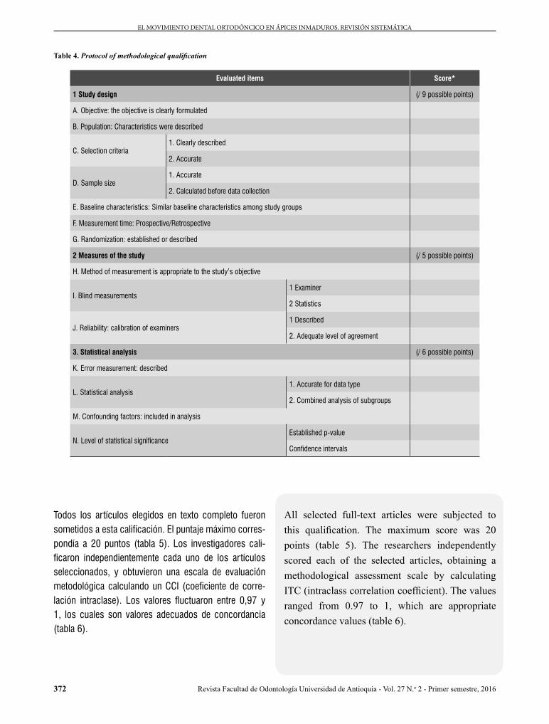
Table 4. Protocol of methodological qualification
Evaluated items Score*
1 Study design (/ 9 possible points)
A. Objective: the objective is clearly formulated
B. Population: Characteristics were described
C. Selection criteria1. Clearly described
2. Accurate
D. Sample size1. Accurate
2. Calculated before data collection
E. Baseline characteristics: Similar baseline characteristics among study groups
F. Measurement time: Prospective/Retrospective
G. Randomization: established or described
2 Measures of the study (/ 5 possible points)
H. Method of measurement is appropriate to the study’s objective
I. Blind measurements 1 Examiner
2 Statistics
J. Reliability: calibration of examiners1 Described
2. Adequate level of agreement
3. Statistical analysis (/ 6 possible points)
K. Error measurement: described
L. Statistical analysis1. Accurate for data type
2. Combined analysis of subgroups
M. Confounding factors: included in analysis
N. Level of statistical significance Established p-value
Confidence intervals
All selected full-text articles were subjected to this qualification. The maximum score was 20 points (table 5). The researchers independently scored each of the selected articles, obtaining a methodological assessment scale by calculating ITC (intraclass correlation coefficient). The values ranged from 0.97 to 1, which are appropriate concordance values (table 6).
Todos los artículos elegidos en texto completo fueron sometidos a esta calificación. El puntaje máximo corres-pondía a 20 puntos (tabla 5). Los investigadores cali-ficaron independientemente cada uno de los artículos seleccionados, y obtuvieron una escala de evaluación metodológica calculando un CCI (coeficiente de corre-lación intraclase). Los valores fluctuaron entre 0,97 y 1, los cuales son valores adecuados de concordancia (tabla 6).
373
ORTHODONTIC TOOTH MOVEMENT IN IMMATURE APICES. A SYSTEMATIC REVIEW
Revista Facultad de Odontología Universidad de Antioquia - Vol. 27 N.o 2 - Primer semestre, 2016
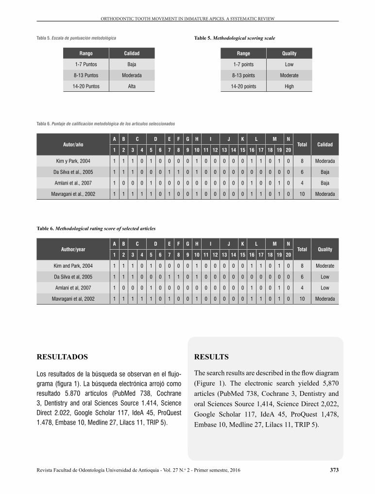
Tabla 5. Escala de puntuación metodológica
Rango Calidad
1-7 Puntos Baja
8-13 Puntos Moderada
14-20 Puntos Alta
Table 5. Methodological scoring scale
Range Quality
1-7 points Low
8-13 points Moderate
14-20 points High
Tabla 6. Puntaje de calificación metodológica de los artículos seleccionados
The search results are described in the flow diagram (Figure 1). The electronic search yielded 5,870 articles (PubMed 738, Cochrane 3, Dentistry and oral Sciences Source 1,414, Science Direct 2,022, Google Scholar 117, IdeA 45, ProQuest 1,478, Embase 10, Medline 27, Lilacs 11, TRIP 5).
RESULTADOS
Los resultados de la búsqueda se observan en el flujo-grama (figura 1). La búsqueda electrónica arrojó como resultado 5.870 artículos (PubMed 738, Cochrane 3, Dentistry and oral Sciences Source 1.414, Science Direct 2.022, Google Scholar 117, IdeA 45, ProQuest 1.478, Embase 10, Medline 27, Lilacs 11, TRIP 5).
374
EL MOVIMIENTO DENTAL ORTODÓNCICO EN ÁPICES INMADUROS. REVISIÓN SISTEMÁTICA
Revista Facultad de Odontología Universidad de Antioquia - Vol. 27 N.o 2 - Primer semestre, 2016
Figura 1. Flujograma
Figure 1. Flowchart
Número de registros o citas identificados en la búsqueda
5.870
Número de registros o citas adicionales identificados en la búsqueda manual
0
Número total de registros o citas duplicados eliminadas 5.816
Número total de registros o citas únicas cribadas
54
Número total de registros o citas duplicadas eliminadas
30
Número total de artículos a texto completo analizados para decidir su elegibilidad
14
Número total de artículos a texto completo excluidos, y razones de su exclusión
10
Número total de estudios incluidos en la síntesis cualitativa de la revisión sistemática
4
Number of records or quotations identified in the search
5,870
Number of additional records or quotations identified in the manual search
0
Total number of duplicate records or quotations eliminated 5,816
Total number of unique records or quotations filtered
54
Total number of duplicate records or quotations eliminated
30
Total number of full-text articles analyzed to decide on their eligibility
14
Total number of excluded full-text articles and reasons for exclusion
10
Total number of included studies in the qualitative synthesis of systematic review
4
375
ORTHODONTIC TOOTH MOVEMENT IN IMMATURE APICES. A SYSTEMATIC REVIEW
Revista Facultad de Odontología Universidad de Antioquia - Vol. 27 N.o 2 - Primer semestre, 2016
La búsqueda manual no arrojó ningún resultado. Luego de revisar los títulos y eliminar los registros y citas dupli-cadas, 54 artículos cumplieron con los criterios de inclu-sión. Después de leer los resúmenes, se seleccionaron 14 artículos para su lectura en texto completo; se exclu-yeron 10 artículos por las siguientes razones: eran estu-dios histológicos, los dientes tratados presentaban trau-ma o tratamiento endodóntico previo, o eran artículos de opinión de expertos o de revisión de la literatura (tabla 7). Finalmente, 4 artículos se seleccionaron para llevar a cabo la evaluación metodológica. Las características de los artículos se encuentran en la tabla 8. Esta muestra no fue suficiente para realizar pruebas de heterogeneidad estadística como la prueba Q de Cochran, I cuadrado o prueba H. Por lo tanto, los resultados se presentarán de forma cualitativa para responder a la siguiente pregunta de investigación: ¿Cuáles son los efectos del movimien-to dental ortodóncico en dientes que no han terminado su formación radicular?
The manual search did not yield any results. After reviewing titles and eliminating duplicate records and quotations, 54 articles met the inclusion criteria. After reading the abstracts, 14 articles were selected for reading in full-text; 10 articles were excluded for the following reasons: they were histological studies, the treated teeth showed trauma or previous endodontic treatment, or were opinion articles by experts or literature reviews (table 7). Finally, 4 articles were selected and the methodological evaluation was conducted. The characteristics of articles are listed in table 8. This sample was not sufficient to run tests of statistical heterogeneity such as the Cochran’s Q test, I2 or H test. Therefore, the results will be presented in a qualitative manner to answer the following research question: What are the effects of orthodontic tooth movement in teeth that have not completed root formation?
Tabla 7. Artículos excluidos
Artículo Autores Motivo
Longitudinal clinical and radiographic evaluation of severely intruded permanent incisors in a pediatric population
Neto Godhin
De CarvalhoGiro42
Se realizó en dientes tratados endodónticamente
Endodontic-orthodontic relationship: a review of integrated treatment planning challenges
HamiltonGutmann38 Estudio in vitro. Pruebas histológicas
A radiographic comparison of apical root resorption after orthodontic treatment with the edgewise and speed appliances
Blake WoodsidePharoah12 Realizado en pacientes con dentición permanente
Quantitative digital subtraction radiography in the assessment of external apical toot resorption induced by orthodontic therapy: A retrospective study Sunku et al43
Los pacientes comenzaron el tratamiento de ortodoncia en dentición mixta tardía y dentición permanente
completa.
Treatment and orthodontic movement of a root-fractured maxillary central incisor with an immature apex: ten-year follow-up
MendozaSolano
Segura-Egea44Los dientes presentaban antecedente de trauma
Orthodontic tooth movement in the mixed dentition: Histological study of a human specimen
Rudzki-JansonPaschosDiedrich45
Es un estudio histológico en un individuo con algún tipo de enfermedad, no cumple con los criterios de
inclusiónDetermination of working length for teeth with wide or immature apices: a
reviewKim y
Chandler46 Realizado en dientes con endodoncia
Risk factors of root resorption after orthodontic treatment. LopatieneDumbravaite14 Es una revisión de la literatura de 2002 a 2007
The effect of fixed orthodontic treatment on developing maxillary incisor root apices Fenn 47 No se encontró el texto completo
The effects of a four-fold increased orthodontic force magnitude on tooth movement and root resorptions. An intra-individual study in adolescents Owman-Moll48 Estudio realizado en premolares
376
EL MOVIMIENTO DENTAL ORTODÓNCICO EN ÁPICES INMADUROS. REVISIÓN SISTEMÁTICA
Revista Facultad de Odontología Universidad de Antioquia - Vol. 27 N.o 2 - Primer semestre, 2016
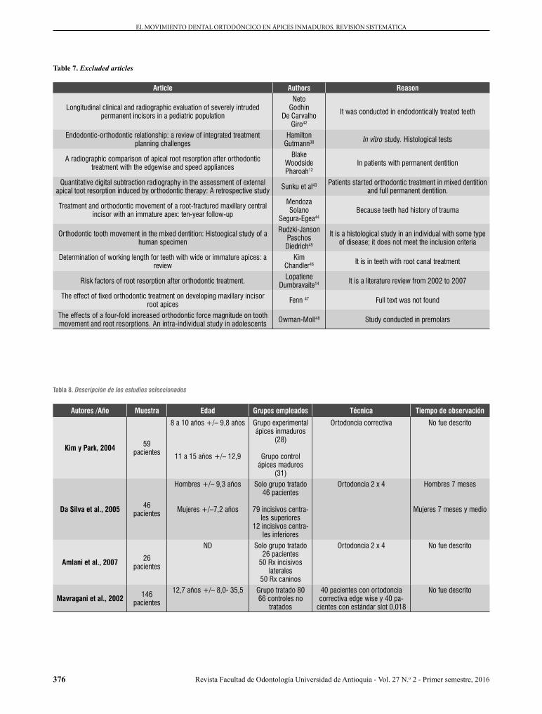
Table 7. Excluded articles
Article Authors Reason
Longitudinal clinical and radiographic evaluation of severely intruded permanent incisors in a pediatric population
Neto Godhin
De CarvalhoGiro42
It was conducted in endodontically treated teeth
Endodontic-orthodontic relationship: a review of integrated treatment planning challenges
HamiltonGutmann38 In vitro study. Histological tests
A radiographic comparison of apical root resorption after orthodontic treatment with the edgewise and speed appliances
BlakeWoodsidePharoah12
In patients with permanent dentition
Quantitative digital subtraction radiography in the assessment of external apical toot resorption induced by orthodontic therapy: A retrospective study Sunku et al43 Patients started orthodontic treatment in mixed dentition
and full permanent dentition.
Treatment and orthodontic movement of a root-fractured maxillary central incisor with an immature apex: ten-year follow-up
MendozaSolano
Segura-Egea44Because teeth had history of trauma
Orthodontic tooth movement in the mixed dentition: Histoogical study of a human specimen
Rudzki-JansonPaschosDiedrich45
It is a histological study in an individual with some type of disease; it does not meet the inclusion criteria
Determination of working length for teeth with wide or immature apices: a review
KimChandler46 It is in teeth with root canal treatment
Risk factors of root resorption after orthodontic treatment. LopatieneDumbravaite14 It is a literature review from 2002 to 2007
The effect of fixed orthodontic treatment on developing maxillary incisor root apices Fenn 47 Full text was not found
The effects of a four-fold increased orthodontic force magnitude on tooth movement and root resorptions. An intra-individual study in adolescents Owman-Moll48 Study conducted in premolars
Tabla 8. Descripción de los estudios seleccionados
Autores /Año Muestra Edad Grupos empleados Técnica Tiempo de observación
Kim y Park, 2004 59 pacientes
8 a 10 años +/– 9,8 años
11 a 15 años +/– 12,9
Grupo experimental ápices inmaduros
(28)
Grupo control ápices maduros
(31)
Ortodoncia correctiva No fue descrito
Da Silva et al., 2005 46 pacientes
Hombres +/– 9,3 años
Mujeres +/–7,2 años
Solo grupo tratado 46 pacientes
79 incisivos centra-les superiores
12 incisivos centra-les inferiores
Ortodoncia 2 x 4 Hombres 7 meses
Mujeres 7 meses y medio
Amlani et al., 2007 26 pacientes
ND Solo grupo tratado 26 pacientes
50 Rx incisivos laterales
50 Rx caninos
Ortodoncia 2 x 4 No fue descrito
Mavragani et al., 2002 146 pacientes
12,7 años +/– 8,0- 35,5 Grupo tratado 80 66 controles no
tratados
40 pacientes con ortodoncia correctiva edge wise y 40 pa-
cientes con estándar slot 0,018
No fue descrito
377
ORTHODONTIC TOOTH MOVEMENT IN IMMATURE APICES. A SYSTEMATIC REVIEW
Revista Facultad de Odontología Universidad de Antioquia - Vol. 27 N.o 2 - Primer semestre, 2016
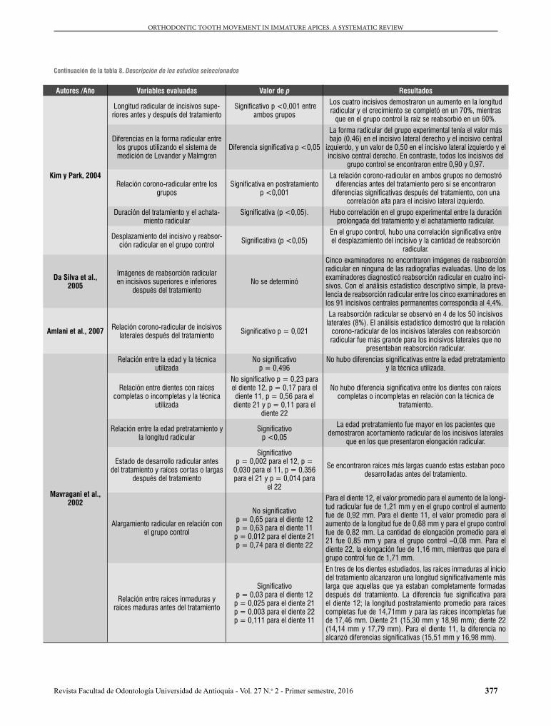
Continuación de la tabla 8. Descripción de los estudios seleccionados
Autores /Año Variables evaluadas Valor de p Resultados
Kim y Park, 2004
Longitud radicular de incisivos supe-riores antes y después del tratamiento
Significativo p <0,001 entre ambos grupos
Los cuatro incisivos demostraron un aumento en la longitud radicular y el crecimiento se completó en un 70%, mientras
que en el grupo control la raíz se reabsorbió en un 60%.
Diferencias en la forma radicular entre los grupos utilizando el sistema de medición de Levander y Malmgren
Diferencia significativa p <0,05
La forma radicular del grupo experimental tenía el valor más bajo (0,46) en el incisivo lateral derecho y el incisivo central
izquierdo, y un valor de 0,50 en el incisivo lateral izquierdo y el incisivo central derecho. En contraste, todos los incisivos del
grupo control se encontraron entre 0,90 y 0,97.
Relación corono-radicular entre los grupos
Significativa en postratamiento p <0,001
La relación corono-radicular en ambos grupos no demostró diferencias antes del tratamiento pero sí se encontraron
diferencias significativas después del tratamiento, con una correlación alta para el incisivo lateral izquierdo.
Duración del tratamiento y el achata-miento radicular
Significativa (p <0,05). Hubo correlación en el grupo experimental entre la duración prolongada del tratamiento y el achatamiento radicular.
Desplazamiento del incisivo y reabsor-ción radicular en el grupo control Significativa (p <0,05)
En el grupo control, hubo una correlación significativa entre el desplazamiento del incisivo y la cantidad de reabsorción
radicular.
Da Silva et al., 2005
Imágenes de reabsorción radicular en incisivos superiores e inferiores
después del tratamientoNo se determinó
Cinco examinadores no encontraron imágenes de reabsorción radicular en ninguna de las radiografías evaluadas. Uno de los examinadores diagnosticó reabsorción radicular en cuatro inci-sivos. Con el análisis estadístico descriptivo simple, la preva-lencia de reabsorción radicular entre los cinco examinadores en los 91 incisivos centrales permanentes correspondía al 4,4%.
Amlani et al., 2007 Relación corono-radicular de incisivos laterales después del tratamiento Significativo p = 0,021
La reabsorción radicular se observó en 4 de los 50 incisivos laterales (8%). El análisis estadístico demostró que la relación
corono-radicular de los incisivos laterales con reabsorción radicular fue más grande para los incisivos laterales que no
presentaban reabsorción radicular.
Mavragani et al., 2002
Relación entre la edad y la técnica utilizada
No significativop = 0,496
No hubo diferencias significativas entre la edad pretratamiento y la técnica utilizada.
Relación entre dientes con raíces completas o incompletas y la técnica
utilizada
No significativo p = 0,23 para el diente 12, p = 0,17 para el diente 11, p = 0,56 para el diente 21 y p = 0,11 para el
diente 22
No hubo diferencia significativa entre los dientes con raíces completas o incompletas en relación con la técnica de
tratamiento.
Relación entre la edad pretratamiento y la longitud radicular
Significativop <0,05
La edad pretratamiento fue mayor en los pacientes que demostraron acortamiento radicular de los incisivos laterales
que en los que presentaron elongación radicular.
Estado de desarrollo radicular antes del tratamiento y raíces cortas o largas
después del tratamiento
Significativop = 0,002 para el 12, p =
0,030 para el 11, p = 0,356 para el 21 y p = 0,014 para
el 22
Se encontraron raíces más largas cuando estas estaban poco desarrolladas antes del tratamiento.
Alargamiento radicular en relación con el grupo control
No significativop = 0,65 para el diente 12p = 0,63 para el diente 11
p = 0,012 para el diente 21p = 0,74 para el diente 22
Para el diente 12, el valor promedio para el aumento de la longi-tud radicular fue de 1,21 mm y en el grupo control el aumento fue de 0,92 mm. Para el diente 11, el valor promedio para el aumento de la longitud fue de 0,68 mm y para el grupo control fue de 0,82 mm. La cantidad de elongación promedio para el 21 fue 0,85 mm y para el grupo control –0,08 mm. Para el diente 22, la elongación fue de 1,16 mm, mientras que para el grupo control fue de 1,71 mm.
Relación entre raíces inmaduras y raíces maduras antes del tratamiento
Significativo p = 0,03 para el diente 12
p = 0,025 para el diente 21p = 0,003 para el diente 22 p = 0,111 para el diente 11
En tres de los dientes estudiados, las raíces inmaduras al inicio del tratamiento alcanzaron una longitud significativamente más larga que aquellas que ya estaban completamente formadas después del tratamiento. La diferencia fue significativa para el diente 12; la longitud postratamiento promedio para raíces completas fue de 14,71mm y para las raíces incompletas fue de 17,46 mm. Diente 21 (15,30 mm y 18,98 mm); diente 22 (14,14 mm y 17,79 mm). Para el diente 11, la diferencia no alcanzó diferencias significativas (15,51 mm y 16,98 mm).
378
EL MOVIMIENTO DENTAL ORTODÓNCICO EN ÁPICES INMADUROS. REVISIÓN SISTEMÁTICA
Revista Facultad de Odontología Universidad de Antioquia - Vol. 27 N.o 2 - Primer semestre, 2016
Table 8. Description of selected studies
Authors / year Sample Age Groups used Technique Time of observation
Kim & Park, 2004 59 patients
8 to 10 years +/–9.8 years
11-15 years +/– 12.9
Experimental group immature apices
(28)
Control group matu-re apices (31)
Corrective orthodontics Not described
Da Silva et al, 2005 46 patients
Males +/–9.3 years
Females +/–7.2 years
Only group treated 46 patients
79 upper central
incisors12 lower central
incisors
Orthodontics 2 x 4 Males 7 months
Females 7 months and a half
Amlani et al, 2007 26 patients
NA Only group treated 26 patients
50 Rx of lateral incisors
50 Rx of canines
Orthodontics 2 x 4 Not described
Mavragani et al, 2002 146 patients
12.7 years +/– 8,0-35.5 Group treated: 8066 untreated
controls
40 patients with edge wise corrective orthodontics and
40 patients with standard slot 0.018
Not described
Table 8 continued. Description of selected studies
Authors / year Evaluated variables p value Results
Kim & Park, 2004
Root length of upper incisors before and after treatment
Significant p <0.001 between both groups
The four incisors showed increased root length and growth was completed in 70%, while in the control group the root was
reabsorbed by 60%.
Differences in root shape among groups using the system of measure-
ment by Levander and MalmgrenSignificant difference p <0.05
Root shape of experimental group had the lowest values (0.46) in the right lateral and left central incisors, and a value of 0.50 in the left lateral and right central incisors. In contrast, all of the in-cisors of the control group were found between 0.90 and 0.97.
Crown-root ratio inter-groups Significant in post-treatment p <0.001
Crown-root ratio in both groups showed no differences before treatment, but there were significant differences after treat-
ment, with a high correlation for left lateral incisors.
Duration of treatment and root flattening Significant (p < 0.05). There was correlation in the experimental group between
duration of extended treatment and root flattening.
Displacement of incisor and root resorption in control group Significant (p < 0.05) In the control group, there was significant correlation between
movement of incisor and amount of root resorption.
Da Silva et al, 2005 Images of root resorption in upper and lower incisors post-treatment Not defined
Five examiners found no root resorption images in any of the evaluated radiographs. One of the examiners diagnosed root resorption in four incisors. The simple descriptive statistical
analysis showed that prevalence of root resorption among five examiners in 91 permanent central incisors was 4.4%.
Amlani et al, 2007 Crown-root ratio of lateral incisors post-treatment Significant p = 0.021
Root resorption occurred in 4 of 50 lateral incisors (8%). The statistical analysis showed that crown-root ratio in lateral
incisors with root resorption was larger for the lateral incisors with no root resorption.
379
ORTHODONTIC TOOTH MOVEMENT IN IMMATURE APICES. A SYSTEMATIC REVIEW
Revista Facultad de Odontología Universidad de Antioquia - Vol. 27 N.o 2 - Primer semestre, 2016
Authors / year Evaluated variables p value Results
Mavragani et al, 2002
Relationship between age and tech-nique used
Non-significantp = 0.496
There were no significant differences between pretreatment age and technique.
Relationship between teeth with complete or incomplete roots and
technique used
Non-significant p = 0.23 for tooth 12, p = 0.17 for tooth 11,
p = 0.56 for tooth 21, and p = 0.11 for tooth 22
There was no significant difference between teeth with com-plete or incomplete roots in relation to treatment technique.
Relationship between pretreatment age and root length Significant p < 0.05 Pretreatment age was higher in patients with root shortening
of lateral incisors than those with root elongation.
State of root development pre-treat-ment and short or long roots
post-treatment
Significantp = 0.002 for 12, p = 0.030 for 11,
p = 0.356 for 21 and p = 0.014 for 22
Longer roots were found when they were poorly developed prior to treatment.
Root lengthening in relation to control group
Non-significantp = 0.65 for tooth 12 p = 0.63 for tooth 11
p = 0.012 for tooth 21 p = 0.74 for tooth 22
For tooth 12, the average value of root length increase was 1.21 mm while this value was 0.92 mm in the control group. For tooth 11, the average value for length increase was 0.68 mm and for the control group was 0.82 mm. The average elongation amount for tooth 21 was 0.85 mm and for the control group 0.08 mm. For tooth 22, elongation was 1.16 mm, while for the control group was 1.71 mm
Relationship between immature and mature roots pre-treatment
Significantp = 0.03 for tooth 12
p = 0.025 for tooth 21 p = 0.003 for tooth 22 p = 0,111 for tooth 11
In three of the studied teeth, immature roots at baseline reached a significantly longer length than those that were already fully formed after treatment. The difference was significant for tooth 12; avera-ge post-treatment length for full roots was 14.71 mm and for in-complete roots 17.46 mm. Tooth 21 (15.30 mm and 18.98 mm); tooth 22 (14,14 mm and 17,79 mm). For tooth 11, the difference did not reach significant differences (15,51 mm and 16,98 mm).
En el año 2005, Da Silva y colaboradores31 encontraron que 5 examinadores no hallaron imágenes de reabsor-ción radicular en ninguna de las radiografías evaluadas. Solo 2 examinadores detectaron pequeños signos de re-absorción radicular en las radiografías periapicales luego de la nivelación. Uno de los examinadores diagnosticó reabsorción radicular en 4 incisivos, y el otro en un solo incisivo que coincidía con uno de los 4 incisivos ya iden-tificados. Con el análisis estadístico descriptivo simple, la prevalencia de reabsorción radicular entre los 5 exa-minadores en los 91 incisivos centrales permanentes correspondía al 4,4%.
Kim y Park en 2004,32 encontraron que los 4 incisivos demostraron un aumento en la longitud radicular y que el crecimiento radicular se completó en un 70% en el grupo experimental, mientras que en el grupo control la raíz se reabsorbió en un 60%. Los cambios en la longitud radicular entre ambos grupos fueron estadísticamente significativos (p <0,001).
In 2005, Da Silva et al31 found out that 5 examiners found no root resorption images in any of the evaluated radiographs. Only 2 examiners detected small signs of root resorption in periapical radiographs after leveling. One of the examiners diagnosed root resorption in 4 incisors, and the other in a single incisor that coincided with one of the 4 incisors already identified. Simple descriptive statistical analysis showed that the prevalence of root resorption among 5 examiners in 91 permanent central incisors was 4.4%.
In 2004, Kim & Park32 found out that 4 incisors showed increased root length and that root growth was completed by 70% in the experimental group, while in the control group the root was reabsorbed by 60%. Changes in root length between the two groups were statistically significant (p < 0.001).
380
EL MOVIMIENTO DENTAL ORTODÓNCICO EN ÁPICES INMADUROS. REVISIÓN SISTEMÁTICA
Revista Facultad de Odontología Universidad de Antioquia - Vol. 27 N.o 2 - Primer semestre, 2016
Mavragani y colaboradores, en 2002,33 encontraron di-ferencias significativas para tres de los incisivos maxi-lares cuando se considera el estado pretratamiento de desarrollo radicular entre raíces acortadas y alargadas. Hallaron raíces más largas cuando estas estaban poco desarrolladas antes del tratamiento (p = 0,002 para el diente 12, p = 0,030 para el 11, p = 0,356 para el 21 y p = 0,014 para el 22).
Edad al inicio del tratamiento
Mavragani y colaboradores, en 2002,33 observaron que la edad pretratamiento fue mayor en los pacientes que demostraron acortamiento radicular de los incisivos la-terales que en los que presentaron elongación radicular (p < 0,05). Para el incisivo central, las diferencias en la edad no fueron significativas. El análisis de regresión reveló que la longitud radicular postratamiento de todos los dientes estudiados estuvo relacionada con la edad al inicio del tratamiento. El estado del desarrollo radicular demostró un coeficiente de correlación significativo solo para el incisivo lateral. En tres de los dientes estudiados, las raíces inmaduras al inicio del tratamiento alcanzaron una longitud significativamente más larga que la de aque-llas que ya estaban completamente formadas después del tratamiento. La diferencia fue significativa para el diente 12, con longitud postratamiento promedio de 14,71 mm para raíces completas y de 17,46 mm para raíces incom-pletas (p = 0,03). Diente 21 (15,30 mm y 18,98 mm; p = 0,025); diente 22 (14,14mm y 17,79 mm; p = 0,003). Para el diente 11, la diferencia no alcanzó diferencias sig-nificativas (15,51 mm, 16,98 mm; p = 0,111).
Longitud radicular pretratamiento
Kim y Park32 encontraron que, en el grupo experimental, la longitud radicular fue más larga antes del tratamiento en 24 incisivos laterales derechos (85,7%), 19 incisivos cen-trales derechos (67,9%), 22 incisivos centrales izquier-dos (78,6%) y 21 incisivos laterales izquierdos (75%). Mientras que en el grupo control la longitud radicular fue más corta en 19 incisivos laterales derechos (61,3%),
In 2002, Mavragani et al33 found significant differences for three of the maxillary incisors when considering the pretreatment state of root development between shortened and elongated roots. They found longer roots when they were poorly developed prior to treatment (p = 0.002 for tooth 12, p = 0.030 for 11, p = 0.356 for 21 and p = 0.014 for 22).
Age of treatment start up
In 2002, Mavragani et al33 observed that pretreatment age was higher in patients with root shortening of lateral incisors compared with patients with root elongation (p < 0,05). For the central incisor, age differences were not significant. Regression analysis showed that post-treatment root length in all studied teeth was related to age at baseline. The state of root development showed significance of correlation coefficient for lateral incisors only. In three of the studied teeth, immature roots at baseline reached a significantly longer length than the ones that were already fully formed after treatment. The difference was significant for tooth 12, with an average post-treatment length of 14,71 mm for complete roots and 17.46 mm for incomplete roots (p = 0.03). Tooth 21 (15.30 mm and 18,98 mm; p = 0.025); tooth 22 (14.14 mm and 17.79 mm; p = 0.003). For tooth 11, the difference did not reach significant differences (15.51 mm, 16.98 mm; p = 0.111).
Pre-treatment root length
Kim & Park32 found out that, in the experimental group, root length was longer before treatment in 24 right lateral incisors (85.7%), 19 right central incisors (67.9%), 22 left central incisors (78.6%), and 21 left lateral incisors (75%). While in the control group root length was shorter in 19 right lateral incisors (61.3%),
381
ORTHODONTIC TOOTH MOVEMENT IN IMMATURE APICES. A SYSTEMATIC REVIEW
Revista Facultad de Odontología Universidad de Antioquia - Vol. 27 N.o 2 - Primer semestre, 2016
23 incisivos centrales derechos (74,2%), 20 incisivos centrales izquierdos (64,5%) y 20 incisivos laterales iz-quierdos (64,5%). La relación corono-radicular entre los dos grupos no demostró una diferencia estadísticamente significativa antes del tratamiento, pero sí fue significa-tiva después del tratamiento, con un alto coeficiente de correlación para el incisivo lateral izquierdo (p <0,001).
Amlani y colaboradores, en 2007,34 encontraron que la reabsorción radicular se observó en 4 de los 50 incisivos laterales (8%). El análisis estadístico demostró que la re-lación corono-radicular de los incisivos laterales fue más grande para los incisivos laterales que no presentaban reabsorción radicular (p = 0,021).
Longitud radicular durante el tratamiento
Mavragani y colaboradores33 encontraron que la evalua-ción de los cambios en la longitud radicular durante el tratamiento reveló un promedio de pérdida de la longitud radicular de 1,86 mm (SE 0,26; rango –3,8 a 6,54 mm) para el diente 12; 1,82 mm (SE 0,26; rango –3,82 a 8,9 mm) para el diente 11; 1,93 mm (SE 0,25; rango –5.7 a 6,27 mm) para el 21 y 1,78 mm (SE 0,33; rango –8.39 a 7,48 mm) para el 22. Se encontraron valores negativos, que indican elongación radicular, en 50 de 280 dientes.
Longitud radicular después del tratamiento
Kim y Park32 encontraron diferencias estadísticamente significativas en la forma radicular, haciendo uso del sistema de medición de Levander y Malmgren, el cual utiliza cinco grados de reabsorción para identificar el ni-vel de reabsorción radicular. El grupo experimental tenía el valor más bajo (0,46) en el incisivo lateral derecho y el incisivo central izquierdo, y un valor de 0,50 en el incisivo lateral izquierdo y el incisivo central derecho. En contraste, todos los incisivos del grupo control se encontraron entre 0,90 y 0,97. La diferencia fue estadís-ticamente significativa para los cuatro incisivos, con un valor p <0,05. La relación corono-radicular en ambos grupos no demostró diferencias antes del tratamiento,
23 right central incisors (74.2%), 20 left central incisors (64.5%) and 20 left lateral incisors (64.5%). Crown-root ratio between the two groups did not show statistically significant differences before treatment, but differences were significant after treatment, with a high coefficient of correlation for left lateral incisors (p < 0.001).
In 2007, Amlani et al34 found root resorption in 4 of 50 lateral incisors (8%). Statistical analysis showed that crown-root ratio in lateral incisor was larger for the lateral incisors with no root resorption (p = 0.021).
Root length during treatment
Mavragani et al33 found that evaluation of changes in root length during treatment showed an average of root length loss of 1.86 mm for tooth 12 (0.26 SE, –3.8 to 6.54 mm range); 1.82 mm for tooth 11 (0.26 SE, –3.82 to 8.9 mm range); 1.93 mm for tooth 21 (0.25 SE, –5.7 to 6.27 mm range) and 1.78 mm for tooth 22 (0.33 SE, –8.39 to 7.48 mm range). Negative values were found in 50 of 280 teeth, suggesting root canal elongation.
Post-treatment root length
Kim & Park32 found statistically significant differences in root shape by means of the Levander & Malmgren measuring system, which uses five resorption degrees to identify levels of root resorption. The experimental group had the lowest value (0.46) in the right lateral and left central incisors, and a value of 0.50 in the left lateral and right central incisors. In contrast, all of the incisors in the control group were found between 0.90 and 0.97. The difference was statistically significant for the four incisors, with <0.05 p value. Crown-root ratio in both groups showed no pre-treatment differences,
382
EL MOVIMIENTO DENTAL ORTODÓNCICO EN ÁPICES INMADUROS. REVISIÓN SISTEMÁTICA
Revista Facultad de Odontología Universidad de Antioquia - Vol. 27 N.o 2 - Primer semestre, 2016
pero sí se encontraron diferencias significativas después del tratamiento, con una correlación alta para el incisivo lateral izquierdo (p <0,001).
Mavragani y colaboradores33 encontraron que las raí-ces alargadas durante el tratamiento tenían una longi-tud radicular similar a aquellas raíces no tratadas en individuos de la misma edad. Para el diente 12, el valor promedio de aumento de la longitud radicular fue de 1,21 mm y en el grupo control el aumento fue de 0,92 mm (p = 0,65). Para el diente 11, el valor promedio de au-mento de la longitud fue de 0,68 mm, y para el grupo control fue de 0,82 mm (p = 0,63). La cantidad de elon-gación promedio para el diente 21 fue de 0,85 mm y para el grupo control fue de –0,08mm (p = 0,012). Para el diente 22, la elongación fue de 1,16 mm, mientras que para el grupo control fue de 1,71 mm (p = 0,74).
Técnica utilizada y la edad antes del tratamiento
Mavragani y colaboradores33 no encontraron diferencias significativas entre la edad pretratamiento y la técnica uti-lizada (p = 0,496). Así mismo, no hubo diferencia signi-ficativa entre los dientes con raíces completas o incom-pletas en relación con la técnica de tratamiento. (p = 0,23 para el diente 12, p = 0,17 para el diente 11, p = 0,56 para el diente 21 y p = 0,11 para el diente 22).
Duración del tratamiento y reabsorción
radicular
Kim y Park32 encontraron correlación en el grupo experimental entre la duración del tratamiento y el achatamiento radicular (p <0,05).
DISCUSIÓN
En la presente revisión sistemática se incluyeron cuatro artículos con un nivel de evidencia entre baja y modera-da. De los cuatro artículos, tres fueron retrospectivos y uno prospectivo. Ninguno calculó el tamaño de la mues-tra; tampoco reportaron aleatorización ni el cegamiento
but there were significant post-treatment differences, with a high correlation for the left lateral incisor (p < 0.001).
Mavragani et al33 found out that roots which elongated during treatment had a root length similar to that of un-treated roots in individuals of the same age. For tooth 12, the average value of root length increase was 1.21 mm and in the control group the increase was 0.92 mm (p= 0.65). For tooth 11, the average value of length increase was 0.68 mm, and for the control group was 0.82 mm (p = 0.63). The amount of average elongation for tooth 21 was 0.85 mm and for the control group was –0.08 mm (p = 0.012). For tooth 22, elongation was 1.16 mm, while for the control group was 1.71 mm (p = 0.74).
Technique used and pre-treatment age
Mavragani et al33 found no significant differences between pretreatment age and technique used (p = 0.496). Similarly, there was no significant difference between teeth with incomplete or complete roots in relation to treatment technique (p = 0.23 for tooth 12, p = 0.17 for tooth 11, p = 0.56 for tooth 21 and p = 0.11 for tooth 22).
Duration of treatment and root resorption
Kim & Park32 found correlation between treatment duration and root flattening (p < 0.05) in the experimental group.
DISCUSSION
This systematic review included four articles with low to moderate levels of evidence. Of the four articles, three were retrospective and one was prospective. None of them calculated sample size, and they did not report randomization nor
383
ORTHODONTIC TOOTH MOVEMENT IN IMMATURE APICES. A SYSTEMATIC REVIEW
Revista Facultad de Odontología Universidad de Antioquia - Vol. 27 N.o 2 - Primer semestre, 2016
del investigador ni del estadístico. Asimismo, no se re-portó la concordancia intraexaminador en ninguno de los estudios y no se establecieron intervalos de confianza. Solo un estudio consideró los factores de confusión, de-terminación del error de medición y calibración intraexa-minador.
Existen pocos estudios acerca del comportamiento ra-dicular en dientes con ápices abiertos en donde el pro-ceso biológico de formación radicular aún se encuentra en proceso. Se presenta controversia sobre el riesgo biológico que tiene el movimiento ortodóncico en ápi-ces inmaduros. Por una parte, algunos autores30, 36, 37 coinciden en que los dientes que no han completado su formación radicular son menos propensos a presentar reabsorción después de inducir el movimiento con apa-ratos fijos. Asimismo, Linge y colaboradores37 y Mavra-gani y colaboradores33 reportan menores daños radicu-lares en ápices con formación radicular incompleta. Sin embargo, Hendrix y colaboradores29 encuentran que los dientes con ápices abiertos que no alcanzan la longitud normal promedio presentan raíces más largas, compa-radas con dientes con ápices cerrados, probablemente por el desplazamiento del ápice durante el tratamiento. Hamilton y Gutmann38 reportan que se produce un cie-rre apical prematuro al aplicar fuerzas ortodóncicas en ápices inmaduros. En consecuencia, la principal variable evaluada en su revisión sistemática fueron los cambios que se presentaron cuando se hicieron movimientos dentales en ápices que no habían terminado su forma-ción radicular.
En cuanto a los cambios en la longitud radicular antes y después del tratamiento, el estudio de Kim y Park32 encuentra una diferencia estadísticamente significativa entre ambos grupos, de modo que se reporta un aumen-to de la longitud radicular que se completa en un 70%, mientras que en el grupo control la raíz se reabsorbe en un 60%. Dicho estudio concluye que el grado de re-absorción es leve, debido a que los participantes en el mismo fueron tratados sin exodoncias y por lo tanto la distancia del desplazamiento y la duración fueron cortas.
researcher or statistician blinding. Also, none of them reported intra-examiner concordance and none established confidence intervals. Only one study considered confounders, measurement error, or intra-examiner calibration.
There are few studies on root behavior in teeth with open apices where the biological process of root formation is still in process. There is controversy over the biological risk of orthodontic movement in immature apices. On the one hand, some authors30, 36, 37
agree that teeth that have not completed root formation are less prone to resorption after inducing movement with fixed appliances. In addition, Linge et al37 and Mavragani et al33 report lower root damage in apices with incomplete root formation. However, Hendrix et al29 found out that teeth with open apices which do not achieve the average standard length usually have longer roots compared to teeth with closed apices, probably due to apex displacement during treatment. Hamilton & Gutmann38 reported that premature apical closure occurs when applying orthodontic forces on immature apices. In consequence, the main variable evaluated in their systematic review was the changes occurring when tooth movements were applied on apices that had not completed root formation.
Concerning changes in root length before and after treatment, the study by Kim & Park32 found statistically significant differences between both groups, reporting increased root length that is complete by 70%, while in the control group the root reabsorbed by 60%. Their study concludes that the resorption degree was low because participants were treated without extractions and therefore both displacement distance and duration were short.
384
EL MOVIMIENTO DENTAL ORTODÓNCICO EN ÁPICES INMADUROS. REVISIÓN SISTEMÁTICA
Revista Facultad de Odontología Universidad de Antioquia - Vol. 27 N.o 2 - Primer semestre, 2016
En cuanto a la proporción corono-radicular, Kim y Park32 no reportan diferencias significativas antes del trata-miento entre las raíces con ápices inmaduros y las que habían completado su desarrollo. Sin embargo, sí hay diferencias significativas entre los dos grupos en el pe-riodo posterior al tratamiento, debido a que las raíces in-maduras tienden a desarrollarse, mientras que las raíces con ápices maduros se reabsorben y se acortan.
Kim y Park32 encuentran diferencias significativas en cuanto a la forma de la raíz después del tratamiento de ortodoncia en ambos grupos, utilizando el sistema de medición descrito por Levander y Malmgren,39 lo cual se puede explicar porque la forma final de los ápices in-maduros fue similar a la forma de los ápices maduros.
De igual manera, el cambio en la forma de la raíz se asoció a la duración del tratamiento: a mayor duración, permitió la completa formación de la raíz, seguida por re-absorción y acortamiento. En conclusión, a pesar de que la posibilidad de reabsorción en ápices inmaduros es re-ducida, y si la duración del tratamiento es más larga, no se deben esperar diferencias con los ápices maduros.
En cuanto al cambio de longitud y forma de la raíz, Kim y Park32 concluyen que, si el tratamiento de ortodoncia se comienza antes de la formación completa de la raíz, será conveniente para la reducción de la reabsorción ra-dicular. Sin embargo, dicho estudio se basa en cambios observados en radiografías de ápices inmaduros, exclu-yendo otros factores como los aspectos histológicos, la historia de trauma o los hábitos.
Por otra parte, Da Silva y colaboradores31 reportan que el riesgo de reabsorción, o cualquier alteración en raíces en formación durante el movimiento inducido en incisi-vos permanentes con ápices abiertos, es prácticamente nulo. De igual forma, Rudolph35 concuerda con que el tratamiento de ortodoncia es menos perjudicial en raíces en formación, es decir, cuando se inicia tempranamente. Sin embargo, dicho resultado se debe analizar cuidado-samente, debido a la metodología empleada (el uso de radiografías panorámicas), pues los autores tomaron como referencia la edad cronológica y no el estadio de formación radicular, lo que provee poca información so-bre el estadio de formación de las raíces.
Concerning crown-root ratio, Kim & Park32 reported no significant differences before treatment between roots with immature apices and those that had completed their development. However, there were significant post-treatment differences between the two groups, since immature roots tend to grow while roots with mature apices are reabsorbed and shorten.
Kim & Park32 found significant differences in terms of root shape after orthodontic treatment in both groups, using the measuring system described by Levander and Malmgren,39 which can be explained because the final shape of immature apices was similar to the shape of mature apices.
Similarly, the change in root shape was associated with treatment duration: longer treatment periods allowed complete root formation, followed by reabsorption and shortening. In conclusion, while the possibility of reabsorption in immature apices is low, in longer treatments one should not expect differences with mature apices.
Concerning changes in root length and shape, Kim & Park32 concluded that starting orthodontic treatment before full root formation is convenient for reducing root resorption. However, their study is based on changes observed in x-rays of immature apices, excluding other factors such as histological aspects, history of trauma, or habits.
On the other hand, Da Silva et al31 reported that the risk of resorption or any other alteration in roots in formation during induced movement in permanent incisors with open apices is practically non-existent. In the same way, Rudolph35 agrees that orthodontic treatment is less damaging in roots in formation, i.e., when it is started early. However, this result should be taken carefully because of the methodology used (panoramic radiographs), since the authors took chronological age as a reference instead of the stage of root formation, thus providing little information on the stage of root formation.
385
ORTHODONTIC TOOTH MOVEMENT IN IMMATURE APICES. A SYSTEMATIC REVIEW
Revista Facultad de Odontología Universidad de Antioquia - Vol. 27 N.o 2 - Primer semestre, 2016
Posteriormente, mediante radiografías periapicales, Lin-ge y colaboradores37 estudiaron la incidencia y extensión de la reabsorción radicular en incisivos superiores, y en-contraron que la reabsorción radicular fue dos veces ma-yor en los ápices que habían completado su formación, en comparación con los ápices inmaduros.
Da Silva y colaboradores31 concluyen que, desde una perspectiva radiográfica, no se evidencia iatrogenia en la integridad radicular durante el movimiento de ortodon-cia en incisivos centrales permanentes con formación incompleta.
Mavragani y colaboradores33 coinciden con lo antes ex-puesto, dado que en su estudio encuentran que los dientes con formación radicular parcial antes del tratamiento al-canzan mayor longitud radicular que los dientes con raíces completamente formadas al inicio del tratamiento. Por lo tanto, concluyen que el tratamiento de ortodoncia no causa reabsorción radicular en dientes con ápices inmaduros.
Con base en los resultados obtenidos, se debe tener en cuenta que la mayoría de los estudios de ortodoncia fueron escasos y presentaron fallas metodológicas. Sin embargo, los hallazgos de la presente revisión coinciden con los de varios autores,30, 36, 32 quienes sugieren que el tratamiento de ortodoncia en dientes con formación radicular incompleta no representa un riesgo biológico para su desarrollo.
CONCLUSIONES
Con base en los resultados del presente estudio, se pue-den sacar las siguientes conclusiones:
1. La longitud radicular final de los incisivos que no han terminado su cierre apical, y que son movidos con ortodoncia fija, no se altera.
2. La técnica utilizada parece no ser relevante en la lon-gitud radicular final.
3. Estos resultados hay que tomarlos con mucha caute-la porque la evidencia resultante fue baja y media.
Subsequently, by means of periapical radiographs, Linge et al37 studied the incidence and extent of root resorption in maxillary incisors, finding out that root resorption was two times higher in apices which had completed their formation, compared with immature apices.
Da Silva et al31 concluded that, from a radiographic perspective, there is no evidence of iatrogenesis in root integrity during orthodontic movement in permanent central incisors with incomplete formation.
Mavragani et al33 agree, as in their study they found out that teeth with partial root formation prior to treatment reach longer root length than teeth with fully formed roots when treatment starts. Therefore, they conclude that orthodontic treatment does not cause root resorption in teeth with immature apices.
Based on the obtained results, we must bear in mind that most orthodontic studies were scarce and had methodological weaknesses. However, the findings of the present review agree with those of several authors,30, 36, 32 who suggest that orthodontic treatment in teeth with incomplete root formation does not represent a biological risk for its development.
CONCLUSIONS
Based on the results of the present study, the following conclusions can be drawn:
1. There are no alterations in the final root length of incisors that have not completed apical closure and which are moved with fixed orthodontics.
2. The technique used does not seem to be relevant in final root length.
3. These results should be taken cautiously because the resultant evidence was low to moderate.
386
EL MOVIMIENTO DENTAL ORTODÓNCICO EN ÁPICES INMADUROS. REVISIÓN SISTEMÁTICA
Revista Facultad de Odontología Universidad de Antioquia - Vol. 27 N.o 2 - Primer semestre, 2016
4. Se necesitan ensayos clínicos aleatorizados a largo plazo, que nos muestren las alteraciones a nivel radi-cular en dientes que fueron intervenidos con fuerzas tempranamente.
CONFLICTO DE INTERESES
Esta revisión sistemática no presenta conflictos de in-terés, no fue patrocinada por ninguna casa comercial ni por la Universidad El Bosque.
No se registró ningún protocolo en las bases de datos para revisiones sistemáticas.
1. Tafur AM, Tuesta O, Raymundo J. Biología del movimiento ortodóntico. Rev Estomatol Hered 2001; 11(1-2): 46-51.
2. Masella RS, Meister M. Current concepts in the biology of orthodontic tooth movement. Am J Orthod Dentofacial Orthop 2006; 129(4): 458-468.
3. Schwarz AM. Tissue changes incident to orthodontic tooth movement. Int J Orthod 1932; (18): 331-352.
4. Melsen B. Biological reaction of alveolar bone to orthodontic tooth movement. Angle Orthod 1999; 69 (2): 151-158.
5. Dolce C, Scott MJ, Wheeler TT. Current concepts in the biology of orthodontic tooth movement. Semin Orthod 2002; 8 (1): 6-12.
6. Zainal S, Yamamoto Z, Zainol I, Abdul R, Zainal Z. Cellular and molecular changes in orthodontic tooth movement. ScientificWorld Journal 2011; (11): 1788-1803.
7. Melsen B. Tissue reaction to orthodontic tooth movement: a new paradigm. Eur J Orthod 2001; 23(6): 671-681.
8. Von-Böhl M, Kuijpers-Jagtman AM. Hyalinization during orthodontic tooth movement: a systematic review on tissue reactions. Eur J Orthod 2009; 31(1): 30-36.
9. Quintero P, Yepes E, Rendón J. Pulp tissue reactions to specific orthodontic movements: a literature review. Angle Orthod 2011; 7(13): 54-60.
10. Ren Y, Maltha JC, Kuijpers-Jagtman AM. Optimum force magnitude for orthodontic tooth movement: a systematic literature review. Angle Orthod 2003; 73(1): 86-92.
11. Brin I, Tulloch JF, Koroluk L, Philips C. External apical root resorption in Class II malocclusion: a retrospective review of 1-versus 2-phase treatment. Am J Orthod Dentofacial Orthop 2003; 124(2): 151-156.
12. Blake M, Woodside DG, Pharoah MJ. A radiographic comparison of apical root resorption after orthodontic
387
ORTHODONTIC TOOTH MOVEMENT IN IMMATURE APICES. A SYSTEMATIC REVIEW
Revista Facultad de Odontología Universidad de Antioquia - Vol. 27 N.o 2 - Primer semestre, 2016
treatment with the edgewise and Speed appliances. Am J Orthod Dentofacial Orthop 1995; 108(1): 76-84.
13. Acar A, Canyürek U, Kocaaga M, Erverdi N. Continuous vs. discontinuous force application and root resorption. Angle Orthod 1999; 69(2): 163-164.
14. Lopatiene K, Dumbravait A. Risk factors of root resorption after orthodontic treatment. Stomatologija 2008; 10(3): 89-95.
15. Costopoulos G, Nanda R. An evaluation of root resorption incident to orthodontic intrusion. Am J Orthod Dentofacial Orthop 1996; 109(5): 543-548.
16. Sameshima GT, Sinclair PM. Predicting and preventing root resorption: Part I. Diagnostic factors. Am J Orthod Dentofacial Orthop 2001; 119(5): 505-510.
17. Mirabella AD, Årtun J. Risk factors for apical root resorption of maxillary anterior teeth in adult orthodontic patients. Am J Orthod Dentofacial Orthop 1995; 108(1): 48-55.
18. Harris EF, Kineret SE, Tolley EA. A heritable component for external apical root resorption in patients treated orthodontically. Am J Orthod Dentofacial Orthop 1997; 111(3): 301-309.
19. Lee RY, Årtun J, Alonzo TA. Are dental anomalies risk factors for apical root resorption in orthodontic patients?. Am J Orthod Dentofacial Orthop 1999; 116(2): 187-195.
20. Mavragani M, Apisariyakul J, Brudvik P, Selvig KA. Is mild dental invagination a risk factor for apical root resorption in orthodontic patients?. Eur J Orthod 2006; 28(4): 307-312.
21. Janson GR, De-Luca-Canto G, Martins DR, Henriques JF, De Freitas MR. A radiographic comparison of apical root resorption after orthodontic treatment with 3 different fixed appliance techniques. Am J Orthod Dentofacial Orthop 2000; 118(3): 262-273.
22. Costopoulos G, Nanda R. An evaluation of root resorption incident to orthodontic intrusion. Am J Orthod Dentofacial Orthop 1996; 109(5): 543-548.
23. Han G, Huang S, Von den Hoff JW, Zeng X, Kuijpers-Jagtman AM. Root resorption after orthodontic intrusion and extrusion: an intra-individual study. Angle Orthod 2005; 75(6): 912-918.
24. Weiland F. Constant versus dissipating forces in orthodontics: the effect on initial tooth movement and root resorption. Eur J Orthod 2003; 25(4): 335-342.
25. Sameshima GT, Sinclair PM. Predicting and preventing root resorption: part II. Treatment factors. Am J Orthod Dentofacial Orthop 2001; 119(5): 511-515.
26. Oppenheim A. Human tissue response to orthodontic intervention of short and long duration. Am J Orthod. 1942; 28(5): 263-301.
27. Phillips, J. Apical Root Resorption Under Orthodontic Therapy. Angle Orthod 1955; 25(1): 1-22.
28. Consolaro A, Ortiz M, Velloso, T, Dentes com rizogênese incompleta e movimento ortodôntico: bases biológicas. R Dental Press Ortodon Ortop Facial 2001; 6(2): 25-30.
29. Hendrix I, Carels C, Kuijpelrs-Jagtman AM, Van‘T-Hof M. A radiographic study of posterior apical root resorption in orthodontic patients. Am J Orthod Dentofacial Orthop 1994; 105(4): 345-349.
30. Lagravere M, Majorb P, Flores C. Long-term skeletal changes with rapid maxillary expansion: a systematic review. Angle Orthod 2005; 75(6): 1046-1052.
31. Da Silva Filho O, Mendez Ode F, Ozawa TO, Ferrari Junior FM, Correa TM. Behavior of partially formed roots of teeth submitted to orthodontic movement. J Clin Pediatr Dent 2005; 28 (2): 147-154
32. Kim H, Park S. The changes of root length and form in immature teeth after orthodontic treatment. Korean J Orthod 2004; 3 (3): 241-251.
33. Mavragani M, Egil O, Wisth B, Selvig K. Changes in root length during orthodontic treatment: advantages for immature teeth. Eur J Orthod 2002; 24(1): 91-97.
34. Amlani MS, Inocencio F, Hatibovic-Kofman S. Lateral incisor root resorption and active orthodontic treatment in the early mixed dentition. Eur J Paediatr Dent 2007; 8(4): 188-192.
35. Rudolph CE. An evaluation of root resorption occurring during orthodontic treatment. J Dent Res 1940; 19(4): 367-371.
36. Reitan K. Initial tissue behavior during apical root resorption. Angle Orthod 1974; 44(1): 68-82.
37. Linge B. Ohm, Linge L. Apical root resorption in upper anterior teeth. Eur J Orthod 1983; 5(3): 173-183.
38. Hamilton RS, Gutmann JL. Endodontic-orthodontic relationships: a review of integrated treatment planning challenges. Int Endod J 1999; 32(5): 343-360.
388
EL MOVIMIENTO DENTAL ORTODÓNCICO EN ÁPICES INMADUROS. REVISIÓN SISTEMÁTICA
Revista Facultad de Odontología Universidad de Antioquia - Vol. 27 N.o 2 - Primer semestre, 2016
39. Levander E, Malmgren O. Evaluation of the risk of root resorption during orthodontic treatment: a study of upper incisors. Eur J Orthod 1988; 10(1): 30-38.
40. Rosenberg M. An evaluation of the incidence and amount of apical root resorption and dilaceration occurring in orthodontically treated teeth having incompletely formed roots at the beginning of Begg treatment. Am J Orthod 1972; 61(5): 524-525.
41. Stenvik A, Ivar A. The effect of experimental tooth intrusion on pulp and dentine. Oral Surg Oral Med Oral Pathol 1971; 32(4): 639-648.
42. Neto JJ, Gondim JO, de-Carvalho FM, Giro EM. Longitudinal clinical and radiographic evaluation of severely intruded permanent incisors in a pediatric population. Dent Traumatol 2009; 25(5): 510-514.
43. Sunku R, Roopesh R, Kancherla P, Perumalla KK, Yudhistar PV, Reddy VS. Quantitative digital subtraction radiography in the assessment of external apical toot resorption induced by orthodontic therapy: A retrospective study. J Contemp Dent Pract 2011; 12(6): 422-428.
44. Mendoza A, Solano E, Segura-Egea J. Treatment and orthodontic movement of a root-fractured maxillary central incisor with an immature apex: 10-year follow-up. Int Endod J 2010; 43(12): 1162-1170.
45. Rudzki-Janson E, Paschos E, Diedrich P. Orthodontic tooth movement in the mixed dentition: Histological study of a human specimen. J Orofac Orthop 2001; 62(3): 177-190.
46. Kim YJ, Chandler NP. Determination of working length for teeth with wide or immature apices: a review. Int Endod J 2013; 46(6): 483-491.
47. Fenn KM. The effect of fixed orthodontic treatment on developing maxillary incisor root apices. Am J Orthod Dentofacial Orthop 1998; 114(5): A1.
48. Owman-Moll P. The effects of a four-fold increased orthodontic force magnitude on tooth movement and root resorptions. An intra-individual study in adolescents. Eur J Orthod 1996; 18 (3): 287-294.