1 THE EMERGING ROLE OF CALRETICULIN IN CANCER CELLS WCRJ 2017; 4 (3): e926 INTRODUCTION The human calreticulin gene (CALR) is located on chromosome 19p13.2 with nine exons. Calre- ticulin promoter region contains several binding sites for reputed transcription factors and many of these factors have been identified as important modulators of CRT expression including NKx2.5, MEF2C, COUP-TF1, GATA6, Evi-1, and PPAR factors 1 . Moreover, calcium (Ca ++ ) depletion and endoplasmatic reticulum (ER) stress were shown to be essential activators of CALR transcription 2 . Recently, studies have also demonstrated that nerve growth factor (NGF) can also upregulate CALR expression in both ovarian cells and neu- ronal differentiation 3,4 . These results suggested an involvement of CALR expression in various bio- logical and pathological processes (Table I). From several years, many studies have shown that calreticulin is involved in the development of different cancers concerning tumor genera- tion and progression. On the other hand, CALR expressed on cell surface represent a phagocytic signal for natural killer cells by immune system. In addition to immunogenicity and tumorigene- sis, interactions throughout cell adhesion between CALR and integrins indicate the primary role of the CALR in cancer metastasis. Corresponding Author: Salvo Di Martino; e-mail: [email protected]Abstract – Background: Recently, Calreticulin (CALR) has been proposed to participate in var- ious physiological and pathological processes in cells. The two major functions of CALR inside the Endoplasmic Reticulum (ER) are protein chaperoning and regulation of Ca++ homeostasis. Further- more, accumulated studies indicate that CALR also regulates important biological functions includ- ing cell adhesion, gene expression, and RNA stability. Upgrading: This review aims to highlight the most biological function of CALR protein in cancer development. In addition, a comprehensive evaluation of the detection method for revelation of low amount of mutant DNA in a large excess of wild type (wt) DNA is proposed and discussed. Conclusions: Acknowledgments about CALR function in oncology field will allow the design of new research project that will get light to relationship between normal physiological function and tumors development mediated by CALR. Based on these fields, in the next future, the oncologists will have new markers to strike a precise diagnosis. KEYWORDS: CALR, Genomic, Mutation, Tissue expression. 1 CETAC Research Center, Caserta, Italy 2 Italian Association of Pharmacogenomics and Molecular Diagnostics, Caserta, Italy 3 GORI “Gruppo Oncologico Ricercatori Italiani” Onlus, Pordenone, Italy 4 Second Division of Neurology, Campania University Luigi Vanvitelli School of Medicine, Caserta, Italy 5 Scientific Directorate, ISCD, Istituto per lo Studio e la Cura del Diabete, Casagiove, Caserta, Italy 6 Scientific Directorate, CETAC Research Center, Caserta, Italy S. DI MARTINO 1 , G. CRESCENTE 2,3 , V. DE LUCIA 4 , M. DI PAOLO 1 , G. MAROTTA 5 , D. DE LUCIA 5 , A. ABBADESSA 6
Transcript
1
THE EMERGING ROLE OF CALRETICULININ CANCER CELLS
WCRJ 2017; 4 (3): e926
INTRODUCTION
The human calreticulin gene (CALR) is located on chromosome 19p13.2 with nine exons. Calre-ticulin promoter region contains several binding sites for reputed transcription factors and many of these factors have been identified as important modulators of CRT expression including NKx2.5, MEF2C, COUP-TF1, GATA6, Evi-1, and PPAR factors1. Moreover, calcium (Ca++) depletion and endoplasmatic reticulum (ER) stress were shown to be essential activators of CALR transcription2. Recently, studies have also demonstrated that nerve growth factor (NGF) can also upregulate
CALR expression in both ovarian cells and neu-ronal differentiation3,4. These results suggested an involvement of CALR expression in various bio-logical and pathological processes (Table I).
From several years, many studies have shown that calreticulin is involved in the development of different cancers concerning tumor genera-tion and progression. On the other hand, CALR expressed on cell surface represent a phagocytic signal for natural killer cells by immune system. In addition to immunogenicity and tumorigene-sis, interactions throughout cell adhesion between CALR and integrins indicate the primary role of the CALR in cancer metastasis.
Abstract – Background: Recently, Calreticulin (CALR) has been proposed to participate in var-ious physiological and pathological processes in cells. The two major functions of CALR inside the Endoplasmic Reticulum (ER) are protein chaperoning and regulation of Ca++ homeostasis. Further-more, accumulated studies indicate that CALR also regulates important biological functions includ-ing cell adhesion, gene expression, and RNA stability.
Upgrading: This review aims to highlight the most biological function of CALR protein in cancer development. In addition, a comprehensive evaluation of the detection method for revelation of low amount of mutant DNA in a large excess of wild type (wt) DNA is proposed and discussed.
Conclusions: Acknowledgments about CALR function in oncology field will allow the design of new research project that will get light to relationship between normal physiological function and tumors development mediated by CALR. Based on these fields, in the next future, the oncologists will have new markers to strike a precise diagnosis.
1CETAC Research Center, Caserta, Italy2Italian Association of Pharmacogenomics and Molecular Diagnostics, Caserta, Italy3GORI “Gruppo Oncologico Ricercatori Italiani” Onlus, Pordenone, Italy4Second Division of Neurology, Campania University Luigi Vanvitelli School of Medicine, Caserta, Italy5Scientific Directorate, ISCD, Istituto per lo Studio e la Cura del Diabete, Casagiove, Caserta, Italy6Scientific Directorate, CETAC Research Center, Caserta, Italy
S. DI MARTINO1, G. CRESCENTE2,3, V. DE LUCIA4, M. DI PAOLO1, G. MAROTTA5, D. DE LUCIA5, A. ABBADESSA6
2
THE EMERGING ROLE OF CALRETICULIN IN CANCER CELLS
evidence that modification of CALR levels affects cell adhesion on extracellular matrix molecules (ECM)6,7. In fact, is noted that CALR plays a role in the control of cell adhesiveness through regula-tion of fibronectin expressions and collagen depo-sition mediated by Ca2++- regulation and c-SRC activity8. In addition, previous studies9 revealed that CALR-mediated cell adhesion might be due to direct interaction between CALR and integrins by binding to the cytoplasmic KXGFFKR motif of the integrin a-subunit. These studies provided evidence that CALR plays a pivotal role in cellu-lar adhesiveness.
Here, we reviewed the biological roles of CALR in cancer development. Also, available methods for low detection amount of mutant DNA in a large excess of wild type (wt) DNA are taken in consideration5.
Mechanisms of Cancer development involving CALR
The cell adhesion is based on the regulation of fo-cal contact via multiple mechanisms and the hy-pothesis that CALR might be involved is based on
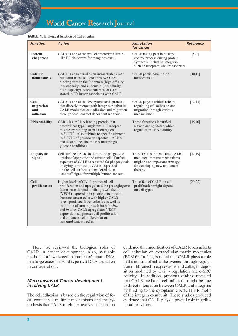
TABLE 1. Biological function of Calreticulin.
Function Action Annotation Reference for cancer
Protein CALR is one of the well characterized lectin- CALR taking part in quality [5-9] chaperone like ER chaperons for many proteins. control process during protein synthesis, including integrins, surface receptors, and transporters.
Calcium CALR is considered as an intracellular Ca2++ CALR participate in Ca2++ [10,11] homeostasis regulator because it contains two Ca2++- homeostasis. binding sites in the P-domain (high-affinity, low-capacity) and C-domain (low affinity, high-capacity). More than 50% of Ca2++
stored in ER lumen associates with CALR.
Cell CALR is one of the few cytoplasmic proteins CALR plays a critical role in [12-14] migration that directly interact with integrin a-subunits. regulating cell adhesion and and CALR modulates cell adhesion and migration migration through various adhesion through focal contact dependent manners. mechanisms. RNA stability CARL is a mRNA binding protein that These functions identified [15,16] destabilizes type I angiotensin II receptor a trans-acting factor, which mRNA by binding to AU-rich region regulates mRNA stability. in 3’-UTR. Also, it binds to specific element in 3’-UTR of glucose transporter-1 mRNA and destabilizes the mRNA under high- glucose conditions.
Phagocytic Cell surface CALR facilitates the phagocytic These results indicate that CALR- [17-19] signal uptake of apoptotic and cancer cells. Surface mediated immune mechanisms exposure of CALR is required for phagocytosis might be an important strategy on dying tumor cells. CALR expressed for developing new anticancer on the cell surface is considered as an therapy. “eat-me” signal for multiple human cancers. Cell Higher levels of CALR promoted cell The effect of CALR on cell [20-22] proliferation proliferation and upregulated the proangiogenic proliferation might depend factor vascular endothelial growth factor on cell types. (VEGF) expression in gastric cancer cells. Prostate cancer cells with higher CALR levels produced fewer colonies as well as inhibition of tumor growth both in vitro and in vivo. CALR upregulates VEGF expression, suppresses cell proliferation and enhances cell differentiation in neuroblastoma cells.
3
THE EMERGING ROLE OF CALRETICULIN IN CANCER CELLS
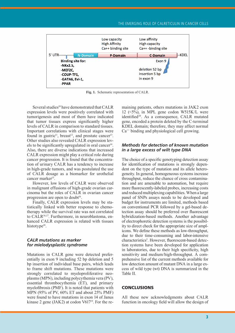
maining patients, others mutations in JAK2 exon 12 (<5%), in MPL gene codon W515K/L were identified20. As a consequence, CALR mutated gene, encoded a protein deleted by the C-terminal KDEL domain; therefore, they may affect normal Ca++ binding and physiological cell growing.
Methods for detection of known mutationin a large excess of wilt type DNA
The choice of a specific genotyping detection assay for identification of mutations is strongly depen-dent on the type of mutation and its allele hetero-geneity. In general, homogeneous systems increase throughput, reduce the chance of cross contamina-tion and are amenable to automation, but require more fluorescently-labeled probes, increasing costs and reduced multiplexing capabilities. When a large panel of SNPs assays needs to be developed and budget for instruments are limited, methods based on conventional PCR followed by a gel-based de-tection assay should be preferred over fluorescent hybridization-based methods. Another advantage of electrophoretic detection systems is the possibil-ity to direct check for the appropriate size of ampl-icons. We define these methods as low-throughput, due to their time-consuming and labor-intensive characteristics5. However, fluorescent-based detec-tion systems have been developed for application in laboratories, due to their high specificity, high sensitivity and medium/high-throughput. A com-prehensive list of the current methods available for low detection amount of mutant DNA in a large ex-cess of wild type (wt) DNA is summarized in the Table II.
CONCLUSIONS
All these new acknowledgments about CALR function in oncology field will allow the design of
Several studies10 have demonstrated that CALR expression levels were positively correlated with tumorigenesis and most of them have indicated that tumor tissues express significantly higher levels of CALR in comparison to standard tissues. Important correlations with clinical stages were found in gastric11, breast12, and prostate cancer13. Other studies also revealed CALR expression lev-els to be significantly upregulated in oral cancer14. Also, there are diverse indications that increased CALR expression might play a critical role during cancer progression. It is found that the concentra-tion of urinary CALR has a tendency to increase in high-grade tumors, and was postulated the use of CALR dosage as a biomarker for urothelial cancer marker15.
However, low levels of CALR were observed in malignant effusions of high-grade ovarian car-cinoma but the roles of CALR in ovarian cancer progression are open to doubt16.
Finally, CALR expression levels may be sta-tistically linked with better response to chemo-therapy while the survival rate was not correlated to CALR16,17. Furthermore, in neuroblastoma, en-hanced CALR expression is related with tissues histotype18.
CALR mutations as marker for mielodysplastic syndrome
Mutations in CALR gene were detected prefer-entially in exon 9 including 52 bp deletion and 5 bp insertion of individual base pairs, which leads to frame shift mutations. These mutations were strongly correlated to myeloproliferative neo-plasms (MPN), including polycythemia vera (PV), essential thrombocythemia (ET), and primary myelofibrosis (PMF). It is noted that patients with MPN (95% of PV, 60% ET and about 25% PMF) were found to have mutations in exon 14 of Janus kinase 2 gene (JAK2) at codon V61719. For the re-
Fig. 1. Schematic representation of CALR.
4
THE EMERGING ROLE OF CALRETICULIN IN CANCER CELLS
REFERENCES
1 Qiu Y, Michalak M. Transcriptional control of the calre-ticulin gene in health and disease. Int J Biochem Cell Biol 2009; 41: 531-538.
2 NguYeN TQ, capra JD, SoNTheiMer rD. Calreticulin is transcriptionally upregulated by heat shock, calcium and heavy metals. Mol Immunol 1996; 33: 379-386.
3 Vera c, Tapia V, kohaN k, gabler F, Ferreira a, SelMaN a, Vega M, roMero c. Nerve growth factor induces the expression of chaperone protein calreticulin in human epi-thelial ovarian cells. Horm Metab Res 2012; 44: 639-643.
4 Shih YY, Nakagawara a, lee h, JuaN hF, JeNg YM, liN DT, YaNg Yl, TSaY Yg, huaNg Mc, paN cY, hSu wM, liao YF. Calreticulin mediates nerve growth factor-in-duced neuronal differentiation. J Mol Neurosci 2012; 47: 571-581.
5 De MoNaco a, D’orTa a, Fierro c, Di paolo M, cileNTi l, Di FraNcia r. Rational selection of PCR-based platforms for pharmacogenomic testing. WCRJ 2014; 1: e391.
new research project that will get a light to rela-tionship between the inflammation, cellular dam-age, and tumors development21.
In addition, promising, we believe that the val-idation of the productive methods able to detect costly either CALR mutations and/or expression allow to integrate this information in a personal-ized approach22. In this way, in order to interpret correctly lab results of the CALR it is necessary an upgrading of the oncologist in genomics field23.
Acknowledgements: The authors wish to thank Dr O. Barletta for bibliography research assistance.
conflict of interest: None declared
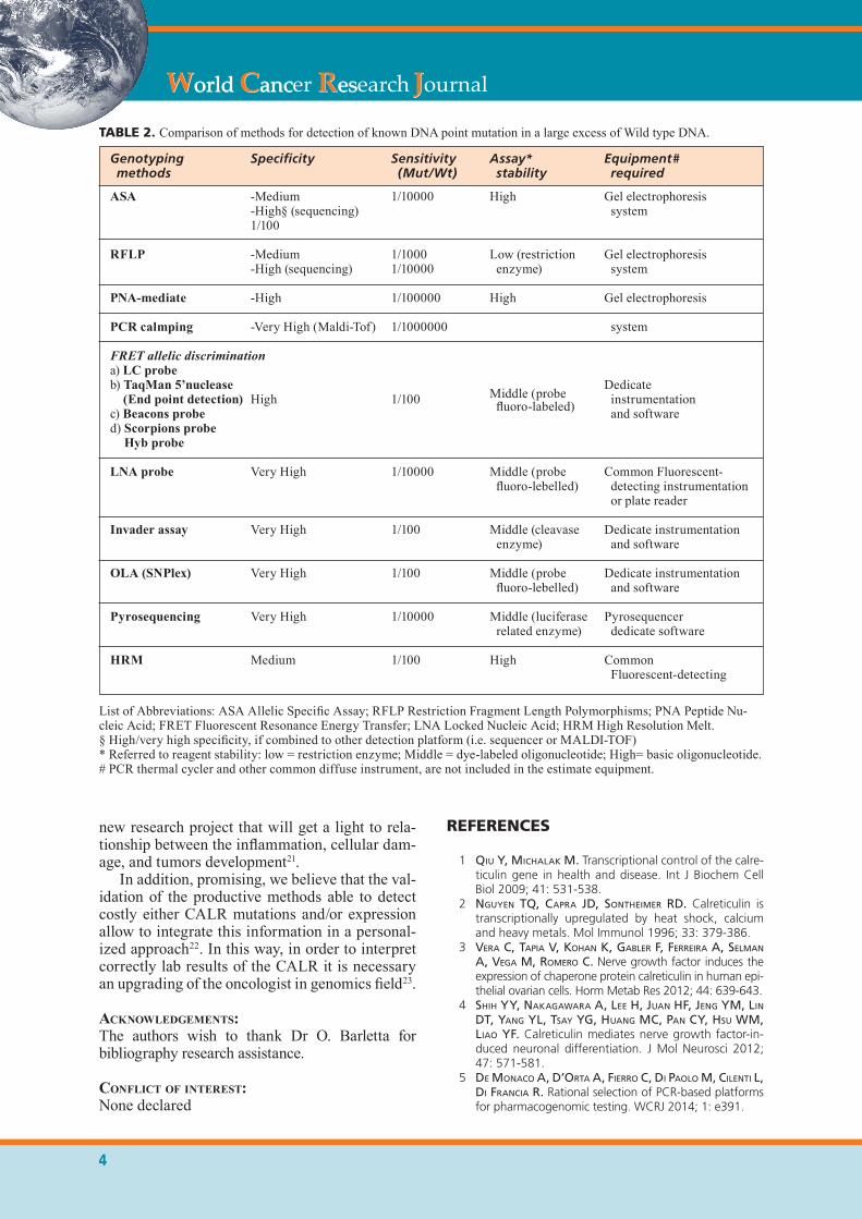
TABLE 2. Comparison of methods for detection of known DNA point mutation in a large excess of Wild type DNA.
List of Abbreviations: ASA Allelic Specific Assay; RFLP Restriction Fragment Length Polymorphisms; PNA Peptide Nu-cleic Acid; FRET Fluorescent Resonance Energy Transfer; LNA Locked Nucleic Acid; HRM High Resolution Melt. § High/very high specificity, if combined to other detection platform (i.e. sequencer or MALDI-TOF) * Referred to reagent stability: low = restriction enzyme; Middle = dye-labeled oligonucleotide; High= basic oligonucleotide.# PCR thermal cycler and other common diffuse instrument, are not included in the estimate equipment.
Genotyping Specificity Sensitivity Assay* Equipment# methods (Mut/Wt) stability required ASA -Medium 1/10000 High Gel electrophoresis -High§ (sequencing) system 1/100
RFLP -Medium 1/1000 Low (restriction Gel electrophoresis -High (sequencing) 1/10000 enzyme) system
PNA-mediate -High 1/100000 High Gel electrophoresis
PCR calmping -Very High (Maldi-Tof) 1/1000000 system FRET allelic discriminationa) LC probe b) TaqMan 5’nuclease Dedicate (End point detection) High 1/100 Middle (probe instrumentationc) Beacons probe fluoro-labeled) and softwared) Scorpions probe Hyb probe
LNA probe Very High 1/10000 Middle (probe Common Fluorescent- fluoro-lebelled) detecting instrumentation or plate reader
Invader assay Very High 1/100 Middle (cleavase Dedicate instrumentation enzyme) and software
OLA (SNPlex) Very High 1/100 Middle (probe Dedicate instrumentation fluoro-lebelled) and software
Pyrosequencing Very High 1/10000 Middle (luciferase Pyrosequencer related enzyme) dedicate software
HRM Medium 1/100 High Common Fluorescent-detecting
5
THE EMERGING ROLE OF CALRETICULIN IN CANCER CELLS
16 VakSMaN o, DaViDSoN b, Tropé c, reich r. Calreticu-lin expression is reduced in high-grade ovarian serous carcinoma effusions compared with primary tumors and solid metastases. Hum Pathol 2013; 44: 2677-2683.
17 De MoNaco a, Faioli D, Di paolo M, caTapaNo o, D’orTa a, Del buoNo M, Del buoNo r, Di FraNcia r. Pharmacogenomics markers for prediction response and toxicity in cancer therapy. WCRJ 2014; 1: e276.
18 chaNg hh, lee h, hu Mk, TSao pN, JuaN hF, huaNg Mc, Shih YY, waNg bJ, JeNg YM, chaNg cl, huaNg SF, TSaY Yg, hSieh FJ, liN kh, hSu wM, liao YF. Notch1 expression predicts an unfavorable prognosis and ser-ves as a therapeutic target of patients with neurobla-stoma. Clin Cancer Res 2010; 16: 4411-4420.
19 kraloVicS r, paSSaMoNTi F, buSer aS, Teo SS, TieDT r, paSSweg Jr, Tichelli a, cazzola M, SkoDa rc. A gain-of-function mutation of JAK2 in myeloproliferative di-sorders. N Engl J Med 2005; 352: 1779-1790.
20 NaNgalia J, MaSSie ce, baxTer eJ, Nice Fl, guNDeM g, weDge Dc, aVezoV e, li J, kollMaNN k, keNT Dg, aziz a, goDFreY al, hiNToN J, MarTiNcoreNa i, VaN loo p, JoNeS aV, guglielMelli p, TarpeY p, harDiNg hp, FiTz-paTrick JD, gouDie cT, orTMaNN ca, loughraN SJ, raiNe k, JoNeS Dr, buTler ap, Teague Jw, o’Meara S, MclareN S, biaNchi M, Silber Y, DiMiTropoulou D, blox-haM D, MuDie l, MaDDiSoN M, robiNSoN b, keohaNe c, MacleaN c, hill k, orcharD k, Tauro S, Du MQ, greaVeS M, boweN D, huNTlY bJp, harriSoN cN, croSS Ncp, roN D, VaNNucchi aM, papaeMMaNuil e, caMpbell pJ, greeN ar. Somatic CALR mutations in myeloprolife-rative neoplasms with nonmutated JAK2. N Engl J Med 2013; 369: 2391-2405.
21 berreTTa M, Di FraNcia r, Tirelli u. Editorial - The new oncologic challenges in the 3rd millennium. WCRJ 2014; 1: e133.
22 cillo M, Di paolo M, puglieSe S, TroiSi a, creSceNTe g, MaroTTa g. Costs and quality of genomics tests in the oncology field. WCRJ 2016; 3: e801.
23 Di FraNcia r, ValeNTe D, puglieSe S, Del buoNo a, ber-reTTa M. What health professions in oncology needs to know about pharmacogenomics? WCRJ 2014; 1: e90.
6 VillagoMez M, Szabo e, poDcheko a, FeNg T, papp S, opaS M. Calreticulin and focal-contact-dependent adhesion. Biochem Cell Biol 2009; 87: 545-556.
7 FaDel Mp, Dziak e, lo cM, Ferrier J, MeSaeli N, Michal-ak M, opaS M. Calreticulin affects focal contact-depen-dent but not close contact-dependent cell-substratum adhesion. J Biol Chem 1999; 274: 15085-15094.
8 papp S, FaDel Mp, kiM h, Mcculloch ca, opaS M. Calreticulin affects fibronectin-based cell-substratum adhesion via the regulation of c-Src activity. J Biol Chem 2007; 282: 16585-16598.
9 coppoliNo M g, DeDhar S. Ligand-specific, transient in-teraction between integrins and calreticulin during cell adhesion to extracellular matrix proteins is dependent upon phosphorylation/dephosphorylation events. Bio-chem J 1999; 340: 41-50.
10 zaMaNiaN M, VeerakuMaraSiVaM a, abDullah S, roSli r. Calreticulin and cancer. Pathol Oncol Res 2013; 19: 149-154.
11 cheN cN, chaNg cc, Su Te, hSu wM, JeNg YM, ho Mc, hSieh FJ, lee ph, kuo Ml, lee h, chaNg kJ. Iden-tification of calreticulin as a prognosis marker and an-giogenic regulator in human gastric cancer. Ann Surg Oncol 2009; 16: 524-533.
12 lwiN zM, guo c, SaliM a, Yip gw, chew FT, NaN J, Thike aa, TaN ph, baY bh. Clinicopathological signifi-cance of calreticulin in breast invasive ductal carcinoma. Mod Pathol 2010; 23: 1559-1566.
13 alaiYa a, roblick u, egeVaD l, carlSSoN a, FraNzéN b, Volz D, huweNDiek S, liNDer S, auer g. Polypeptide ex-pression in prostate hyperplasia and prostate adenocar-cinoma. Anal Cell Pathol 2000; 21: 1-9.
14 chiaNg wF, hwaNg Tz, hour Tc, waNg lh, chiu cc, cheN hr, wu YJ, waNg cc, waNg lF, chieN cY, cheN Jh, hSu cT, cheN JY. Calreticulin, an endoplasmic reti-culum-resident protein, is highly expressed and essen-tial for cell proliferation and migration in oral squamous cell carcinoma. Oral Oncol 2013; 49: 534-541.
15 kageYaMa S, iSoNo T, MaTSuDa S, uShio Y, SaToMura S, Terai a, arai Y, kawakiTa M, okaDa Y, YoShiki T. Urinary calreticulin in the diagnosis of bladder urothelial carcinoma. Int J Urol 2009; 16: 481-486.