Ultrastructure and motility pattern of the spermatozoa of Aleochara curtula (Coleoptera, Staphylinidae) Michael Werner a , Thomas Tscheulin b , Thomas Speck c , Dieter Zissler a , Klaus Peschke a, * a Institut fu ¨r Biologie I (Zoologie), Hauptstr. 1, D-79104 Freiburg i.Br., Germany b Department of Biology, Imperial College of Science, Technology and Medicine, Silwood Park, Ascot, Berkshire SL5 7PY, UK c Plant Biomechanics Group, Botanischer Garten der Albert-Ludwigs-Universita ¨t Freiburg, Scha ¨nzlestr. 1, D-79104 Freiburg, Germany Received 4 July 2002; accepted 29 August 2002 Abstract Ultrastructure and motility pattern of spermatozoa of the rove beetle Aleochara curtula were examined using electron and light microscopic methods. The spermatozoon is about 100 mm long and filiform. The head piece comprises a 5 mm long triple layered acrosome and 10 mm long nucleus. The flagellum consists of a 9þ 9þ 2 axoneme, two accessory bodies and two mitochondrial derivatives about equal in size but of different shape in their cross sections. In both derivatives there are paracrystalline inclusions. The flagellum is attached to the head by a 2 mm long centriole adjunct which is characterized by its electron dense material that forms a three layered folded lamellar structure. When liberated in buffer solution the sperm flagella assume a coiled hook-like form with the excentric stiff head protruding in front. The spermatozoa are driven through the medium by a small helicoidal wave of high frequency superimposed to the bent flagella. The maximum speed measured was 15.2 mm/s. The sperm architecture of A. curtula is similar to that of other Aleochara species but differs in total length and dimensions of the mitochondrial derivatives. For that reason Aleochara sperm can certainly prove useful to study the effect of the mitochondrial derivatives on sperm motility. q 2002 Published by Elsevier Science Ltd. Keywords: Insect spermatozoa; Sperm motility; Mitochondrial derivatives; Centriole adjunct 1. Introduction Sperm ultrastructure in the vast insect order Coleoptera has been investigated in many different families and was found to match basically the classical pterygote type (Jamieson, 1987; Jamieson et al., 1999), which is charac- terized by the presence of a 9þ 9þ 2 axoneme, two accessory bodies and two mitochondrial derivatives. Variation in sperm architecture has been studied mostly to resolve phylogenetic issues in beetles (Baccetti and Daccordi, 1988; Burrini et al., 1988) as well as in other insect species (Baccetti, 1972; Dallai, 1979; Carcupino et al., 1995; Dallai and Afzelius, 1995; Jamieson, 1987). Obser- vations and interpretations of sperm motility, however, are scarce, in animals in general (Brokaw et al., 1970), and in insects as well (Baccetti et al., 1973a,b; Phillips, 1974). The 9þ 9þ 2 insect spermatozoa in motion generally display a cylindrical-helicoidal type of wave (Baccetti, 1972). A much more complicated double wave motion pattern has been described for the coleopteran Tenebrio molitor (Baccetti et al., 1973a; Phillips, 1974), the hemipteran Lygaeus (Phillips, 1974) and the phasmid Bacillus rossius (Baccetti et al., 1973b). In this motion pattern, according to Baccetti et al., (1973a), so called ‘large waves’ which arise from behind the sperm head spreading towards the posterior tail tip are superimposed by small amplitude waves (‘small waves’) of higher frequency spreading in the same direction. According to Baccetti and Afzelius (1976), such small waves superimposed on large waves are supposed to be restricted to occur in spermatozoa, which possess two accessory bodies endowed with ATPase activity. Besides the accessory bodies, however, the mitochondrial derivatives have been considered to be 1467-8039/02/$ - see front matter q 2002 Published by Elsevier Science Ltd. PII: S1467-8039(02)00046-4 Arthropod Structure & Development 31 (2002) 243–254 www.elsevier.com/locate/asd * Corresponding author. Tel.: þ 49(0) 761-203-2546; fax: þ 49(0) 761- 203-2546. E-mail address: [email protected] (K. Peschke).
Transcript
Ultrastructure and motility pattern of the spermatozoa
of Aleochara curtula (Coleoptera, Staphylinidae)
Michael Wernera, Thomas Tscheulinb, Thomas Speckc, Dieter Zisslera, Klaus Peschkea,*
aInstitut fur Biologie I (Zoologie), Hauptstr. 1, D-79104 Freiburg i.Br., GermanybDepartment of Biology, Imperial College of Science, Technology and Medicine, Silwood Park, Ascot, Berkshire SL5 7PY, UK
involved in insect sperm motility. They may impart stiffness
to the flagellar beat, affecting the form of the flagellar wave
(Phillips, 1974) and establish a particular motility pattern
specific to each insect species (Tokuyasu, 1974). Attempt-
ing to analyse the role of the mitochondrial derivatives in
sperm motility the rove beetle genus Aleochara
probably can prove useful, since variations in sperm
structure can be found mainly in total sperm length and
dimensions of the mitochondrial derivatives. Video
analyses of sperm movement in buffer solution of the
rove beetles Aleochara bilineata and A. tristis showed
that they move through the medium in a corkscrew-like
motion (Werner et al., 2000, 2001). A helical wave of high
frequency and low amplitude runs along the helically coiled
tail and propels the sperm through the medium, which is, on
first sight, similar to the double wave system described by
Baccetti et al. (1973a,b) for Tenebrio and Bacillus.
In this study, a detailed description of ultrastructure as well
as a hypothesis for the observed motility pattern is given for
spermatozoa of Aleochara curtula to provide additional
information for further comparative investigations of sperm
structure and function amongst different Aleochara species.
2. Materials and methods
2.1. Beetle culture and dissection
All beetles used in this survey were taken from our own
laboratory stock cultures, reared according to Fuldner
(1968) on blow fly pupae (Calliphora vicina ) as hosts for
the parasitoid beetle larvae. Females were kept individually
and males in groups of ten. To obtain spermatozoa from the
spermatheca, females were allowed to mate at least one day
before dissection. Spermathecae were dissected in the same
buffer solution that was used for further processing or sperm
observation (see later).
2.2. Sperm length measurements
Spermatozoa were liberated in a 0.1 M Phosphate
buffer (PB) containing 6 mM of the fluorochrome
diamidinophenylindole (DAPI), which stains DNA.
Combined fluorescence and phase contrast images
were taken with a Zeiss Axiocam mounted onto a Zeiss
Axioskop. Images were imported into image analysis
software (Optimas 6.51, Media Cybernetics). Acrosomes,
nuclei and sperm tails were measured by tracing these
elements with the cursor.
2.3. Transmission electron microscopy
Positive staining. Spermathecae from inseminated
females were cut into two or three parts and fixed
immediately in a 2% glutaraldehyde solution in 0.1 M PB
(pH 7.4) containing 1% tannic acid and 1.8% sucrose.
After rinsing in water several times the spermathecae were
block-stained in 1% uranyl acetate in distilled water
according to Afzelius (1988) and rinsed in water again.
Samples were dehydrated in a graded series of acetone and
embedded in Epon. Ultrathin sections were cut on a Reichert
OMU-3 ultramicrotome and stained with uranyl acetate and
lead citrate using a Leica Ultrostain. The sections were
examined in a Zeiss EM-9S2 and EM10 transmission
electron microscope operating at 60 and 80 kV, respectively.
Negative staining. Spermatozoa from the spermatheca
were liberated into a drop of distilled water by breaking or
puncturing the chitinized wall of the spermatheca. The
sperm suspension was then transferred onto slot grids
covered with formvar and negatively stained with 2%
phosphotungstate acid and 0.4% sucrose (pH 7.4).
2.4. Observations of sperm motility
For all live recordings of sperm we used a diphosphate
buffered saline (DPBS, pH 7.4) containing 52.02 mM NaCl,
39.70 mM KCl, 0.54 mM CaCl2, 1.18 mM MgCl2 £ 6H2O,
1.22 mM MgSO4 £ 7H2O, 1 mM glucose and 58.43 mM
succrose in 0.05 M PB. After dissection spermathecae were
placed in a drop of buffer solution on a slide and sperm were
liberated. Samples were examined in dark field mode using
a Zeiss Axioskop at 30 8C. The microscope stage was
equipped with a MINITUP HT 200 heating system for exact
temperature control. Sperm movements were recorded on an
S-VHS videotape with a co-recorded stop watch signal.
Single frames or sequences from this tape were digitalized
for further computer image analysis. Swimming velocity
was estimated using the ‘motion analysis’ macro of the
image analysis program. Using this tool, we followed the
trajectory of a spermatozoan head obtained from a
digitalized sequence with a frame interval of 0.5 s. The
relative coordinates of the sperm heads were recorded and
the straight line velocities were calculated. High-speed
recordings of sperm movement were made with a Red
Lake Motion Scope digital camera working at 250 or
500 frames/s. Digitalized sequences were read out from the
camera’s internal buffer and recorded directly on S-VHS
videotape at rates of 5 or 10 frames/s again with a co-
recorded stop watch signal. Flagella undulation frequencies
were measured from these recordings and calculated back to
their original recording speed.
3. Results
3.1. Sperm architecture
Morphology. Light microscopical examination of A.
curtula spermatozoa revealed them to be long and slender
cells with an overall length of 100.2 ^ 12.8 mm (�x ^ s:d:;
n ¼ 67). The headpiece, consisting of acrosome and
nucleus, is an elongated and stiff structure that is tapered
M. Werner et al. / Arthropod Structure & Development 31 (2002) 243–254244
off towards the tip. The lengths of acrosome and nucleus
were determined to be 4.9 ^ 0.2 mm (n ¼ 19) and
10.5 ^ 0.4 mm (n ¼ 158) respectively, such that the head-
piece comprises about 15% the of total sperm length.
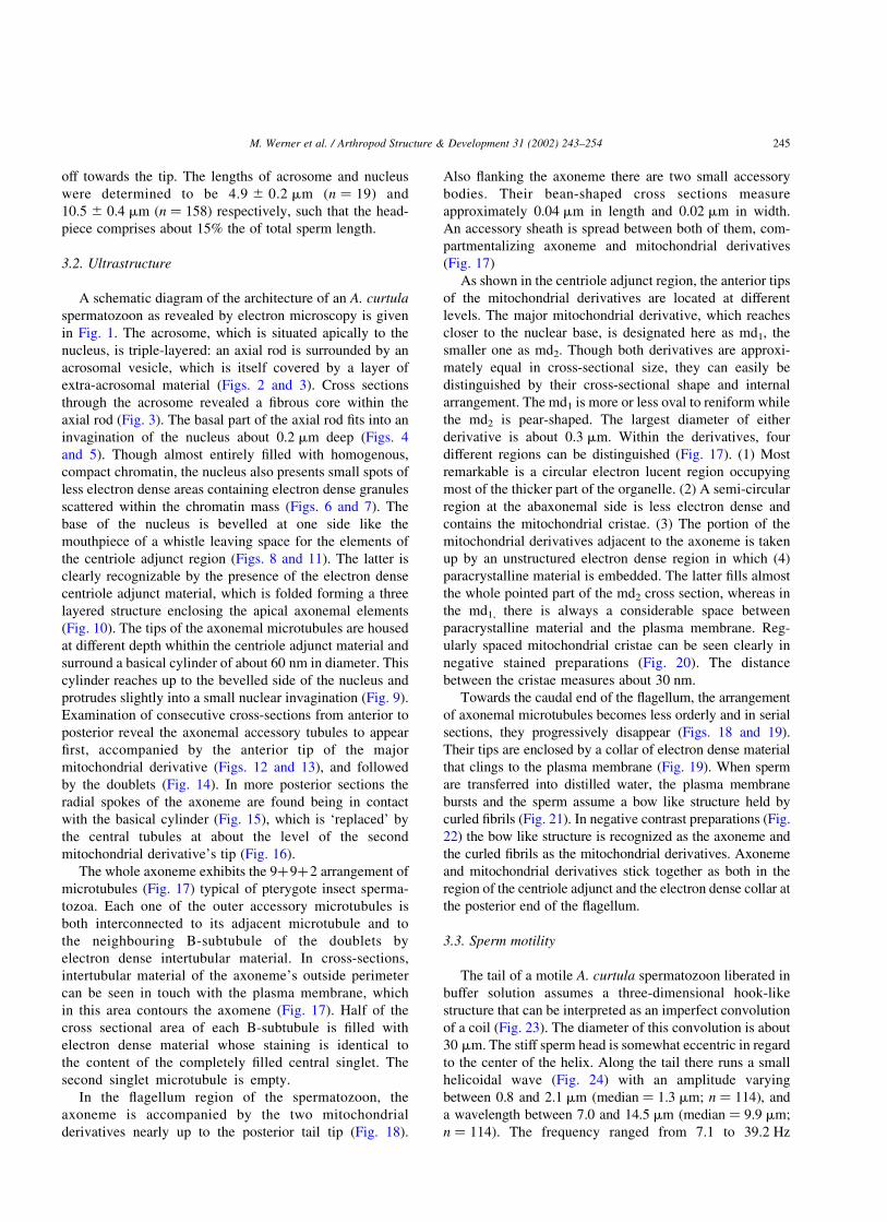
3.2. Ultrastructure
A schematic diagram of the architecture of an A. curtula
spermatozoon as revealed by electron microscopy is given
in Fig. 1. The acrosome, which is situated apically to the
nucleus, is triple-layered: an axial rod is surrounded by an
acrosomal vesicle, which is itself covered by a layer of
extra-acrosomal material (Figs. 2 and 3). Cross sections
through the acrosome revealed a fibrous core within the
axial rod (Fig. 3). The basal part of the axial rod fits into an
invagination of the nucleus about 0.2 mm deep (Figs. 4
and 5). Though almost entirely filled with homogenous,
compact chromatin, the nucleus also presents small spots of
less electron dense areas containing electron dense granules
scattered within the chromatin mass (Figs. 6 and 7). The
base of the nucleus is bevelled at one side like the
mouthpiece of a whistle leaving space for the elements of
the centriole adjunct region (Figs. 8 and 11). The latter is
clearly recognizable by the presence of the electron dense
centriole adjunct material, which is folded forming a three
layered structure enclosing the apical axonemal elements
(Fig. 10). The tips of the axonemal microtubules are housed
at different depth whithin the centriole adjunct material and
surround a basical cylinder of about 60 nm in diameter. This
cylinder reaches up to the bevelled side of the nucleus and
protrudes slightly into a small nuclear invagination (Fig. 9).
Examination of consecutive cross-sections from anterior to
posterior reveal the axonemal accessory tubules to appear
first, accompanied by the anterior tip of the major
mitochondrial derivative (Figs. 12 and 13), and followed
by the doublets (Fig. 14). In more posterior sections the
radial spokes of the axoneme are found being in contact
with the basical cylinder (Fig. 15), which is ‘replaced’ by
the central tubules at about the level of the second
mitochondrial derivative’s tip (Fig. 16).
The whole axoneme exhibits the 9þ9þ2 arrangement of
microtubules (Fig. 17) typical of pterygote insect sperma-
tozoa. Each one of the outer accessory microtubules is
both interconnected to its adjacent microtubule and to
the neighbouring B-subtubule of the doublets by
electron dense intertubular material. In cross-sections,
intertubular material of the axoneme’s outside perimeter
can be seen in touch with the plasma membrane, which
in this area contours the axomene (Fig. 17). Half of the
cross sectional area of each B-subtubule is filled with
electron dense material whose staining is identical to
the content of the completely filled central singlet. The
second singlet microtubule is empty.
In the flagellum region of the spermatozoon, the
axoneme is accompanied by the two mitochondrial
derivatives nearly up to the posterior tail tip (Fig. 18).
Also flanking the axoneme there are two small accessory
bodies. Their bean-shaped cross sections measure
approximately 0.04 mm in length and 0.02 mm in width.
An accessory sheath is spread between both of them, com-
partmentalizing axoneme and mitochondrial derivatives
(Fig. 17)
As shown in the centriole adjunct region, the anterior tips
of the mitochondrial derivatives are located at different
levels. The major mitochondrial derivative, which reaches
closer to the nuclear base, is designated here as md1, the
smaller one as md2. Though both derivatives are approxi-
mately equal in cross-sectional size, they can easily be
distinguished by their cross-sectional shape and internal
arrangement. The md1 is more or less oval to reniform while
the md2 is pear-shaped. The largest diameter of either
derivative is about 0.3 mm. Within the derivatives, four
different regions can be distinguished (Fig. 17). (1) Most
remarkable is a circular electron lucent region occupying
most of the thicker part of the organelle. (2) A semi-circular
region at the abaxonemal side is less electron dense and
contains the mitochondrial cristae. (3) The portion of the
mitochondrial derivatives adjacent to the axoneme is taken
up by an unstructured electron dense region in which (4)
paracrystalline material is embedded. The latter fills almost
the whole pointed part of the md2 cross section, whereas in
the md1, there is always a considerable space between
paracrystalline material and the plasma membrane. Reg-
ularly spaced mitochondrial cristae can be seen clearly in
negative stained preparations (Fig. 20). The distance
between the cristae measures about 30 nm.
Towards the caudal end of the flagellum, the arrangement
of axonemal microtubules becomes less orderly and in serial
sections, they progressively disappear (Figs. 18 and 19).
Their tips are enclosed by a collar of electron dense material
that clings to the plasma membrane (Fig. 19). When sperm
are transferred into distilled water, the plasma membrane
bursts and the sperm assume a bow like structure held by
curled fibrils (Fig. 21). In negative contrast preparations (Fig.
22) the bow like structure is recognized as the axoneme and
the curled fibrils as the mitochondrial derivatives. Axoneme
and mitochondrial derivatives stick together as both in the
region of the centriole adjunct and the electron dense collar at
the posterior end of the flagellum.
3.3. Sperm motility
The tail of a motile A. curtula spermatozoon liberated in
buffer solution assumes a three-dimensional hook-like
structure that can be interpreted as an imperfect convolution
of a coil (Fig. 23). The diameter of this convolution is about
30 mm. The stiff sperm head is somewhat eccentric in regard
to the center of the helix. Along the tail there runs a small
helicoidal wave (Fig. 24) with an amplitude varying
between 0.8 and 2.1 mm (median ¼ 1.3 mm; n ¼ 114), and
a wavelength between 7.0 and 14.5 mm (median ¼ 9.9 mm;
n ¼ 114). The frequency ranged from 7.1 to 39.2 Hz
M. Werner et al. / Arthropod Structure & Development 31 (2002) 243–254 245
(median ¼ 19.2 Hz; n ¼ 84). This wave, originating behind
the sperm head and spreading towards the tail tip,
propagates the sperm through the medium (Fig. 25). The
trajectory is rectilinear and velocities measured varied
between 3.7 and 15.2 mm/s (median ¼ 8.4 mm/s; n ¼ 63).
During propagation the whole sperm rotates in a counter
clockwise direction in the course of which the eccentric
sperm head describes a helicoidal path (Fig. 23).
Fig. 1. Schematic diagram of an A. curtula spermatozoon. ab, accessory body; ar, axial rod; av, acrosomal vesicle; ax, axoneme; bc, basical cylinder; ca,
centriole adjunct; edc, electron dense collar; el, extra-acrosomal layer; md1, major mitochondrial derivative; md2, minor mitochondrial derivative; n, nucleus.
M. Werner et al. / Arthropod Structure & Development 31 (2002) 243–254246
4. Discussion
4.1. Sperm architecture
The spermatozoon of A. curtula exhibits all features
typical of pterygote insect sperm: it is filiform, has a
triple layered acrosomal complex, an elongated cylind-
rical nucleus, two mitochondrial derivatives, two
accessory bodies and a 9þ9þ2 axoneme (Jamieson,
1987). In the genus Aleochara, spermatozoan ultra-
structure has been investigated in A. tristis and A.
clavicornis (Werner et al., 2001) and, more intensively,
in A. bilineata (Werner et al., 1999). All three species
share the same sperm architecture, which is character-
ized by a prominent centriole adjunct region, two small
accessory bodies and two mitochondrial derivatives of
unequal size. At a length of 100 mm, the sperm of A.
curtula are shorter then those of the three other species.
Sperm length in A. bilineata was found to be 1000 mm
(Werner et al., 1999) and in A. tristis and A.
clavicornis, it was 400 and 500 mm, respectively
(Werner et al., 2001).
The triple layered structure of the acrosome is found in
almost all pterygote insects (Baccetti, 1972) and has been
described in Coleoptera for T. molitor (Baccetti et al.,
1973a), some Curculionoidea (Burrini et al., 1988), and
Chrysomelidae (Baccetti and Daccordi, 1988), the clerid
Divales bipustulatus (Mazzini, 1976), and the staphylinid A.
bilineata (Werner et al., 1999).
The nucleus of an A. curtula sperm is not completely
homogeneous, as has been described for the majority of
insect sperm (Baccetti, 1972), but it presents small electron
lucent areas filled with electron dense granules. Similar
structures were also found in the sperm nucleus of the
hymenopteran Bephratelloides pomorum (Lino-Neto et al.,
1999). The granules within this areas could possibly be
interpreted as polyribosomes as described by Baccetti et al.
(1973b) for the phasmid B. rossius, where polyribosomes
were found in an extranuclear space in the center of the
nucleus.
A centriole adjunct at the nucleus flagellum transition
region has been described for many insects (Cantacu-
zene, 1970). This structure has also been reported for
other coleopterans from the family Bathysciinae
Figs. 2–9. Sperm Head. Fig. 2: Negative contrast image of the anterior tip of the acrosome. The three layered structure is clearly visible in this preparation. Fig.
3: Cross section of the acrosome. The axial rod contains a fibrous inner core. Fig. 4: Longitudinal section of the acrosome-nucleus transition. The axial rod fits
into a nuclear invagination. Fig. 5: Cross section of the acrosome-nucleus transition. Fig. 6: Longitudinal section of the nucleus. Scattered within the condensed
chromatin mass there are less electron dense areas containing electron dense granules (arrows). Fig. 7: Cross section of the nucleus. Fig. 8: Negative contrast
preparation of the nucleus-flagellum transition region. The posterior end of the nucleus is bevelled like the mouthpiece of a whistle. Fig. 9: Cross section of the
nuclear base. The anterior tip of the basical cylinder extends into a nuclear invagination. bc, basical cylinder; ar, axial rod; av, acrosomal vesicle; el, extra-
acrosomal layer; md1, major mitochondrial derivative; n, nucleus.
M. Werner et al. / Arthropod Structure & Development 31 (2002) 243–254 247
(Juberthie-Jupeau et al., 1983), Bruchidae (Acantho-
marginalis and Colymbetes fuscus, Mackie and Walker,
1974; Acilius sulcatus, Werner, 1976), Lucanidae
(Aegus lavicollis, Kubo-Irie et al., 2000) and Staphyli-
nidae (A. bilineata, Werner et al., 1999). The structure
of the centriole adjunct in A. curtula is most peculiar. It
enfolds a complicated three-dimensional structure, in
which the anterior parts of the axoneme and the
mitochondrial derivatives are housed, quite similar to
the multilayered centriolar adjunct found in Locusta
migratoria (Cantacuzene, 1970) and in the grasshopper
Melanoplus differentialis (Lindsey and Biesele, 1974).
In contrast to A. curtula, the centriole adjunct of A.
bilineata is made of a compact mass of electron dense
material (Werner et al., 1999). In both species the
centriole adjunct extends along the initial segment of
the axoneme, whereas in the stag beetle Aegus
lavicollis, it covers the outside of the two mitochondrial
derivatives and extends almost half way along the
length of the whole flagellum (Kubo-Irie et al., 2000).
The authors suggested that the centriole adjunct is
Figs. 10–16. Centriole adjunct region. Fig. 10: Longitudinal section of the centriole adjunct region. The three layered electron dense material of the centriole
adjunct surrounds the basical cylinder. Figs. 11–16: Consecutive cross sections of the centriolar adjunct (all to the same scale). Fig. 11: The centriolar adjunct
extends into the space given free by the bevelled posterior top of the nucleus. Figs. 12 and 13: The folded three layered structure of the centriole adjunct
material encloses the anterior tips of some accessory tubules as well as the major mitochondrial derivative. Figs. 14 and 15: Further posterior, the axonemal
doublets (arrows) appear and came into contact with the basical cylinder via the radial spokes (arrowhead). Fig. 16: The centriole adjunct material is displaced
by the fully expressed axoneme and the second mitochondrial derivative. ab, accessory body; at, accessory tubule; ax, axoneme; bc, basical cylinder; ca,
centriole adjunct; md1, major mitochondrial derivative; md2, minor mitochondrial derivative; n, nucleus.
M. Werner et al. / Arthropod Structure & Development 31 (2002) 243–254248
involved in the movement of the flagellum in this
species. Baccetti (1972) also proposed a mechanical role
of this structure in the initiation of flagellar movements.
From the position of the centriole adjunct, Breland et al.
(1966), Phillips (1970) and Lindsey and Biesele (1974)
concluded that it serves to secure the flagellum to the
sperm head. Yasuzumi et al. (1970) supposed a nutritive
function for the developing axoneme during
spermiogenesis.
In pterygote insect flagella, the 9þ9þ2 axoneme is
usually flanked by two accessory bodies (Baccetti, 1972),
rarely by one (Werner, 1965) or three (Bawa and Kanwar,
1975). In the Coleoptera the accessory bodies are often of
considerable size relative to the axoneme. The largest ones
were found in the clerid D. bipustulatus (Mazzini, 1976). In
the Phasmatodea, the accessory bodies replace the mito-
chondrial derivatives during spermiogenesis, and they are
the most prominent organelles in the mature sperm
flagellum (Baccetti et al., 1973b). Some accessory bodies
showed ATPase and UTPase activity; they were considered
to maintain the large wave of the motile spermatozoa
(Baccetti et al., 1973a,b). However, in A. curtula, the
accessory bodies are very small and a mechanical role of
these organelles in sperm motility seems unlikely. The
same is true for A. bilineata, A. tristis and A. clavicornis
(Werner et al., 1999, 2001).
Although A. curtula sperm have two mitochondrial
derivatives of nearly equal cross sectional size, the major
Figs. 17–20. Flagellum region. Figs. 17–19. Cross sections of the flagellum (all to the same scale). Fig. 17: The mitochondrial derivatives are separated from
the axoneme by an accessory sheath that is spread between the accessory bodies. The mitochondrial derivatives show a central less electron dense region , the
region of the cristae , an unstructured electron dense matrix and the paracrystalline inclusion . Fig. 18: At the end of the tail the axonemal arrangement
of microtubules becomes disordered. Electron dense material can be seen between the accessory tubules and the plasma membrane (arrows). Fig. 19:
The plasma membrane at the very end of the flagellum is lined up with electron dense material which forms a collar-like structure around the remaining
microtubules. Fig. 20: Negative contrast image of the major mitochondrial derivative. The cristae are regularly spaced. Paracrystalline material is made of
and Andre, 1970) gave rise to various speculations about
their function within the mature spermatozoon. Besides
from their role in energy metabolism, mitochondrial
derivatives have been ascribed a mechanical function in
the modulation of axonemal movements (Phillips, 1974;
Tokuyasu, 1974). Specifically the crystalline content of the
derivatives was suggested to exhibit elastic properties
similar to that of the parergins in mammalian sperm
(Baccetti et al., 1977). Such mechanical effects are obvious
when the derivatives are of enormous size that they modify
the axonemal movements in such a way that, in artificial
media, even complete immobility results (D. bipustulatus,
Mazzini, 1976; A. clavicornis, Werner et al., 2001), though
locomotion in narrow ducts in the female genital tract may
still be enabled by the observed feeble axonemal motility.
4.2. Sperm motility pattern
Based on investigations on T. molitor (Baccetti et al.,
1973a) and B. rossius (Baccetti et al., 1973b), a complex
spermatozoa movement pattern which is characterized by
the simultaneous presence of two kinds of waves has been
described. A small amplitude wave of high frequency is
superimposed on a wave with higher amplitude and lower
frequency. These waves were termed as ‘small wave’ and
‘large wave’ by Baccetti et al. (1973a) and ‘minor wave’
and ‘major wave’ by Phillips (1974). According to Baccetti
et al. (1973a,b), the double wave system is a product of
two different flagellar components: the small wave is
propagated and maintained by the axoneme motor, and the
large wave by the ATPase and UTPase-rich accessory
bodies. However, Phillips (1974) described the same
motility pattern for the milkweed bug, Lygaeus, which
does not have any accessory bodies (Jamieson, 1987).
When liberated in buffer solution, the A. curtula
sperm tail assumes a helically coiled form which is
superimposed by a helical wave of high frequency and
low amplitude. The action of the latter propels the
sperm through the medium generating a corkscrew
motion of the sperm. At first sight, this motility pattern
resembles the two-wave system of movement described
for Tenebrio (Baccetti et al., 1973a). However, Baccetti
Figs. 21 and 22. Spermatozoa in distilled water. Fig. 21: Phase contrast image of a dissociated spermatozoon in distilled water. The bow-like structure
represents the axoneme the string-like structure both mitochondrial derivatives. Fig. 22: The same preparation in negative contrast. The curled mitochondrial
derivatives can be clearly distinguished from the axoneme. ax, axoneme; md, mitochondrial derivatives; md1, major mitochondrial derivative; md2, minor
mitochondrial derivative; sh, sperm head.
Figs. 23–25. A. curtula spermatozoon in movement. Fig. 23: Computer reconstruction of the three-dimensional bent sperm. The arrow indicates the rotation
during forward progression. Fig. 24: Single frame from a high speed recording showing the small amplitude, high frequency wave (arrow) superimposed to the
bent flagellum. Fig. 25: Sequence of a real time video recording showing a spermatozoon swimming in buffer solution. The frames are at 0.2 s intervals. fl,
flagellum; sh, sperm head. See also the electronic annex under www.elsevier.com for videoclips.
M. Werner et al. / Arthropod Structure & Development 31 (2002) 243–254250