XMAP215 activity sets spindle length by controlling thetotal mass of spindle microtubulesSimone B. Reber1, Johannes Baumgart2, Per O. Widlund1, Andrei Pozniakovsky1, Jonathon Howard1,3,Anthony A. Hyman1,4 and Frank Jülicher2,4
Metaphase spindles are microtubule-based structures that use amultitude of proteins to modulate their morphology and function.Today, we understand many details of microtubule assembly, therole of microtubule-associated proteins, and the action of molec-ular motors1,2. Ultimately, the challenge remains to understandhow the collective behaviour of these nanometre-scale processesgives rise to a properly sized spindle on the micrometre scale. Bysystematically engineering the enzymatic activity of XMAP215,a processive microtubule polymerase3,4, we show that Xenopuslaevis spindle length increases linearly with microtubule growthvelocity, whereas other parameters of spindle organization, suchas microtubule density, lifetime and spindle shape, remainconstant. We further show that mass balance can be used tolink the global property of spindle size to individual microtubuledynamic parameters. We propose that spindle length is setby a balance of non-uniform nucleation and global microtubuledisassembly in a liquid-crystal-like arrangement of microtubules.
The biological importance of organelle size has recently attracted asurge of interest5–7. In the case of the metaphase or postanaphase Bspindle, length is particularly important as it determines the distanceover which chromosomes are segregated. Although a few generalprinciples of spindle length control are beginning to emerge, the relativecontribution of microtubule dynamics, nucleation and transport tospindle size remains elusive8–11.Dynamic spindles assembled in Xenopus egg extracts are an excellent
model system to study spindle organization10–12. The egg extract is anopen system that permits biochemical manipulation and quantitativekinetic studies. It is void of cortical restrictions and spindle material isnot limited13, which allows us to study mechanisms of length controlintrinsic to the spindle. Xenopus spindles are self-organizing ensemblesof microtubules, which nucleate by different pathways1, turn overrapidly14, slide poleward15 and are short when compared with the
1Max Planck Institute of Molecular Cell Biology and Genetics, Pfotenhauer Str. 108, 01307 Dresden, Germany. 2Max Planck Institute for the Physics of ComplexSystems, Nöthnitzer Str. 38, 01187 Dresden, Germany. 3Present address: Department of Molecular Biophysics and Biochemistry, Yale University, New Haven,Connecticut 06520-8114, USA.4Correspondence should be addressed to A.A.H. or F.J. (e-mail: [email protected] or [email protected])
Received 31 October 2012; accepted 24 July 2013; published online 25 August 2013; DOI: 10.1038/ncb2834
overall length of the spindle11. Although all of these processes are likelyto influence spindle assembly, their interplay makes it challenging toestablish direct links between individual parameters of microtubuledynamics and the overall spindle length.Proteins of the XMAP215/Dis1 family are major regulators of
microtubule polymerization16. Supporting the role of XMAP215 as amicrotubule growth promoter, its depletion leads to shorter spindlesor defects in spindle morphology in Schizosaccharomyces pombe,Caenorhabditis elegans, Xenopus egg extracts, Drosophila melanogasterand HeLa cells17. Members of the XMAP215/Dis1 family containvarying numbers of TOG (tumour overexpressed gene) domains thatfunction as αβ-tubulin-binding modules17. Recent in vitro studiesshow that XMAP215 acts as a processive polymerase4 and that itspolymerase activity depends on tubulin binding to multiple TOGdomains18. Mutation of conserved residues in different TOG domainsreduces tubulin binding while concomitantly reducing the maximalgrowth-promoting activity of the polymerase. Combining mutations indifferent TOG domains (Fig. 1a) enables the construction of XMAP215mutants characterized by significantly different maximum growth rates.This collection of engineered polymerases provides a valuable tool andallows us to study the contribution of microtubule polymerizationdynamics to spindle morphology.To determine the quantitative relation between polymerase activity
and spindle length, we depleted endogenous XMAP215 from Xenopusegg extracts, added back green fluorescent protein (GFP)-tagged pointmutants of XMAP215 to the endogenous concentration (Fig. 1b) andassembled spindles. All mutants, although having different affinitiestowards the tubulin heterodimer, localize to spindle microtubules(Supplementary Fig. S1a,b) and interact with XTACC3 (Xenopustransforming acidic coiled-coil protein 3) (Supplementary Fig. S1c), aspreviously reported for the wild-type protein19. These findings confirmthe in vitro data suggesting that the introduced point mutations alterthe polymerase activity of XMAP215 but no other of its characteristics.
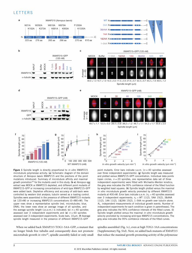
Figure 1 Spindle length is directly proportional to in vitro XMAP215microtubule polymerase activity. (a) Schematic diagram of the domainstructure of Xenopus laevis XMAP215 and the positions of the pointmutations introduced. Summary of microtubule affinity and maximalgrowth promotion18 for the mutants used in this study. (b–e) Xenopus eggextract was MOCK or XMAP215 depleted, and different point mutants ofXMAP215–GFP or increasing concentrations of wild-type XMAP215–GFPwere added back. Depletion efficiency and accuracy of add-back werecontrolled by western blot analysis; tubulin served as a loading control.Spindles were assembled in the presence of different XMAP215 mutants(at 120nM) or increasing XMAP215 concentrations (0–480nM). Theupper rows show a representative spindle (red, microtubules; blue,DNA), the lower rows show an average image of all spindles, andthe average spindle length (±s.e.m.) is indicated; (c) n = 40 spindlesassessed over 3 independent experiments and (e) n = 60 spindlesassessed over 3 independent experiments. Scale bars, 10 µm. (f) Averagespindle length measured in the presence of different XMAP215–GFP
point mutants. Error bars indicate s.e.m. (n = 40 spindles assessedover three independent experiments). (g) Spindle length was measuredand plotted versus XMAP215–GFP concentration. Individual data points(open circles, n = 20 spindles, one representative data set of threeindependent experiments) were fitted with Michaelis–Menten kinetics;the grey area indicates the 95% confidence interval of the fitted functionby weighted least squares. (h) Spindle length plotted versus the maximalin vitro microtubule growth velocity promoted by different XMAP215mutants at 400nM. Error bars indicate s.e.m. (ny =40 spindles assessedover 3 independent experiments; nx =WT 11(2), 5AA 15(2), 3&4AA11(2), 1AA 11(2), 1&2AA 15(2), 1–5AA no growth over tubulin alone.Nx , independent measurements of individual growth events. Number ofindependent experiments for each condition is given in parentheses). Thegrey area indicates the 95% confidence intervals of the fitted curves. (i)Spindle length plotted versus the maximal in vitro microtubule growthvelocity promoted by increasing wild-type XMAP215 concentrations. Thegrey area indicates the 95% confidence intervals of the fitted curves.
When we added back XMAP215 TOG1-5AA–GFP, a mutant thatno longer binds free tubulin and consequently does not promotemicrotubule growth in vitro18, spindle assembly failed or only small
spindles assembled (Fig. 1c), even at high TOG1-5AA concentrations(Supplementary Fig. S1d). Next, we added back mutants of XMAP215that reduce the maximal growth-promoting activity of the polymerase
Figure 2 Spindle length scales with spindle microtubule growth velocity.(a) Spindle assembled in Xenopus egg extract (red: Cy3–tubulin, green:EB1–GFP). Scale bar, 10 µm. (b) All EB1–GFP tracks detected in a singletime-lapse sequence (time resolution 0.3 s) of a wild-type Xenopus spindle.Total number of tracks: 2,752. The heat map colour codes for microtubulegrowth, the circle represents the start of the track, and the line its length.(c,d) Histograms of the distance (c) and time (d) EB1 tracks travelled ina spindle assembled at 240nM (green, upper row), 120nM (blue, middlerow) and 60nM (purple, bottom row) XMAP215. (e) The EB1–GFP trackswere binned by time and the average distance for each group was evaluated.
The weighted linear fit without a constant to this evaluated distance versustime data provides the velocity at which the EB1–GFP tracks travel. Errorbars indicate s.e.m., which is based on the corresponding bins shown ind. The total number of tracks for the representative spindle assembled at60nM XMAP215 (green): 138 tracks; at 120nM XMAP215 (blue): 2,688tracks; and at 240nM XMAP215 (purple): 3,223 tracks. (f) Individual datapoints for spindle length versus EB1–GFP track velocity (circle) and fluxvelocity (square). The colour code indicates the XMAP215 concentration atwhich spindles were assembled. The grey area indicates the 95% confidenceinterval of the fitted curve.
over a 5.5-fold range, assembled spindles and measured their length(Fig. 1f). At the endogenous concentration, these mutants promotedassembly of spindles at lengths proportional to their polymerase activity.Similarly, spindle length increased with increasing wild-type XMAP215concentrations (Fig. 1d,e). However, when adding more than 240 nMXMAP215–GFP, spindle length plateaued (Fig. 1g). We correlatedspindle length with the maximum microtubule growth promotionof XMAP215 determined in total-internal-reflection fluorescence
microscopy assays (Supplementary Fig. S1e,f). In both, the pointmutant (Fig. 1h) and the dose–response (Fig. 1i) assay, spindlelength is proportional to the maximal growth rate promoted by thepolymerase XMAP215.Next, we wanted to correlate spindle length withmicrotubule growth
rates measured in spindles. Therefore, we added EB1–GFP, a (+) TIP-binding protein that tracks growing microtubule ends20,21 and imagedspindles over time (Fig. 2a). In each spindle, microtubule plus ends
were automatically detected (Fig. 2b), microtubule growth events weretracked over consecutive frames and track length (Fig. 2c), duration(Fig. 2d) and growth velocities (Fig. 2e) were evaluated. The analysisof EB1–GFP tracks measured in control spindles revealed an averagegrowth velocity of 12.8±0.9 µmmin−1 (s.e.m., n= 6 spindles). Thisvelocity, however, does not represent microtubule growth velocitiesper se but the combined effect of microtubule flux15, which occurs at arate of approximately 2 µmmin−1, and growth velocity (Fig. 2f). Whenwe increased XMAP215 activity in the extract, we observed that themean track velocity increased with spindle length whereas microtubuleflux velocity, determined by speckle microscopy, was independentof XMAP215 concentration and always smaller than the measuredEB1–GFP track velocity. We thus conclude that spindle lengthcorrelates linearly withmicrotubule growth velocities in spindles.Studies in Xenopus eggs and early embryos suggest an upper limit to
spindle length22. To determine whether the upper length of the spindleis limited by the polymerase activity of XMAP215, we performed adose–response assay for spindle length in the presence of differentXMAP215 concentrations for the wild-type protein and two engineeredmutants, TOG1&2AA–GFP and TOG3&4AA–GFP (Fig. 3a). Forall proteins, spindle length initially correlated with XMAP215concentration but subsequently plateaued (Fig. 3b). Interestingly, thespindles plateaued at different lengths depending on the polymeraseactivity of the mutants. However, for all proteins, spindles approachtheir maximal length at approximately 200 nM protein. This is theconcentration at which XMAP215 works at its maximum rate in vitrobecause it saturates microtubule plus ends18. On the basis of our results,we propose that spindle length approaches its maximal length whenmicrotubule plus ends are saturated with the polymerase, and thatthe upper limit to spindle length is determined, at least in part, by themaximal growth velocity of microtubules.XMAP215 was previously reported to also affect other parame-
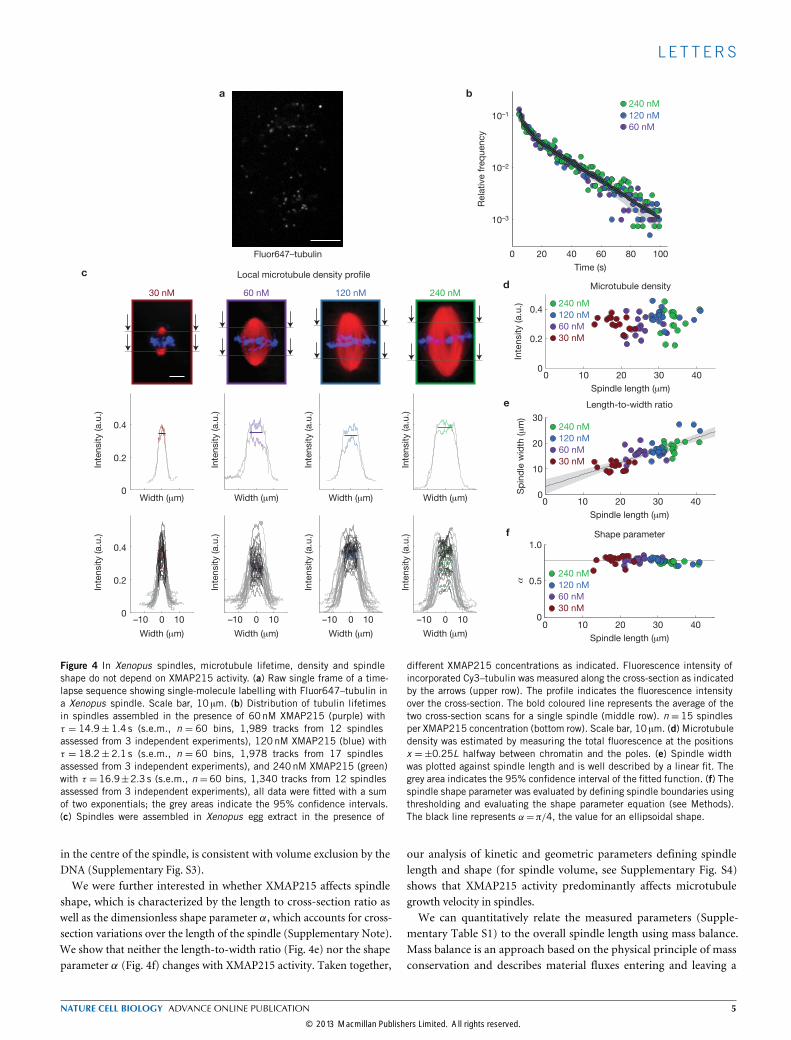
ters of microtubule dynamics, such as microtubule lifetime23 andnucleation24–26. So far, the best approximation for microtubule lifetimein spindles can be obtained by using single-molecule imaging14. We as-sembled spindles in the presence of Fluor647–tubulin at concentrationswhere approximately 1 in 100,000 tubulin molecules is labelled and im-aged spindles over time (Fig. 4a). The assumption is that the appearanceof a single fluorescent tubulin particle reflects a tubulin molecule be-coming incorporated into a growingmicrotubule, and its disappearancereflects the release duringmicrotubule depolymerization.We thus referto the total amount of time between the appearance and disappearanceof a tubulin molecule as its lifetime. We determined a mean micro-tubule lifetime of 18.2±2.1 s (s.e.m. n= 60 bins, 17 spindles), whichis comparable to published data14. However, when we determined themicrotubule lifetime for spindles assembled in the presence of differentXMAP215 concentrations, we indeed saw that neither the lifetimedistribution nor the lifetime itself changed significantly with XMAP215activity (Fig. 4b). As a control, we depleted XKCM1 (Xenopus kinesincatastrophe modulator-1), a microtubule-depolymerizing kinesin-13(ref. 16), fromXenopus egg extracts and show that we can detect changesin microtubule lifetime (Supplementary Fig. S2a,b). Therefore, we con-clude that XMAP215 does not affectmicrotubule lifetime in spindles.Our data show that modulation of XMAP215 activity does not
significantly affect microtubule lifetime within spindles, consistentwith other in vitro data that show that XMAP215 has little effect
WT
1&2AA
3&4AA
Sp
ind
le len
gth
(μ
m)
XMAP215 (nM)
XMAP215–GFP (nM)
MOCK 0
Spindle length (μm)
5.6 ± 0.327.9 ± 0.4
0 100 200 300 400 5000
5
10
15
20
25
30
35
40
WT
1&2AA
3&4AA
120 24060 480
25.9 ± 0.5 28.0 ± 0.714.7 ± 0.5 29.9 ± 0.6
14.9 ± 0.5 22.5 ± 0.99.7 ± 0.6 23.4 ± 0.7
10.9 ± 0.5 12.1 ± 0.48.5 ± 0.8 14.0 ± 0.5
b
a
Figure 3 XMAP215 activity limits upper spindle length. (a) Xenopus eggextract was MOCK or XMAP215 depleted, different concentrations ofrecombinant wild-type (WT) or mutant XMAP215-GFP were added back andspindles were assembled (red: microtubules, blue: DNA). Scale bar, 10 µm.Average spindle length is indicated (±s.e.m., n = 30 spindles assessedover three independent experiments). (b) Dose–response curve for spindlelength at indicated XMAP215 concentrations for wild-type XMAP215(blue), and TOG3&4AA (purple) and TOG1&2AA (red) mutants fitted withMichaelis–Menten kinetics. The dashed line indicates the concentrationat which all XMAP215 mutants saturate the microtubule plus end in vitro.Individual data points (open circles, n =30 spindles assessed over threeindependent experiments) and the average (filled circles) are shown; thegrey areas indicate the 95% confidence intervals of the fitted functions.
on the catastrophe rate27. This is not in contradiction with previousin vitro studies that show that microtubule lifetime increases withgrowth velocity28–30. In these studies, microtubule growth velocity wasincreased by changing tubulin concentration and not by modulatingthe activity of XMAP215, as in our work.Next, we measured microtubule density over the cross-section
at two points within each spindle, halfway between the chromatinand either pole (Fig. 4c, upper row). This measurement resulted intwo intensity profiles, from which we evaluated the average localmicrotubule density for a given spindle (Fig. 4c, middle row). For eachXMAP215 concentration, we summarize the density profiles and therespective mean of 15 spindles (Fig. 4c, bottom row), which is roughlyconstant at any given concentration. Similarly, the total amount ofmicrotubule intensity is independent of XMAP215 activity (Fig. 4d).The variation in intensity along the longitudinal spindle axis, especially
Figure 4 In Xenopus spindles, microtubule lifetime, density and spindleshape do not depend on XMAP215 activity. (a) Raw single frame of a time-lapse sequence showing single-molecule labelling with Fluor647–tubulin ina Xenopus spindle. Scale bar, 10 µm. (b) Distribution of tubulin lifetimesin spindles assembled in the presence of 60nM XMAP215 (purple) withτ = 14.9± 1.4 s (s.e.m., n = 60 bins, 1,989 tracks from 12 spindlesassessed from 3 independent experiments), 120nM XMAP215 (blue) withτ = 18.2± 2.1 s (s.e.m., n = 60 bins, 1,978 tracks from 17 spindlesassessed from 3 independent experiments), and 240nM XMAP215 (green)with τ = 16.9±2.3 s (s.e.m., n = 60 bins, 1,340 tracks from 12 spindlesassessed from 3 independent experiments), all data were fitted with a sumof two exponentials; the grey areas indicate the 95% confidence intervals.(c) Spindles were assembled in Xenopus egg extract in the presence of
different XMAP215 concentrations as indicated. Fluorescence intensity ofincorporated Cy3–tubulin was measured along the cross-section as indicatedby the arrows (upper row). The profile indicates the fluorescence intensityover the cross-section. The bold coloured line represents the average of thetwo cross-section scans for a single spindle (middle row). n = 15 spindlesper XMAP215 concentration (bottom row). Scale bar, 10 µm. (d) Microtubuledensity was estimated by measuring the total fluorescence at the positionsx =±0.25L halfway between chromatin and the poles. (e) Spindle widthwas plotted against spindle length and is well described by a linear fit. Thegrey area indicates the 95% confidence interval of the fitted function. (f) Thespindle shape parameter was evaluated by defining spindle boundaries usingthresholding and evaluating the shape parameter equation (see Methods).The black line represents α=π/4, the value for an ellipsoidal shape.
in the centre of the spindle, is consistent with volume exclusion by theDNA (Supplementary Fig. S3).We were further interested in whether XMAP215 affects spindle
shape, which is characterized by the length to cross-section ratio aswell as the dimensionless shape parameter α, which accounts for cross-section variations over the length of the spindle (Supplementary Note).We show that neither the length-to-width ratio (Fig. 4e) nor the shapeparameter α (Fig. 4f) changes with XMAP215 activity. Taken together,
our analysis of kinetic and geometric parameters defining spindlelength and shape (for spindle volume, see Supplementary Fig. S4)shows that XMAP215 activity predominantly affects microtubulegrowth velocity in spindles.We can quantitatively relate the measured parameters (Supple-
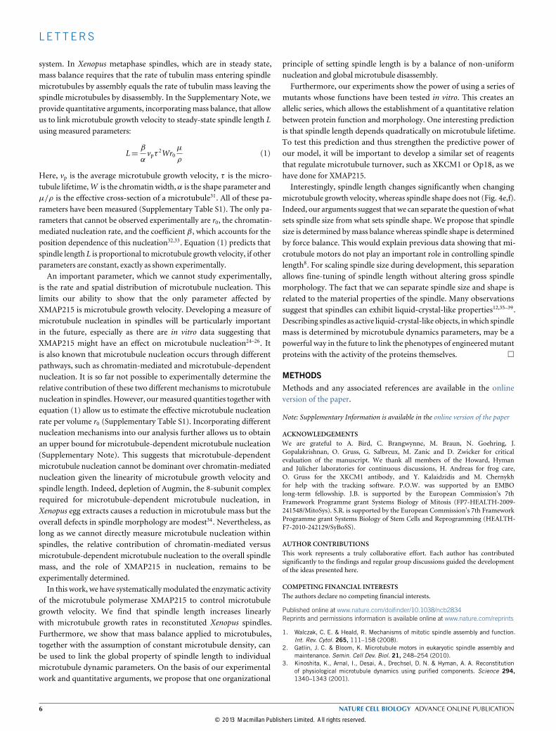
mentary Table S1) to the overall spindle length using mass balance.Mass balance is an approach based on the physical principle of massconservation and describes material fluxes entering and leaving a
system. In Xenopus metaphase spindles, which are in steady state,mass balance requires that the rate of tubulin mass entering spindlemicrotubules by assembly equals the rate of tubulin mass leaving thespindle microtubules by disassembly. In the Supplementary Note, weprovide quantitative arguments, incorporating mass balance, that allowus to link microtubule growth velocity to steady-state spindle length Lusing measured parameters:
L=β
αvpτ 2Wr0
µ
ρ(1)
Here, vp is the average microtubule growth velocity, τ is the micro-tubule lifetime,W is the chromatin width, α is the shape parameter andµ/ρ is the effective cross-section of a microtubule31. All of these pa-rameters have been measured (Supplementary Table S1). The only pa-rameters that cannot be observed experimentally are r0, the chromatin-mediated nucleation rate, and the coefficient β, which accounts for theposition dependence of this nucleation32,33. Equation (1) predicts thatspindle length L is proportional tomicrotubule growth velocity, if otherparameters are constant, exactly as shown experimentally.An important parameter, which we cannot study experimentally,
is the rate and spatial distribution of microtubule nucleation. Thislimits our ability to show that the only parameter affected byXMAP215 is microtubule growth velocity. Developing a measure ofmicrotubule nucleation in spindles will be particularly importantin the future, especially as there are in vitro data suggesting thatXMAP215 might have an effect on microtubule nucleation24–26. Itis also known that microtubule nucleation occurs through differentpathways, such as chromatin-mediated and microtubule-dependentnucleation. It is so far not possible to experimentally determine therelative contribution of these two different mechanisms to microtubulenucleation in spindles. However, ourmeasured quantities together withequation (1) allow us to estimate the effective microtubule nucleationrate per volume r0 (Supplementary Table S1). Incorporating differentnucleation mechanisms into our analysis further allows us to obtainan upper bound for microtubule-dependent microtubule nucleation(Supplementary Note). This suggests that microtubule-dependentmicrotubule nucleation cannot be dominant over chromatin-mediatednucleation given the linearity of microtubule growth velocity andspindle length. Indeed, depletion of Augmin, the 8-subunit complexrequired for microtubule-dependent microtubule nucleation, inXenopus egg extracts causes a reduction in microtubule mass but theoverall defects in spindle morphology are modest34. Nevertheless, aslong as we cannot directly measure microtubule nucleation withinspindles, the relative contribution of chromatin-mediated versusmicrotubule-dependent microtubule nucleation to the overall spindlemass, and the role of XMAP215 in nucleation, remains to beexperimentally determined.In this work, we have systematicallymodulated the enzymatic activity
of the microtubule polymerase XMAP215 to control microtubulegrowth velocity. We find that spindle length increases linearlywith microtubule growth rates in reconstituted Xenopus spindles.Furthermore, we show that mass balance applied to microtubules,together with the assumption of constant microtubule density, canbe used to link the global property of spindle length to individualmicrotubule dynamic parameters. On the basis of our experimentalwork and quantitative arguments, we propose that one organizational
principle of setting spindle length is by a balance of non-uniformnucleation and global microtubule disassembly.Furthermore, our experiments show the power of using a series of
mutants whose functions have been tested in vitro. This creates anallelic series, which allows the establishment of a quantitative relationbetween protein function and morphology. One interesting predictionis that spindle length depends quadratically on microtubule lifetime.To test this prediction and thus strengthen the predictive power ofour model, it will be important to develop a similar set of reagentsthat regulate microtubule turnover, such as XKCM1 or Op18, as wehave done for XMAP215.Interestingly, spindle length changes significantly when changing
microtubule growth velocity, whereas spindle shape does not (Fig. 4e,f).Indeed, our arguments suggest that we can separate the question ofwhatsets spindle size from what sets spindle shape. We propose that spindlesize is determined bymass balance whereas spindle shape is determinedby force balance. This would explain previous data showing that mi-crotubule motors do not play an important role in controlling spindlelength8. For scaling spindle size during development, this separationallows fine-tuning of spindle length without altering gross spindlemorphology. The fact that we can separate spindle size and shape isrelated to the material properties of the spindle. Many observationssuggest that spindles can exhibit liquid-crystal-like properties12,35–39.Describing spindles as active liquid-crystal-like objects, inwhich spindlemass is determined by microtubule dynamics parameters, may be apowerful way in the future to link the phenotypes of engineeredmutantproteins with the activity of the proteins themselves. �
METHODSMethods and any associated references are available in the onlineversion of the paper.
Note: Supplementary Information is available in the online version of the paper
ACKNOWLEDGEMENTSWe are grateful to A. Bird, C. Brangwynne, M. Braun, N. Goehring, J.Gopalakrishnan, O. Gruss, G. Salbreux, M. Zanic and D. Zwicker for criticalevaluation of the manuscript. We thank all members of the Howard, Hymanand Jülicher laboratories for continuous discussions, H. Andreas for frog care,O. Gruss for the XKCM1 antibody, and Y. Kalaidzidis and M. Chernykhfor help with the tracking software. P.O.W. was supported by an EMBOlong-term fellowship. J.B. is supported by the European Commission’s 7thFramework Programme grant Systems Biology of Mitosis (FP7-HEALTH-2009-241548/MitoSys). S.R. is supported by the European Commission’s 7th FrameworkProgramme grant Systems Biology of Stem Cells and Reprogramming (HEALTH-F7-2010-242129/SyBoSS).
AUTHOR CONTRIBUTIONSThis work represents a truly collaborative effort. Each author has contributedsignificantly to the findings and regular group discussions guided the developmentof the ideas presented here.
COMPETING FINANCIAL INTERESTSThe authors declare no competing financial interests.
Published online at www.nature.com/doifinder/10.1038/ncb2834Reprints and permissions information is available online at www.nature.com/reprints
1. Walczak, C. E. & Heald, R. Mechanisms of mitotic spindle assembly and function.Int. Rev. Cytol. 265, 111–158 (2008).
2. Gatlin, J. C. & Bloom, K. Microtubule motors in eukaryotic spindle assembly andmaintenance. Semin. Cell Dev. Biol. 21, 248–254 (2010).
3. Kinoshita, K., Arnal, I., Desai, A., Drechsel, D. N. & Hyman, A. A. Reconstitutionof physiological microtubule dynamics using purified components. Science 294,1340–1343 (2001).
4. Brouhard, G. J. et al. XMAP215 is a processive microtubule polymerase. Cell 132,79–88 (2008).
5. Levy, D. L. & Heald, R. Mechanisms of intracellular scaling. Annu. Rev. Cell Dev.Biol. 28, 113–135 (2012).
6. Chan, Y-H. M. & Marshall, W. F. How cells know the size of their organelles. Science337, 1186–1189 (2012).
7. Goehring, N. W. & Hyman, A. A. Organelle growth control through limiting pools ofcytoplasmic components. Curr. Biol. 22, R330–R9 (2012).
8. Goshima, G., Wollman, R., Stuurman, N., Scholey, J. M. & Vale, R. D. Length controlof the metaphase spindle. Curr. Biol. 15, 1979–1988 (2005).
9. Burbank, K. S., Mitchison, T. J. & Fisher, D. S. Slide-and-cluster models for spindleassembly. Curr. Biol. 17, 1373–1383 (2007).
10. Loughlin, R., Wilbur, J. D., McNally, F. J., Nédélec, F. J. & Heald, R. Katanincontributes to interspecies spindle length scaling in Xenopus. Cell 147,1397–1407 (2011).
11. Brugués, J., Nuzzo, V., Mazur, E. & Needleman, D. J. Nucleation and transportorganize microtubules in metaphase spindles. Cell 149, 554–564 (2012).
12. Shimamoto, Y., Maeda, Y. T., Ishiwata, S., Libchaber, A. J. & Kapoor, T. M.Insights into the micromechanical properties of the metaphase spindle. Cell 145,1062–1074 (2011).
13. Mitchison, T. J. T. et al. Roles of polymerization dynamics, opposed motors, and atensile element in governing the length of Xenopus extract meiotic spindles. Mol.Biol. Cell 16, 3064–3076 (2005).
14. Needleman, D. J. et al. Fast microtubule dynamics in meiotic spindles measured bysingle molecule imaging: evidence that the spindle environment does not stabilizemicrotubules. Mol. Biol. Cell 21, 323–333 (2010).
15. Yang, G. G. et al. Architectural dynamics of the meiotic spindle revealed bysingle-fluorophore imaging. Nat. Cell Biol. 9, 1233–1242 (2007).
16. Howard, J. & Hyman, A. A. Microtubule polymerases and depolymerases. Curr. Opin.Cell Biol. 19, 31–35 (2007).
17. Al-Bassam, J. & Chang, F. Regulation of microtubule dynamics by TOG-domainproteins XMAP215/Dis1 and CLASP. Trends Cell Biol. 21, 604–614 (2011).
18. Widlund, P. O. et al. XMAP215 polymerase activity is built by combining multipletubulin-binding TOG domains and a basic lattice-binding region. Proc. Natl Acad.Sci. 108, 2741–2746 (2011).
19. Kinoshita, K. Aurora A phosphorylation of TACC3/maskin is required forcentrosome-dependent microtubule assembly in mitosis. J. Cell Biol. 170,1047–1055 (2005).
20. Zanic, M., Stear, J. H., Hyman, A. A. & Howard, J. EB1 recognizes the nucleotidestate of tubulin in the microtubule lattice. PLoS ONE 4, e7585 (2009).
21. Maurer, S. P., Fourniol, F. J., Bohner, G., Moores, C. A. & Surrey, T. EBs recognizea nucleotide-dependent structural cap at growing microtubule ends. Cell 149,371–382 (2012).
22. Wuehr, M. et al. Evidence for an upper limit to mitotic spindle length. Curr. Biol. 18,1256–1261 (2008).
23. Hamada, T., Itoh, T. J., Hashimoto, T., Shimmen, T. & Sonobe, S. GTP is requiredfor the microtubule catastrophe-inducing activity of MAP200, a tobacco homolog ofXMAP215. Plant Physiol. 151, 1823–1830 (2009).
24. Popov, A. V., Severin, F. & Karsenti, E. XMAP215 is required for the microtubule-nucleating activity of centrosomes. Curr. Biol. 12, 1326–1330 (2002).
25. Groen, A. C., Maresca, T. J., Gatlin, J. C., Salmon, E. D. & Mitchison, T. J. Functionaloverlap of microtubule assembly factors in chromatin-promoted spindle assembly.Mol. Biol. Cell 20, 2766–2773 (2009).
26. Slep, K. C. & Vale, R. D. Structural basis of microtubule plus end tracking byXMAP215, CLIP-170, and EB1. Mol. Cell 27, 976–991 (2007).
27. Vasquez, R. J., Gard, D. L. & Cassimeris, L. XMAP from Xenopus eggs promotesrapid plus end assembly of microtubules and rapid microtubule polymer turnover.J. Cell Biol. 127, 985–993 (1994).
28. Walker, R. A. et al. Dynamic instability of individual microtubules analyzed byvideo light microscopy: rate constants and transition frequencies. J. Cell Biol. 107,1437–1448 (1988).
29. Verde, F., Dogterom, M., Stelzer, E., Karsenti, E. & Leibler, S. Control of microtubuledynamics and length by cyclin A- and cyclin B-dependent kinases in Xenopus eggextracts. J. Cell Biol. 118, 1097–1108 (1992).
30. Dogterom, M. & Leibler, S. Physical aspects of the growth and regulation ofmicrotubule structures. Phys. Rev. Lett. 70, 1347–1350 (1993).
31. Loughlin, R., Heald, R. & Nedelec, F. A computational model predicts Xenopusmeiotic spindle organization. J. Cell Biol. 191, 1239–1249 (2010).
32. Hyman, A. & Karsenti, E. The role of nucleation in patterning microtubule networks.J. Cell Sci. 111, 2077–2083 (1998) Pt 15.
33. Gruss, O. J. The mechanism of spindle assembly: functions of Ran and its targetTPX2. J. Cell Biol. 166, 949–955 (2004).
34. Petry, S., Pugieux, C., Nédélec, F. J. & Vale, R. D. Augmin promotes meiotic spindleformation and bipolarity in Xenopus egg extracts. Proc. Natl Acad. Sci. USA 108,14473–14478 (2011).
35. Gatlin, J. C., Matov, A., Danuser, G., Mitchison, T. J. & Salmon, E. D. Directlyprobing the mechanical properties of the spindle and its matrix. J. Cell Biol. 188,481–489 (2010).
36. Itabashi, T. et al. Probing the mechanical architecture of the vertebrate meioticspindle. Nat. Methods 6, 167–172 (2009).
37. Inoué, S. Microtubule dynamics in cell division: exploring living cells with polarizedlight microscopy. Annu. Rev. Cell Dev. Biol. 24, 1–28 (2008).
38. Mitchison, T. J. et al. Bipolarization and poleward flux correlate during Xenopusextract spindle assembly. Mol. Biol. Cell 15, 5603–5615 (2004).
39. Isenberg, I. On the theory of the nematic phase and its possible relation to the mitoticspindle structure. Bull. Math. Biophys. 16, 83–96 (1954).
METHODSSpindle assembly in Xenopus egg extract. Cytostatic factor (CSF) extracts wereprepared from Xenopus eggs arrested in metaphase of meiosis II as describedpreviously40. To promote bipolar spindle formation, demembranated sperm nucleiwere added to the CSF extract, cycling to interphase was induced by the additionof CaCl2 to 0.6mM and after 90min the system was rearrested in M-phase usingone volume of CSF extract. Spindles were assembled as described above in eitherMOCK-depleted extracts or in XMAP215-depleted extracts supplemented withXMAP215 buffer, XMAP215–GFP or recombinant XMAP215–GFP mutants to120 nM or indicated concentrations. For each experiment, 15–60 spindles assessedover three different extracts were analysed (Figs 1b–e, 2a–e, 3a and 4a,c andSupplementary Figs S1a–d and S2a) .
Depletion/add-back experiments and pulldown assays. CSF extract (100 µl)was either MOCK-, XKCM1- or XMAP215-depleted by incubating the CSF extracttwice with 20 µl Dynal Protein A beads (Invitrogen) coupled to unspecific rabbitserum IgG, a carboxy-terminal XMAP215 antibody or an antibody against XKCM1,respectively. Depletion efficiency was monitored by functional assays and westernblot analysis. The pulldown assay was performed by crosslinking 0.5 µg anti-GFPantibody per microlitre of Dynal Protein G beads (Invitrogen) in the presence ofDMP (SIGMA). Recombinant, GFP-tagged proteins or GFP alone was added to CSFextracts and incubated with the anti-GFP beads at 4 ◦C for 40min. The beads werewashed extensively and eluted with CSF-XB (without sucrose) at pH 7.7, 1M KCl.The elutionwas TCAprecipitated, separated by SDS gel electrophoresis and analysedby western blotting.
Antibodies. The C-terminal antibody used for XMAP215 depletion from Xenopusegg extracts was raised against a peptide containing the last 15 amino acids ofXMAP215 and affinity purified against this peptide as described before24. Forwestern blot analysis, anti-XMAP215 was used at 5 µgml−1 final concentration.Anti-Xenopus TACC3 antibodies were raised and purified against the GST fusionprotein that contained an amino-terminal fragment of TACC3 (amino acids 7–208)as described previously19. For western blot analysis, anti-TACC3 was used at3 µgml−1 final concentration. A monoclonal anti-α-tubulin (SIGMA, T5168, CloneB-5-1-2) was used in western blots as a loading control (working dilution 1:5,000).Goat anti-GFP was raised against His-tagged full-length EGFP, affinity-purifiedwith GST-tagged full-length EGFP and used at 50 ngml−1 final concentration. Theanti-XKCM1 antibody was a gift from O. Gruss (ZMBH, Germany) and was raisedagainst full-length SUMO-tagged XKCM1. XKCM1 serum (250 µl) was coupled to100 µl Protein A beads; 2×40 µl beads were used to deplete 100 µl CSF extract.
Purification of XMAP215– and EB1–GFP. Wild-type XMAP215–GFP andmutants were expressed in SF+ cells using the Bac-to-Bac system (Invitrogen) andpurified as described previously18. EB1–GFPwas expressed and purified as describedpreviously20.
Porcine tubulin: preparation, labelling and polymerization with GMPCPP.Labelling of cycled tubulin with Cy3 (GEHealthcare) or AlexaFluor488 (Invitrogen)was performed as described previously41. Porcine HiLyte Fluor647 tubulin waspurchased from Cytoskeleton (# TL670M). GMPCPP microtubules were grown asdescribed previously42.
Image acquisition and data processing. The total-internal-reflection fluores-cence imaging was performed with a set-up described previously4,18. Microtubulegrowth measurements (Fig. 1h,i and Supplementary Fig. S1e,f) were performed inMetamorph (Universal Imaging).
To measure spindle length, shape and microtubule density, the assembledspindles were fixed in the presence of 0.2mgml−1 Cy3-labelled tubulin and DAPIin squash-fixed samples. Stacks of fixed spindles were acquired in 1-µm steps with aZeiss LSM700 laser scanningmicroscope and aZeiss LCI Plan-Neofluar×63, 1.3NAimmersion correction objective using ZEN software. Quantitative analysis of spindlelength (Figs 1c,e and 3a,b and Supplementary Fig. S1d) was carried out using acustom algorithm as described before43. For quantifying the fluorescence intensity ofincorporated Cy3–tubulin, labelled tubulin was added to the extract before the startof the experiment to ensure equal Cy3–tubulin concentrations in controls and allconditions. Images were captured at the same camera setting and further processed
using ImageJ. Using the maximum-intensity projection we created an output imagefrom the acquired stack, in which we manually set the threshold level to define theregion of interest corresponding to the spindle contours. Using the defined regionof interest, the acquired stack and the image processing toolbox in Matlab (Matlab8.0, The MathWorks), the overall intensity in each plane was evaluated. For furtherevaluation, the plane with the highest value was selected. The longitudinal spindleaxis was determined on the basis of the intensity map. At first, the centroid wascomputed by weighting with the intensity in the region of interest. With respectto this centroid, the second moments of area, also weighted with the intensity, wereevaluated and the first principal axis was taken as the longitudinal and the second asthe normal axis. The density profile along the longitudinal axis was then extracted byaveraging in the normal direction of this slice. To determine the width of the alignedchromosomes, we stainedDNAwithDAPI (Supplementary Fig. S3) and analysed theintensity profile in the same coordinate system as the tubulin staining. The spindleshape parameter (Fig. 4f) was evaluated using the boundaries of the region of interestand integrating Supplementary Equation S9 with the trapezoidal rule.
To measure microtubule growth velocities in spindles (Fig. 2), we addedEB1–GFP to assembled spindles and imaged every 0.3 s using an Olympus IX71inverted spinning-disc confocal microscope equipped with a Yokogawa CSU10 scanhead, a 488 nm solid-state coherent sapphire (75mW) laser line, an Andor iXonEM+ DU-897 BV back-illuminated electron-multiplying CCD (charge-coupleddevice) camera, an Olympus UPlan FluarN 60× 0.9 objective, and the Andorsoftware. Detection and tracking of EB1–GFP was performed using the MotionTracking software package (http://motiontracking.mpi-cbg.de; ref 44), written inthe Pluk development environment45. The coordinates of detected EB1–GFP trackswere used to calculate the end-to-end distance and duration of every track. Then, theEB1–GFP tracks were binned by time and the average distance for each group wasevaluated. Theweighted linear fit to this evaluated distance versus time data providesthe velocity at which the EB1–GFP comets grow in a spindle (Fig. 2c–e).
To measure tubulin turnover and microtubule flux (Figs 4b and 2f), we usedtubulin single-molecule imaging14,46. Briefly, spindle reactions were supplementedwith Fluor647–tubulin to achieve a final concentration of 100 pM and movies wereacquired using an Olympus IX81 inverted stand spinning-disc confocal microscopewith a Yokogawa CSU-X1 scan head, a 640 nm solid-state laser line (100mW),an Andor iXon EM+ DU-897 BV back-illuminated electron-multiplying CCDcamera, an Olympus UPlanSApo ×60 1.35 NA oil objective, and Andor software.To evaluate the flux velocities, detection and tracking of tubulin speckles as well asthe calculations were performed as described for the EB1–GFP tracks. The durationof the individual tracks was used to estimate the lifetime distribution. The lifetimedata were fitted, using the fitting toolbox in Matlab (Matlab 8.0, The MathWorks),with a double-exponential function, which corresponds to a case where the numberof rescues per microtubule is small11,47,48. To control that photobleaching is notsignificant under our imaging conditions, we analysed the number of objects,their integral intensity and the number of tracks over time (data not shown).
40. Hannak, E. & Heald, R. Investigating mitotic spindle assembly and function in vitrousing Xenopus laevis egg extracts. Nat. Protoc. 1, 2305–2314 (2006).
41. Hyman, A. et al. Preparation of modified tubulins. Methods Enzymol. 196,478–485 (1991).
42. Gell, C. et al. Purification of tubulin from porcine brain. Methods Mol. Biol. 777,15–28 (2011).
43. Reber, S., Over, S., Kronja, I. & Gruss, O. J. CaM kinase II initiates meioticspindle depolymerization independently of APC/C activation. J. Cell Biol. 183,1007–1017 (2008).
44. Rink, J., Ghigo, E., Kalaidzidis, Y. & Zerial, M. Rab conversion as a mechanism ofprogression from early to late endosomes. Cell 122, 735–749 (2005).
45. Kalaidzidis, L. Y., Gavrilov, A. V., Zaitsev, P. V., Kalaidzidis, A. L. & Korolev, E. V.PLUK—an environment for software development. Prog. Comput. Softw. 23,206–211 (1997).
46. Mirny, L. A. & Needleman, D. J. Quantitative characterization of filament dynamicsby single-molecule lifetime measurements.Methods Cell Biol. 95, 583–600 (2010).
47. Wilde, A. et al. Ran stimulates spindle assembly by altering microtubule dynamicsand the balance of motor activities. Nat. Cell Biol. 3, 221–227 (2001).
48. Brown, K. S. et al. Xenopus tropicalis egg extracts provide insight into scaling of themitotic spindle. J. Cell Biol. 176, 765–770 (2007).
Figure S1 XMAP215 TOG1-5AA binds to spindle microtubules and XTACC3 (a) Xenopus egg extract was MOCK or XMAP215 depleted, recombinant GFP-tagged XMAP215 wildtype (WT) or TOG1-5AA protein was added back to 120 nM, which is the endogenous concentration. Tubulin was used as a loading control. (b) A spindle assembled in XMAP215-depleted Xenopus egg extract in the presence of recombinant XMAP215-GFP (WT) or TOG1-5AA-GFP to 120 nM. Although the full mutant XMAP215 TOG1-5AA no longer binds tubulin, it still localizes to spindle microtubules. Note that spindles assembled in the presence of TOG1-5AA are much smaller than wildtype spindles, also at high concentrations. Scale bar: 10 µm (c) Pulldown of recombinant proteins from Xenopus egg extract on anti GFP-beads, beads eluate was analyzed by Western Blot for the presence of XTACC3. (d) Spindles were assembled in the presence of XMAP215 or the full mutant XMAP215 TOG1-5AA at the indicated concentrations. The upper row shows representative spindles (red: microtubules, blue: DNA), the lower row shows
an average image of all spindles (n=40, assessed over three independent experiments). Scale bar: 10 µm (e) Spindle length (Fig. 1c) plotted versus the in vitro microtubule growth velocities promoted by different XMAP215 mutants at 100 nM (as compared to the maximum growth promotion at 400 nM, shown in Fig. 1h). Error bars indicate SE (ny=40 spindles assessed over 3 independent experiments; nx = WT 11(2), 5AA 15(2), 3&4AA 11(2), 1AA 11 (2), 1&2AA 15 (2), 1-5 AA no growth. Nx=independent measurements of individual growth events. Number of independent experiments for each condition is given in parentheses.). Grey area indicates the 95% confidence interval of the fitted linear function. (f) Microtubule growth velocities measured at different XMAP215 concentrations in a TIRF assay. The mean growth velocity is indicated by open circles, individual data points by dots (n=10 independent measurements of individual growth events), which were fitted with Michaelis-Menten kinetics. Grey area indicates the 95% confidence interval of the fitted function.
Figure S2 Depleting XKCM1 from Xenopus egg extracts increases microtubule lifetime. (a) CSF-extract was immuno-depleted of XKCM1 (2ndD), one volume of fresh CSF-extract was added back to obtain 50% of endogenous XKCM1 concentration. (b) Spindles were assembled in the presence of Fluor647-tubulin and imaged with an Olympus IX81 spinning disc confocal microscope. Distribution of tubulin lifetimes in spindles assembled in control extracts
(blue) with (SE, n=60 bins, 13 745 tracks from 12 spindles assessed from 3 independent experiments) and XKCM1-depleted extracts (orange) with (SE, n=60 bins, 63 583 tracks from 13 spindles assessed from 3 independent experiments). All data were fitted with a sum of two exponentials, the grey areas indicate the 95% confidence intervals. Note that the setup of the microscope used to determine the lifetimes in Figure 4b was different (see Methods).
Figure S3 Reduction in microtubule density at the spindle centre is due to volume exclusion by DNA. Microtubule density (bold line) along the longitudinal spindle axis was quantified by the incorporated Cy3-tubulin fluorescence. The variation in intensity along the axis is due to volume
exclusion by the DNA, as quantified by the DAPI signal (thin line). The graphs show an average fluorescence intensity of 15 spindles per XMAP215 concentration. The chromatin width was estimated by fitting a Gaussian to the DAPI intensity profiles (SE, n=15 spindles per concentration).
Figure S4 Spindle volume (a) Individual data points of spindle volume versus spindle length at different XMAP215 concentrations fitted by a power law (exponent: 1.96 ± 0.18 (SE, n=60 spindles assessed over 3 different experiments). (b) Individual data points of spindle height versus spindle
length at different XMAP215 concentrations fitted by a linear function. (c) Spindle volume versus microtubule growth velocity by combing fits from Fig. S4a and 2f. Grey areas indicate the 95% confidence interval of the fitted functions.
Parameter Symbol Value (SE) Unit ReferenceSpindle length L 39.1 ± 2.2 µm This study, Figure 2fSpindle shape parameter α 0.785 - This study, Figure 4fNucleation profile coefficient β 2.24 - This study, Figure S3Width of nucleation zone W 8.3 ± 1.3 µm This study, Supplementary NoteMicrotubule cross-section* a 0.0025 µm2 Loughlin et al., 201010
Microtubule lifetime τ 18.2 ± 2.1 s This study, Figure 4bMicrotubule growth velocity vp 12.8 ± 0.9 µm·min-1 This study, Figure 2fNucleation rate per volume r0 9.3 ± 2.7 µm-3·s-1 This study, Equation (1)
* a = μ/ρ denotes the average area per microtubule in the spindle cross-section.
XMAP215 activity sets spindle length by controlling the total mass of spindle microtubules
Simone B Reber, Johannes Baumgart, Per O Widlund, Andrei Pozniakovsky, Jonathon Howard, Anthony A Hyman and Frank Jülicher
Mass balance links microtubule dynamics to spindle length
Mass balance allows us to express the total mass ! of spindle microtubules in terms of microtubule kinetic parameters that we measured. Spindle volume ! follows from spindle mass assuming constant mass density ! . The spindle geometry is known from the experiments. The only parameter that has not been measured so far is the nucleation rate. In the following we discuss how spindle length ! can be related to microtubule kinetic parameters and nucleation rates using mass balance.
Mass balance
In metaphase spindles, mass balance implies that the tubulin mass !!" that enters the spindle balances the tubulin mass that accumulates within the spindle and the mass !!"# that leaves the spindle as
d!d! =
d!!"
d! −d!!"#
d! , (S1)
where d!!" d! denotes tubulin influx, d!!"# d! tubulin outflux, and ! denotes time. Because Xenopus metaphase spindles are in steady state, the rate of tubulin mass assembling into spindle microtubules equals the rate of tubulin mass leaving the spindle by disassembly. Therefore, we can represent the microtubule mass balance of a metaphase spindle as
d!!"
d! =d!!"#
d! . (S2)
The tubulin in- and outfluxes are related to microtubule assembly and disassembly as is described in the following.
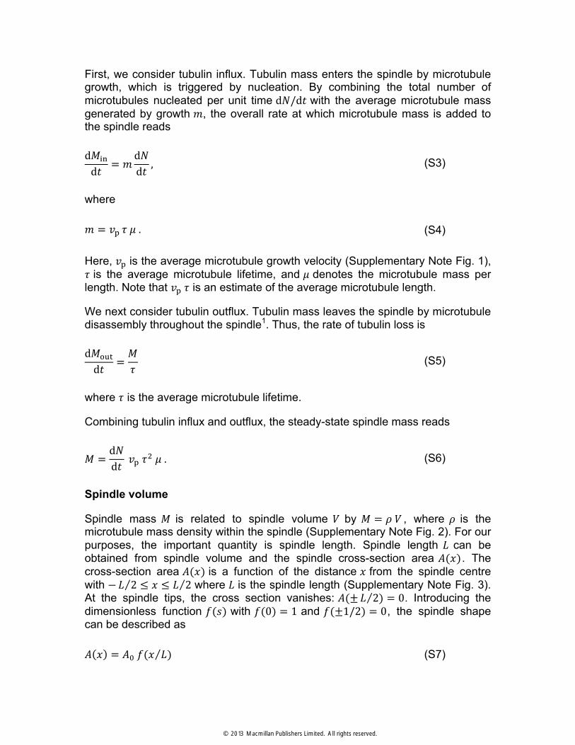
First, we consider tubulin influx. Tubulin mass enters the spindle by microtubule growth, which is triggered by nucleation. By combining the total number of microtubules nucleated per unit time d!/d! with the average microtubule mass generated by growth !, the overall rate at which microtubule mass is added to the spindle reads
d!!"
d! = !d!d! ,
(S3)
where
! = !! ! ! . (S4)
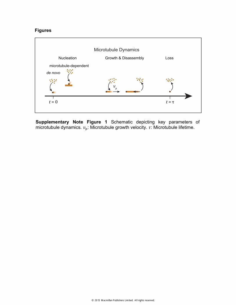
Here, !! is the average microtubule growth velocity (Supplementary Note Fig. 1), ! is the average microtubule lifetime, and ! denotes the microtubule mass per length. Note that !! ! is an estimate of the average microtubule length.
We next consider tubulin outflux. Tubulin mass leaves the spindle by microtubule disassembly throughout the spindle1. Thus, the rate of tubulin loss is
d!!"#
d! =!!
(S5)
where ! is the average microtubule lifetime.
Combining tubulin influx and outflux, the steady-state spindle mass reads
! =d!d! !! !
! ! . (S6)
Spindle volume
Spindle mass ! is related to spindle volume ! by ! = ! ! , where ! is the microtubule mass density within the spindle (Supplementary Note Fig. 2). For our purposes, the important quantity is spindle length. Spindle length ! can be obtained from spindle volume and the spindle cross-section area !(!). The cross-section area !(!) is a function of the distance ! from the spindle centre with − ! 2 ≤ ! ≤ ! 2 where ! is the spindle length (Supplementary Note Fig. 3). At the spindle tips, the cross section vanishes: !(± ! 2) = 0. Introducing the dimensionless function !(!) with !(0) = 1 and !(±1/2) = 0, the spindle shape can be described as
with ! = ! ! and !! denoting the cross-section area in the spindle centre. The spindle volume is given by
! = d! ! ! = ! !! !! !
!! ! (S8)
where
! = d! !(!)! !
!! ! (S9)
is a dimensionless number that characterizes spindle shape, independent of the aspect ratio of the spindle, which is defined as !/ !!.
Chromatin-dependent microtubule nucleation
The rate of chromatin-dependent microtubule nucleation in the spindle can be written as
d!!d! = d! ! ! ! ! .
! !
!! ! (S10)
Where !(!) is the nucleation rate per unit volume at distance ! from the centre. Microtubules nucleate predominantly in the vicinity of chromatin, which provides a signalling gradient2,3. For simplicity, we choose localized nucleation with a Gaussian profile of local nucleation rate per volume
! ! = !! exp −2 !!
!! . (S11)
The characteristic width of the nucleation zone is denoted !. The profile (S11) describes effects of a nucleation-promoting signal near the chromatin, where !! is the maximal nucleation rate per volume. The rate of chromatin-mediated nucleation can be written as
is a coefficient that depends on the spindle shape and the nucleation profile.
Spindle length
We first ignore the effects of microtubule-dependent microtubule nucleation d!/d! = d!!/d! . Using the expressions (S6) and (S8) together with the nucleation rate (S12), the steady-state spindle length is
! = !! ! !! !! !!
!! . (S14)
Here, ! ! is the average area per microtubule in the spindle cross section4.
Equation (S14) predicts that spindle length is directly proportional to the microtubule growth velocity !!, to the width ! of the region in which microtubules nucleate, and to the nucleation rate !! . Using the measured parameters (Supplementary Table 1), we can estimate the effective nucleation rate per volume !! = 9.3 ± 2.7 µμm!! · s!! . Furthermore equation (S14) predicts that spindle length ! depends quadratically on the average microtubule lifetime !. This strong dependence on lifetime results from the fact that an increased microtubule lifetime increases both the incorporated microtubule mass per nucleation event and the time over which this mass contributes to spindle mass.
Microtubule-dependent microtubule nucleation
Next, we incorporate effects of microtubule-dependent microtubule nucleation. A single chromatin-mediated nucleation event is followed by subsequent microtubule-dependent nucleation events, creating on average ! =!! !! !! additional microtubules. Here, !! is the rate of microtubule-dependent nucleation per unit microtubule length. Since subsequent nucleations create new microtubules that themselves can serve as nucleators and so on, the total nucleation rate becomes d!/d! = 1+ ! + !! +⋯ d!!/d! . This can be expressed as d!/d! = 1− ! !! d!!/d! or
d!d! =
11− !! !! !!
d!!d! (S15)
combining this with equations (S6), (S8), and (S12) we find
which has the same form as equation (S14), but with an effective nucleation rate per volume
!!"" = !!
1− !! !! !! , (S17)
which accounts for the effects of both chromatin-mediated and microtubule-dependent nucleation. Note that !!"" depends on the polymerization velocity !!. Thus, spindle length as described by equation (S16) could become a nonlinear function of !! if microtubule-dependent nucleation occurred at a significant rate !!.
The experimentally observed linear relationship, however, provides an upper bound on the value of the microtubule-dependent nucleation rate !! ≪ 1/(!! !!) and implies that chromatin-mediated nucleation dominates !!"" ≈ !!. Fitting the relation (S16) to the observed spindle length as a function of !! (Supplementary Note Fig. 4) provides an upper bound on the value of the microtubule-dependent nucleation rate per unit length !! ≤ 0.0004 µμm!! · s!! and a corresponding chromatin-mediated nucleation rate per volume !! = 8.5 ± 2.6 µμm!! · s!!.
Supplementary Note Figure 2 Due to the liquid-like properties of spindle material microtubule density ! is constant. On a molecular level this is driven by intermolecular interactions by cross-linking proteins and motors. (e) Microtubule density along the spindle pole-to-pole axis measured by Cy3-tubulin incorporation. Brown line shows the average of 15 spindles, the grey area indicates the interval of two standard deviations.
Supplementary Note Figure 3 (a) The spindle volume ! is defined by ! ! !!, with a dimensionless coefficient ! (equation S9), spindle length ! and the spindle cross-section !! . The nucleation volume !! ! is described by the spindle cross-section !! and the width ! of the nucleation zone. Nucleation occurs predominately in the vicinity of chromatin at a given rate per volume !!. (b) Width of the nucleation zone as determined by fluorescent DAPI staining. Green line shows the average of 15 spindles, the grey area indicates the interval of two standard deviations. (c) Shape and nucleation profile parameters ! and ! (equation S9 and S13) of simple geometries. Parameters representing the shape and the nucleation profile for different geometries. Black diamond indicates ! !⁄ measured for a typical Xenopus wildtype spindle with ! !⁄ = 0.21 and an elliptical shape.
Supplementary Note Figure 4 Fitting the spindle length versus microtubule growth data with equation S16 (orange line) provides an upper bound on the value of the microtubule-dependent nucleation rate per unit length. For comparison the fit with confidence intervals is also given for the model without microtubule-dependent nucleation (equation S14, dashed black line).