Development of female genital systems Reproductive block …………………………………………………………………. Objectives : ✓ Describe the development of gonads (indifferent& different stages) ✓ Describe the development of the female gonad (ovary). ✓ Describe the development of the internal genital organs (uterine tubes, uterus & vagina). ✓ Describe the development of the external genitalia. ✓ List the main congenital anomalies. Resources : ✓ 435 embryology (males & females) lectures. ✓ BRS embryology Book. ✓ The Developing Human Clinically Oriented Embryology book. Color Index: ✓ EXTRA ✓ Important ✓ Day, Week, Month Team leaders : Afnan AlMalki & Helmi M AlSwerki. Helpful video Focus on female genital system
Transcript
Development of female genital systems Reproductive block
………………………………………………………………….
Objectives : ✓ Describe the development of gonads (indifferent& different
stages) ✓ Describe the development of the female gonad (ovary). ✓ Describe the development of the internal genital organs
(uterine tubes, uterus & vagina). ✓ Describe the development of the external genitalia. ✓ List the main congenital anomalies.
INTRODUCTION Sex Determination - Chromosomal and genetic sex is established at fertilization and depends upon
the presence of Y or X chromosome of the sperm. - Development of female phenotype requires two X chromosomes. - The type of sex chromosomes complex established at fertilization determine the
type of gonad differentiated from the indifferent gonad - The Y chromosome has testis determining factor (TDF) testis determining factor.
One of the important result of fertilization is sex determination.
- The primary female sexual differentiation is determined by the presence of the X chromosome , and the absence of Y chromosome and does not depend on hormonal effect.
- The type of gonad determines the type of sexual differentiation in the Sexual Ducts and External Genitalia.
- The Female reproductive system development comprises of : Gonad (Ovary) , Genital Ducts ( Both male and female embryo have two pair of genital ducts , They do not depend on ovaries or hormones ) and External genitalia.
DEVELOPMENT OF THE GONADS (ovaries) - Is Derived From Three Sources (Male Slides)
- Genital (Gonadal) Ridge pic.A : Appears during the 5th week as a pair of longitudinal ridges (from the intermediate mesoderm), on the medial side of the Mesonephros pic.B (nephrogenic cord).
- In the 6th week, the Primordial germ cells pic.A (which appear early in the 4th week among the Endodermal cell in the wall of the yolk sac near origin of the allantois) migrate to the Gonadal Ridges. The primordial germ cells have an
(mesodermal epithelium ) lining the posterior abdominal wall
underlying embryonic connective tissue
appear among the Endodermal cell s in the wall of the yolk sac).
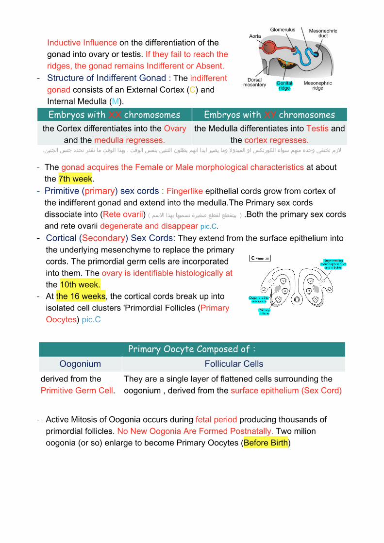
Inductive Influence on the differentiation of the gonad into ovary or testis. If they fail to reach the ridges, the gonad remains Indifferent or Absent.
- Structure of Indifferent Gonad : The indifferent gonad consists of an External Cortex (C) and Internal Medulla (M).
- The gonad acquires the Female or Male morphological characteristics at about the 7th week.
- Primitive (primary) sex cords : Fingerlike epithelial cords grow from cortex of the indifferent gonad and extend into the medulla.The Primary sex cords dissociate into (Rete ovarii) ( بيتقطع لقطع صغيرة نسميها بهذا االسم ) .Both the primary sex cords and rete ovarii degenerate and disappear pic.C.
- Cortical (Secondary) Sex Cords: They extend from the surface epithelium into the underlying mesenchyme to replace the primary cords. The primordial germ cells are incorporated into them. The ovary is identifiable histologically at the 10th week.
- At the 16 weeks, the cortical cords break up into isolated cell clusters 'Primordial Follicles (Primary Oocytes) pic.C
- Active Mitosis of Oogonia occurs during fetal period producing thousands of primordial follicles. No New Oogonia Are Formed Postnatally. Two milion oogonia (or so) enlarge to become Primary Oocytes (Before Birth)
Embryos with XX chromosomes Embryos with XY chromosomesthe Cortex differentiates into the Ovary
and the medulla regresses.the Medulla differentiates into Testis and
the cortex regresses.الزم تختفي وحده منهم سواء الكورتكس او الميدوال وما يصير ابدا انهم يظلون الثنتين بنفس الوقت ، بهذا الوقت ما نقدر نحدد جنس الجنين.
Primary Oocyte Composed of : Oogonium Follicular Cells
derived from the Primitive Germ Cell.
They are a single layer of flattened cells surrounding the oogonium , derived from the surface epithelium (Sex Cord)
DEVELOPMENT OF THE FEMALE DUCT SYSTEM
The Paramesonephric Ducts - They form most of the female genital tract. - Derivatives Of Paramesonephric Ducts :
- The endometrial stroma and myometrium are derived from the splanchinic mesoderm.
Postnatal Changes of the Ovary : 1- Surface Epithelium: Flattened into a single layer and separated from follicles in the cortex by a thin tunica albuginea.
2- The ovaries descend from the posterior abdominal wall into the pelvis, just inferior to the pelvic brim.
regress due to absence of the testosterone hormone. for development of male reproductive syste
develop due to absence of MIS (Müllerian Inhibiting Substance). for development of female reproductive system.
1- Uterine Tubes: 2. Uterovaginal Primordium:develop from the cranial
unfused parts of the ducts. It differentiates into:
Uterus (Body and Cervix) Superior Portion of Vagina.
تتبعو الكالم بالصفحة التالیة واربطوه بالصور
الصورة مكبرة باخر صفحة
Development of Lower Portion of Vagina
Differentiation of Vagina - The lining of the entire vagina is derived from the Vaginal Plate (urogenital
sinus). - The lumen of vagina is separated from the urogenital sinus by the Hymen which
remains as a thin fold of mucous membrane just within the vaginal orifice. Pic.F - Paramesonephric Ducts → Upper part of vagina - urogenital sinus → lower part of vagina
Paramesonephric Ducts develop lateral to the gonads and mesonephric ducts. Pic. ATheir funnel-shaped cranial ends open into the peritoneal cavity.
Forms FALLOPIAN
TUBES
They pass caudally parallel to mesonephric ducts to reach the future pelvic region. Pic. BThey Cross ventral to the mesonephric ducts & approach each other in the median plane and fuse to form the Y shaped Uterovaginal Primordial (which opens into the dorsal wall of the urogenital sinus and produces Paramesonephric (müllerian) Tubercle.
Pic. C
It is derived from the Urogenital Sinus Pic.CThe contact of the uterovaginal primordium with the urogenital sinus induces formation of SinoVaginal Bulbs.
The bulbs proliferate and fuse to form a solid Vaginal Plate. Pic.C&DThe central cells of the vaginal plate break down to form the lumen of the vagina. -
�
EXTERNAL GENITALIA (NEED HORMONE )
Development of Female External Genitalia In 7th weak in both (female & male)
- Proliferation of Mesenchyme at the Cranial end and Sides of the Cloacal Membrane, forms: 1. Genital Tubercle. 2. Urogenital Folds (Urethral Folds) 3. Labioscrotal Swellings (Genital Swellings)
Feminization of External Genitalia ( in 9th weak in female )
- Estrogen produced by both the placenta and the fetal ovaries has a role in feminization of the external genitalia.
In females, the genital tubercle becomes the clitoris, the genital swellings become the labia majora, and the genital folds become the labia minora. In males, the genital tubercle becomes the glans penis, the genital swellings fuse to become the scrotum, the genital folds elongate and fuse to form the shaft of the penis and the penile urethra, and the prostate forms in the wall of the urogenital sinus.
External GenitaliaAre Similar in both sexes up to the 7th week (indifferent stage).
Begin to differentiate in the 9th week
Fully differentiated by the 12th week.نقدر نحدد جنس الجنين
1-The Genital Tubercle 2- Urethral Folds 3-Labioscrotal Foldsproliferates to form the Primordial Phalls → The phalls elongates slightly to form the Clitoris.
do not fuse and form the Labia Minora.
form the Labia Majora , they fuse to form the Posterior& the Anterior Labial Commissures.
Female Sex Glands
Congenital AnomaliesVarious types of anomalies can result due to: 1. Arrest of development of the uterovaginal primordium during the 8th week. 2. Incomplete development of the paramesonephric ducts. 3. Incomplete fusion of the paramesonephric ducts. 4. Failure of parts of one or both paramesonephric ducts to develop. 5. Incomplete canalization.
1- Urethral & Paraurethral Glands:
2. Greater VestibularGglands (Bartholin glands)
grow as buds from the urethra, they are corresponding to the Prostate Gland of the male.
outgrowths of the urogenital sinus, they are corresponding to the Bulbourethral Glands of the male.
Vaginal Plate The lining of the entire vaginaUterus Didelphys Due to failure of fusion of inferior parts of the
paramesonephric ducts.Bicornuate uterus The duplication involves the superior segment.
Unicornuate Uterus: One paramesonephric duct fails to develop. Cervical Atresia incomplete development of the upper vagina or lower uterus.
Transversely septate vagina
Results from faulty canalization of the fused müllerian ducts.
Summary
1-The external genitalia begin to differentiate duringA. week 3 of development B. week 5 of development C. week 7 of development D. week 9 of development E. week 12 of development
2- The external genitalia are fully differentiated during A. week 3 of development B. week 5 of development C. week 7 of development D. week 12 of development E. week 20 of development
3- which of the following is a correct answer about Uterus Didelphys ? A. failure of fusion of inferior parts of the paramesonephric ducts. B. One paramesonephric duct fails to develop. C. Only associated with a double vagina D. Only associated with a singlevagina
4- The labia minora arise embryologically from which of the following structures? A. Phallus B. Labioscrotal swellings C. Sinovaginal bulbs D. Urogenital folds E. Paramesonephric duct
5- which one of the following duct will regress in the absence of testosterone ? A. paramesonephric duct B. mesonephric duct C. nephric duct
6- Phallus elongated to form which one of the following ? A. labia minora B. labia majora C. clitoris