differentiated from immunodeficiencies in which the non- specific mediators of innate immunity, such as phagocytes or complement, are impaired. Immunodeficiencies are conve- niently categorized by the type or the developmental stage of the cells involved. Figure 19-1 reviews the overall cellular de- velopment in the immune system, showing the locations of defects that give rise to primary immunodeficiencies. As Chapter 2 explained, the two main cell lineages important to immune function are lymphoid and myeloid. Most defects that lead to immunodeficiencies affect either one or the other. The lymphoid cell disorders may affect T cells, B cells, or, in combined immunodeficiencies, both B and T cells. The myeloid cell disorders affect phagocytic function. Most of the primary immunodeficiencies are inherited, and the precise molecular variations and the genetic defects that lead to many of these dysfunctions have been determined (Table 19-1 and Figure 19-2). In addition, there are immunodefi- ciencies that stem from developmental defects that impair proper function of an organ of the immune system. The consequences of primary immunodeficiency depend on the number and type of immune system components in- volved. Defects in components early in the hematopoietic de- velopmental scheme affect the entire immune system. In this category is reticular dysgenesis, a stem-cell defect that affects the maturation of all leukocytes; the resulting general failure of immunity leads to susceptibility to infection by a variety of microorganisms. Without aggressive treatment, the affected individual usually dies young from severe infection. In the chapter 19 ■ Primary Immunodeficiencies ■ AIDS and Other Acquired or Secondary Immunodeficiencies AIDS and Other Immunodeficiencies L - , immune system is subject to failure of some or all of its parts. This failure can have dire consequences. When the system loses its sense of self and begins to attack host cells and tissues, the result is autoimmunity, which is described in Chapter 20. When the system errs by failing to protect the host from disease-causing agents or from malig- nant cells, the result is immunodeficiency, which is the sub- ject of this chapter. A condition resulting from a genetic or developmental de- fect in the immune system is called a primary immunodefi- ciency. In such a condition, the defect is present at birth although it may not manifest itself until later in life. Sec- ondary immunodeficiency, or acquired immunodeficiency, is the loss of immune function and results from exposure to various agents. By far the most common secondary immun- odeficiency is acquired immunodeficiency syndrome, or AIDS, which results from infection with the human immun- odeficiency virus 1 (HIV-1). In the year 2000, AIDS killed ap- proximately 3 million persons, and HIV infection continues to spread to an estimated 15,000 persons per day. AIDS pa- tients, like other individuals with severe immunodeficiency, are at risk of infection with so-called opportunistic agents. These are microorganisms that healthy individuals can har- bor with no ill consequences but that cause disease in those with impaired immune function. The first part of this chapter describes the common pri- mary immunodeficiencies, examines progress in identifying the genetic defects that underlie these disorders, and consid- ers approaches to their treatment, including innovative uses of gene therapy. Animal models of primary immunodefi- ciency are also described. The rest of this chapter describes acquired immunodeficiency, with a strong focus on HIV in- fection, AIDS, and the current status of therapeutic and prevention strategies for combating this fatal acquired im- munodeficiency. Primary Immunodeficiencies A primary immunodeficiency may affect either adaptive or innate immune functions. Deficiencies involving compo- nents of adaptive immunity, such as T or B cells, are thus Nude Mouse (nu/nu)
Transcript
differentiated from immunodeficiencies in which the non-specific mediators of innate immunity, such as phagocytes orcomplement, are impaired. Immunodeficiencies are conve-niently categorized by the type or the developmental stage ofthe cells involved. Figure 19-1 reviews the overall cellular de-velopment in the immune system, showing the locations ofdefects that give rise to primary immunodeficiencies. AsChapter 2 explained, the two main cell lineages important toimmune function are lymphoid and myeloid. Most defectsthat lead to immunodeficiencies affect either one or theother. The lymphoid cell disorders may affect T cells, B cells,or, in combined immunodeficiencies, both B and T cells. Themyeloid cell disorders affect phagocytic function. Most of theprimary immunodeficiencies are inherited, and the precisemolecular variations and the genetic defects that lead tomany of these dysfunctions have been determined (Table 19-1 and Figure 19-2). In addition, there are immunodefi-ciencies that stem from developmental defects that impairproper function of an organ of the immune system.
The consequences of primary immunodeficiency dependon the number and type of immune system components in-volved. Defects in components early in the hematopoietic de-velopmental scheme affect the entire immune system. In thiscategory is reticular dysgenesis, a stem-cell defect that affectsthe maturation of all leukocytes; the resulting general failureof immunity leads to susceptibility to infection by a variety ofmicroorganisms. Without aggressive treatment, the affectedindividual usually dies young from severe infection. In the
chapter 19
■ Primary Immunodeficiencies
■ AIDS and Other Acquired or SecondaryImmunodeficiencies
AIDS and OtherImmunodeficiencies
L - ,
immune system is subject to failure of some or allof its parts. This failure can have dire consequences.
When the system loses its sense of self and begins to attackhost cells and tissues, the result is autoimmunity, which isdescribed in Chapter 20. When the system errs by failing toprotect the host from disease-causing agents or from malig-nant cells, the result is immunodeficiency, which is the sub-ject of this chapter.
A condition resulting from a genetic or developmental de-fect in the immune system is called a primary immunodefi-ciency. In such a condition, the defect is present at birthalthough it may not manifest itself until later in life. Sec-ondary immunodeficiency, or acquired immunodeficiency,is the loss of immune function and results from exposure tovarious agents. By far the most common secondary immun-odeficiency is acquired immunodeficiency syndrome, orAIDS, which results from infection with the human immun-odeficiency virus 1 (HIV-1). In the year 2000, AIDS killed ap-proximately 3 million persons, and HIV infection continuesto spread to an estimated 15,000 persons per day. AIDS pa-tients, like other individuals with severe immunodeficiency,are at risk of infection with so-called opportunistic agents.These are microorganisms that healthy individuals can har-bor with no ill consequences but that cause disease in thosewith impaired immune function.
The first part of this chapter describes the common pri-mary immunodeficiencies, examines progress in identifyingthe genetic defects that underlie these disorders, and consid-ers approaches to their treatment, including innovative usesof gene therapy. Animal models of primary immunodefi-ciency are also described. The rest of this chapter describesacquired immunodeficiency, with a strong focus on HIV in-fection, AIDS, and the current status of therapeutic and prevention strategies for combating this fatal acquired im-munodeficiency.
Primary ImmunodeficienciesA primary immunodeficiency may affect either adaptive orinnate immune functions. Deficiencies involving compo-nents of adaptive immunity, such as T or B cells, are thus
Nude Mouse (nu/nu)
more restricted case of defective phagocytic function, themajor consequence is susceptibility to bacterial infection.Defects in more highly differentiated compartments of theimmune system have consequences that are more specific
432 P A R T I V The Immune System in Health and Disease
and usually less severe. For example, an individual with selec-tive IgA deficiency may enjoy a full life span, troubled only bya greater than normal susceptibility to infections of the respi-ratory and genitourinary tracts.
V I S U A L I Z I N G C O N C E P T S
FIGURE 19-1 Congenital defects that interrupt hematopoiesisor impair functioning of immune-system cells result in variousimmunodeficiency diseases. (Orange boxes � phagocytic defi-
ciencies, green � humoral deficiencies, red � cell-mediated defi-ciencies, and purple � combined immunodeficiencies, defectsthat affect more than one cell lineage.)
Neutrophil
Plasma cell
MatureB cell
Monocyte
Stem cell
Lymphoidprogenitorcell
Pre–B cell Pre–T cell
Memory B cell
Mature T cell
Myeloidprogenitorcell
Thymus
Severecombinedimmuno-deficiency
X-linkedagammaglobulinemia
Reticular dysgenesis
Severe combinedimmunodeficiency(SCID)
Congenitalagranulocytosis
Leukocyte-adhesiondeficiency
Bare-lymphocytesyndrome
Selective immunoglobulin deficiency
Common variablehypogammaglobulinemia
X-linkedhyper-IgM syndrome
DiGeorgesyndrome
Chronicgranulomatousdisease
Wiskott-Aldrichsyndrome
Lymphoid Immunodeficiencies May Involve B Cells, T Cells, or BothThe combined forms of lymphoid immunodeficiency affectboth lineages and are generally lethal within the first fewyears of life; these arise from defects early in developmentalpathways. They are less common than conditions, usually lesssevere, that result from defects in more highly differentiatedlymphoid cells.
B-cell immunodeficiency disorders make up a diversespectrum of diseases ranging from the complete absence ofmature recirculating B cells, plasma cells, and immuno-globulin to the selective absence of only certain classes of
AIDS and Other Immunodeficiencies C H A P T E R 19 433
immunoglobulins. Patients with these disorders usually aresubject to recurrent bacterial infections but display normalimmunity to most viral and fungal infections, because the T-cell branch of the immune system is largely unaffected. Mostcommon in patients with humoral immunodeficiencies areinfections by such encapsulated bacteria as staphylococci,streptococci, and pneumococci, because antibody is criticalfor the opsonization and clearance of these organisms.
Because of the central role of T cells in the immune sys-tem, a T-cell deficiency can affect both the humoral and thecell-mediated responses. The impact on the cell-mediatedsystem can be severe, with a reduction in both delayed-typehypersensitive responses and cell-mediated cytotoxicity.
TABLE 19-1 Some primary human immunodeficiency diseases and underlying genetic defects
Immunodeficiency Inheritance Chromosomaldisease Specific defect Impaired function mode* defect
Severe combined RAG-1/RAG-2 deficiency No TCR or Ig gene AR 11p13immunodeficiency rearrangement(SCID)
ADA deficiency Toxic metabolite in T AR 20q13PNP deficiency and B cells AR 14q13
JAK-3 deficiency Defective signals from AR 19p13IL-2R�-deficiency IL-2, 4, 7, 9, 15, XL Xq13
ZAP-70 deficiency Defective signal from AR 2q12TCR
Bare lymphocyte Defect in MHC class II No class II MHC AR 16p13syndrome gene promoter molecules
Wiskott-Aldrich Cytoskeletal protein (CD43) Defective T cells and XL Xp11syndrome (WAS) platelets
Interferon gamma IFN-�–receptor defect Impaired immunity to AR 6q23receptor mycobacteria
DiGeorge syndrome Thymic aplasia T- and B-cell development AD 22q11
Ataxia telangiectasia Defective cell-cycle kinase Low IgA, IgE AR 11q22
Gammaglobulinemias X-linked Bruton’s tyrosine kinase XL Xq21agammaglobulinemia (Btk); no mature B cells
Common variable Low IgG, IgA; variable Compleximmunodeficiency IgM
Selective IgA deficiency Low or no IgA Complex
Chronic granulomatous Cyt p91phox XL Xp21disease Cyt p67phox No oxidative burst AR 1q25
Cyt p22phox for bacterial killing AR 16q24
Chediak-Higashi syndrome Defective intracellular Inability to lyse bacteria AR 1q42transport protein (LYST)
Leukocyte-adhesion defect Defective integrin �2 Leukocyte extravasation AR 21q22(CD18)
*AR � autosomal recessive; AD � autosomal dominant; XL � X linked; “Complex” indicates conditions for which precise
genetic data are not available and that may involve several interacting loci.
}
} }
}
} }
Immunoglobulin deficiencies are associated primarily withrecurrent infections by extracellular bacteria, but those af-fected have normal responses to intracellular bacteria, as wellas viral and fungal infections. By contrast, defects in the cell-mediated system are associated with increased suscepti-bility to viral, protozoan, and fungal infections. Intracellularpathogens such as Candida albicans, Pneumocystis carinii,and Mycobacteria are often implicated, reflecting the impor-tance of T cells in eliminating intracellular pathogens. Infec-tions with viruses that are rarely pathogenic for the normalindividual (such as cytomegalovirus or even an attenuatedmeasles vaccine) may be life threatening for those with im-paired cell-mediated immunity. Defects that cause decreasedT-cell counts generally also affect the humoral system, be-cause of the requirement for TH cells in B-cell activation. Gen-erally there is some decrease in antibody levels, particularly inthe production of specific antibody after immunization.
As one might expect, combined deficiencies of the humoraland cell-mediated branches are the most serious of the im-munodeficiency disorders. The onset of infections begins earlyin infancy, and the prognosis for these infants is early death un-less therapeutic intervention reconstitutes their defective im-mune system.As described below, there are increasing numbersof options for the treatment of immunodeficiencies.
The immunodeficiencies that affect lymphoid functionhave in common the inability to mount or sustain a complete
immune response against specific agents. A variety of failurescan lead to such immunodeficiency. Defective intercellularcommunication may be rooted in deleterious mutations ofgenes that encode cell-surface receptors or signal-transductionmolecules; defects in the mechanisms of gene rearrangementand other functions may prevent normal B- or T-cell re-sponses. Figure 19-3 is an overview of the molecules involvedin the more well-described interactions among T cells and B cells that give rise to specific responses, with a focus on pro-teins in which defects leading to immunodeficiency havebeen identified.
SEVERE COMBINED IMMUNODEFICIENCY (SCID)
The family of disorders termed SCID stems from defects inlymphoid development that affect either T cells or both Tand B cells. All forms of SCID have common features despitedifferences in the underlying genetic defects. Clinically, SCIDis characterized by a very low number of circulating lympho-cytes. There is a failure to mount immune responses medi-ated by T cells. The thymus does not develop, and the fewcirculating T cells in the SCID patient do not respond tostimulation by mitogens, indicating that they cannot prolif-erate in response to antigens. Myeloid and erythroid (red-blood-cell precursors) cells appear normal in number andfunction, indicating that only lymphoid cells are depleted inSCID.
SCID results in severe recurrent infections and is usuallyfatal in the early years of life. Although both the T and B lin-eages may be affected, the initial manifestation of SCID in in-fants is almost always infection by agents, such as fungi orviruses, that are normally dealt with by T-cell immunity. TheB-cell defect is not evident in the first few months of the af-fected infant’s life because antibodies are passively obtainedfrom transplacental circulation or from mother’s milk. SCIDinfants suffer from chronic diarrhea, pneumonia, and skin,mouth, and throat lesions as well as a host of other oppor-tunistic infections. The immune system is so compromisedthat even live attenuated vaccines (such as the Sabin poliovaccine) can cause infection and disease. The life span of aSCID patient can be prolonged by preventing contact with allpotentially harmful microorganisms, for example by con-finement in a sterile atmosphere. However, extraordinary ef-fort is required to prevent direct contact with other personsand with unfiltered air; any object, including food, thatcomes in contact with the sequestered SCID patient mustfirst be sterilized. Such isolation is feasible only as a tempo-rary measure, pending treatment.
The search for defects that underlie SCID has revealedseveral different causes for this general failure of immunity. Asurvey of 141 patients by Rebecca Buckley indicated that themost common cause (64 cases) was deficiency of the com-mon gamma chain of the IL-2 receptor (IL-2R�; see Figure12-7). Defects in this chain impede signaling throughreceptors for IL-4, -7, -9, and -15 as well as the IL-2 receptor,because the chain is present in receptors for all of these cy-tokines. Deficiency in the kinase JAK-3, which has a similar
434 P A R T I V The Immune System in Health and Disease
FIGURE 19-2 Several X-linked immunodeficiency diseases resultfrom defects in loci on the X chromosome. [Data from the Natl. Cen-ter for Biotechnology Information Web site.]
phenotype because the IL receptors signal through this mol-ecule, accounted for 9 of the cases (see Figure 12-10). A raredefect found in only 2 of the patients involved the IL-7 recep-tor; these patients have impaired T and B cells but normalNK cells. Another common defect is the adenosine deami-nase or ADA deficiency found in 22 patients. Adenosinedeaminase catalyzes conversion of adenosine to inosine, andits deficiency results in accumulation of adenosine, which in-terferes with purine metabolism and DNA synthesis. The remaining cases included single instances of reticular dysge-nesis and cartilage hair dysplasia or were classified as autoso-mal recessive defects not related to known IL-2R� or JAK-3mutations. Thirteen of the 141 cases were of unknown ori-gin, with no apparent genetic defect or family history of im-munodeficiency.
There are other known defects that give rise to SCID. Thereis a defect characterized by depletion of CD8� T cells that in-volves the tyrosine kinase ZAP-70, an important element in T-cell signal transduction (see Figures 10-11 and 10-12). In-fants with defects in ZAP-70 may have normal levels of im-munoglobulin and CD4� lymphocytes, but their CD4� Tcells are nonfunctional. A deficiency in the enzyme purine
nucleoside phosphorylase (PNP) causes immunodeficiencyby a mechanism similar to the ADA defect. As described inChapters 5 and 9, both immunoglobulin and T-cell receptorgenes undergo rearrangement to express the active forms ofthese molecules. A defect in the genes that encode mediatorsof the rearrangement processes (recombination-activatingproteins RAG-1 and RAG-2) precludes development of B andT cells with functional receptors and leads to SCID.
A defect leading to general failure of immunity similar toSCID is failure to transcribe the genes that encode class IIMHC molecules. Without these molecules, the patient’s lym-phocytes cannot participate in cellular interactions with Thelper cells. This type of immunodeficiency is also called thebare-lymphocyte syndrome. Molecular studies of a class IIMHC deficiency revealed a defective interaction between a 5�promoter sequence of the gene for the class II MHC moleculeand a DNA-binding protein necessary for gene transcription.Other patients with SCID-like symptoms lack class I MHCmolecules. This rare variant of immunodeficiency was ascribed to mutation in the TAP genes that are vital to anti-gen processing by class I MHC molecules (see Clinical FocusChapter 8). This defect causes a deficit in CD8-mediated
AIDS and Other Immunodeficiencies C H A P T E R 19 435
FIGURE 19-3 Defects in cell interaction and signaling can lead tosevere immunodeficiency. The interaction of T cell and B cell isshown here with a number of the components important to the intra-and extracellular signaling pathways. A number of primary immuno-deficiencies are rooted in defects in these interactions. SCID may re-sult from defects in (1) the recombination-activating genes (RAG-1and -2) required for synthesis of the functional immunoglobulins andT-cell receptors that characterize mature B and T cells; (2) the � chain
of receptors for IL-2, 4, 7, 9, and 15 (IL-R�); (3) JAK-3, which trans-duces signals from the gamma chain of the cytokine receptor; or (4)expression of the class II MHC molecule (bare lymphocyte syn-drome). XLA results from defective transduction of activating signalsfrom the cell-surface IgM by Bruton’s tyrosine kinase (Btk). XHM re-sults from defects in CD40L that preclude normal maturation of Bcells. [Adapted from B. A. Smart and H. D. Ochs, 1997, Curr. Opin. Pediatr. 9:570.]
IL-2, IL-4, IL-7, IL-9,IL-15
IL-Rγ IL-Rγ
Ag
IgM
Ig
B7CD28
CD4
Class II MHC
CD40L CD40
TCR
T cell B cell
Btk
RAG-1/2RAG-1/2
JAK-3
Deficiencyin JAK-3pathway
Defect inCD40L(XHM)
Defect inBruton'styrosinekinase (XLA)
Defect inrecombination-activating genes(RAG-1/2)
Defectiveexpressionof Class II MHC(bare lymphocytesyndrome)
Defect in γ chainof receptors forIL-2, 4, 7, 9, 15
immunity, characterized by susceptibility to viral infection. Arecent case of SCID uncovered a defect in the gene for thecell-surface phosphatase CD45. Interestingly, this defectcaused lack of �� T-cells but spared the �� lineage.
WISKOTT-ALDRICH SYNDROME (WAS)
The severity of this X-linked disorder increases with age andusually results in fatal infection or lymphoid malignancy. Ini-tially, T and B lymphocytes are present in normal numbers.WAS first manifests itself by defective responses to bacterialpolysaccharides and by lower-than-average IgM levels. Otherresponses and effector mechanisms are normal in the earlystages of the syndrome. As the WAS sufferer ages, there are re-current bacterial infections and a gradual loss of humoral andcellular responses. The syndrome includes thrombocytopenia(lowered platelet count; the existing platelets are smaller thanusual and have a short half-life), which may lead to fatal bleed-ing. Eczema (skin rashes) in varying degrees of severity mayalso occur, usually beginning around one year of age. The de-fect in WAS has been mapped to the short arm of the X chro-mosome (see Table 19-1 and Figure 19-2) and involves acytoskeletal glycoprotein present in lymphoid cells calledsialophorin (CD43). The WAS protein is required for assemblyof actin filaments required for the formation of microvesicles.
INTERFERON-GAMMA–RECEPTOR DEFECT
A recently described immunodeficiency that falls into themixed-cell category involves a defect in the receptor for in-terferon gamma (IFN-�, see Chapter 12). This deficiency wasfound in patients suffering from infection with atypical my-cobacteria (intracellular organisms related to the bacteriathat cause tuberculosis and leprosy). Most of those carryingthis autosomal recessive trait are from families with a historyof inbreeding. The susceptibility to infection with mycobac-teria is selective in that those who survive these infections arenot unusually susceptible to other agents, including other in-tracellular bacteria. This immunodeficiency points to a spe-cific role for IFN-� and its receptor in protection frominfection with mycobacteria.
Whereas SCID and the related combined immunodefi-ciencies affect T cells or all lymphoid cells, other primary im-munodeficiencies affect B-cell function and result in thereduction or absence of some or all classes of immunoglobu-lins. While the underlying defects have been identified forsome of these, little information exists concerning the exactcause of some of the more common deficiencies, such as com-mon variable immunodeficiency and selective IgA deficiency.
X-LINKED AGAMMAGLOBULINEMIA
A B-cell defect called X-linked agammaglobulinemia (XLA)or Bruton’s hypogammaglobulinemia is characterized by ex-tremely low IgG levels and by the absence of other im-munoglobulin classes. Individuals with XLA have noperipheral B cells and suffer from recurrent bacterial infec-tions, beginning at about nine months of age. A palliative
treatment for this condition is periodic administration ofimmunoglobulin, but patients seldom survive past theirteens. There is a defect in B-cell signal transduction in thisdisorder, due to a defect in a transduction molecule calledBruton’s tyrosine kinase (Btk), after the investigator who de-scribed the syndrome. B cells in the XLA patient remain inthe pre-B stage with H chains rearranged but L chains in theirgerm-line configuration. (The Clinical Focus in Chapter 11describes the discovery of this immunodeficiency and its un-derlying defect in detail.)
X-LINKED HYPER-IgM SYNDROME
A peculiar immunoglobulin deficiency first thought to resultfrom a B-cell defect has recently been shown to result insteadfrom a defect in a T-cell surface molecule. X-linked hyper-IgM (XHM) syndrome is characterized by a deficiency ofIgG, IgA, and IgE, and elevated levels of IgM, sometimes ashigh as 10 mg/ml (normal IgM concentration is 1.5 mg/ml).Although individuals with XHM have normal numbers of Bcells expressing membrane-bound IgM or IgD, they appearto lack B cells expressing membrane-bound IgG, IgA, or IgE.XHM syndrome is generally inherited as an X-linked reces-sive disorder (see Figure 19-2), but some forms appear to beacquired and affect both men and women. Affected individ-uals have high counts of IgM-secreting plasma cells in theirperipheral blood and lymphoid tissue. In addition, XHM pa-tients often have high levels of autoantibodies to neutrophils,platelets, and red blood cells. Children with XHM suffer re-current infections, especially respiratory infections; these aremore severe than expected for a deficiency characterized bylow levels of immunoglobulins.
The defect in XHM is in the gene encoding the CD40 lig-and (CD40L), which maps to the X chromosome. TH cellsfrom patients with XHM fail to express functional CD40L ontheir membrane. Since an interaction between CD40 on theB cell and CD40L on the TH cell is required for B-cell activa-tion, the absence of this co-stimulatory signal inhibits the B-cell response to T-dependent antigens (see Figures 19-3 and11-10). The B-cell response to T-independent antigens, how-ever, is unaffected by this defect, accounting for the produc-tion of IgM antibodies. As described in Chapter 11, classswitching and formation of memory B cells both requirecontact with TH cells by a CD40–CD40L interaction. The ab-sence of this interaction in XHM results in the loss of classswitching to IgG, IgA, or IgE isotypes and in a failure to pro-duce memory B cells. In addition, XHM individuals fail toproduce germinal centers during a humoral response, whichhighlights the role of the CD40–CD40L interaction in thegeneration of germinal centers.
COMMON VARIABLE IMMUNODEFICIENCY (CVI)
CVI is characterized by a profound decrease in numbers ofantibody-producing plasma cells, low levels of most im-munoglobulin isotypes (hypogammaglobulinemia), and re-current infections. The condition is usually manifested later
436 P A R T I V The Immune System in Health and Disease
in life than other deficiencies and is sometimes called late-onset hypogammaglobulinemia or, incorrectly, acquired hypogammaglobulinemia. However, CVI has a genetic component and is considered a primary immunodeficiency,although the exact pattern of inheritance is not known. Be-cause the manifestations are very similar to those of acquiredhypogammaglobulinemia, there is some confusion betweenthe two forms (see below). Infections in CVI sufferers aremost frequently bacterial and can be controlled by adminis-tration of immunoglobulin. In CVI patients, B cells fail tomature into plasma cells; however in vitro studies show thatCVI B cells are capable of maturing in response to appropri-ate differentiation signals. The underlying defect in CVI isnot known, but must involve either an in vivo blockage of thematuration of B cells to the plasma-cell stage or their inabil-ity to produce the secreted form of immunoglobulins.
HYPER-IgE SYNDROME (JOB SYNDROME)
A primary immunodeficiency characterized by skin abcesses,recurrent pneumonia, eczema, and elevated levels of IgE ac-companies facial abnormalities and bone fragility. Thismulti-system disorder is autosomal dominant and has vari-able expressivity. The gene for hyper IgE syndrome, or HIES,maps to chromosome 4. HIES immunologic signs include re-current infection and eosinophilia in addition to elevated IgElevels.
SELECTIVE DEFICIENCIES OF IMMUNOGLOBULIN CLASSES
A number of immunodeficiency states are characterized bysignificantly lowered amounts of specific immunoglobulinisotypes. Of these, IgA deficiency is by far the most common.There are family-association data showing that IgA defi-ciency prevails in the same families as CVI, suggesting a rela-tionship between these conditions. The spectrum of clinicalsymptoms of IgA deficiency is broad; many of those affectedare asymptomatic, while others suffer from an assortment ofserious problems. Recurrent respiratory and genitourinarytract infections resulting from lack of secreted IgA on mu-cosal surfaces are common. In addition, problems such as in-testinal malabsorption, allergic disease, and autoimmunedisorders may also be associated with low IgA levels. The rea-sons for this variability in the clinical profile of IgA deficiencyare not clear but may relate to the ability of some, but not all,patients to substitute IgM for IgA as a mucosal antibody. Thedefect in IgA deficiency is related to the inability of IgA B cellsto undergo normal differentiation to the plasma-cell stage.IgG2 and IgG4 may also be deficient in IgA-deficient pa-tients. No causative defect in IgA genes has been identified,and the surface IgA molecules on these patients’ B cells ap-pear to be expressed normally. A gene outside of the im-munoglobulin gene complex is suspected to be responsiblefor this fairly common syndrome.
Other immunoglobulin deficiencies have been reported,but these are rarer. An IgM deficiency has been identified asan autosomal recessive trait. Victims of this condition are
subject to severe infection by agents such as meningococcus,which causes fatal disease. IgM deficiency may be accompa-nied by various malignancies or by autoimmune disease. IgGdeficiencies are also rare. These are often not noticed untiladulthood and can be effectively treated by administration ofimmunoglobulin.
ATAXIA TELANGIECTASIA
Although not classified primarily as an immunodeficiency,ataxia telangiectasia is a disease syndrome that includes defi-ciency of IgA and sometimes of IgE. The syndrome is charac-terized by difficulty in maintaining balance (ataxia) and bythe appearance of broken capillaries (telangiectasia) in theeyes. The primary defect appears to be in a kinase involved inregulation of the cell cycle. The relationship between the im-mune deficiency and the other defects in ataxia telangiectasiaremains obscure.
IMMUNE DISORDERS INVOLVING THE THYMUS
Several immunodeficiency syndromes are grounded in fail-ure of the thymus to undergo normal development. Thymicmalfunction has a profound effect on T-cell function; allpopulations of T cells, including helper, cytolytic, and regula-tory varieties, are affected. Immunity to viruses and fungi is especially compromised in those suffering from these conditions.
DiGeorge syndrome, or congenital thymic aplasia, in itsmost severe form is the complete absence of a thymus. Thisdevelopmental defect, which is associated with the dele-tion in the embryo of a region on chromosome 22, causesimmunodeficiency along with characteristic facial abnor-malities, hypoparathyroidism, and congenital heart disease(Figure 19-4). The stage at which the causative developmen-tal defect occurs has been determined, and the syndrome issometimes called the third and fourth pharyngeal pouch syn-drome to reflect its precise embryonic origin. The immunedefect includes a profound depression of T-cell numbers andabsence of T-cell responses. Although B cells are present innormal numbers, affected individuals do not produce anti-body in response to immunization with specific antigens.Thymic transplantation is of some value for correcting the T-cell defects, but many DiGeorge patients have such severeheart disease that their chances for long-term survival arepoor, even if the immune defects are corrected.
Whereas the DiGeorge syndrome results from an in-trauterine or developmental anomaly, thymic hypoplasia, orthe Nezelof syndrome, is an inherited disorder. The mode ofinheritance for this rare disease is not known and its presen-tation varies, making it somewhat difficult to diagnose. Asthe name implies, thymic hypoplasia is a defect in which avestigial thymus is unable to serve its function in T-cell de-velopment. In some patients, B cells are normal, whereas inothers a B-cell deficiency is secondary to the T-cell defect. Af-fected individuals suffer from chronic diarrhea, viral andfungal infections, and a general failure to thrive.
AIDS and Other Immunodeficiencies C H A P T E R 19 437
Immunodeficiencies of the Myeloid Lineage Affect Innate ImmunityImmunodeficiencies of the lymphoid lineage affect adaptiveimmunity. By contrast, defects in the myeloid cell lineage af-fect the innate immune functions (see Figure 19-1). Most ofthese defects result in impaired phagocytic processes that aremanifested by recurrent microbial infection of greater orlesser severity. There are several stages at which the phago-cytic processes may be faulty; these include cell motility, ad-herence to and phagocytosis of organisms, and killing bymacrophages.
REDUCTION IN NEUTROPHIL COUNT
As described in Chapter 2, neutrophils are circulating granu-locytes with phagocytic function. Quantitative deficiencies inneutrophils can range from an almost complete absence ofcells, called agranulocytosis, to a reduction in the concentra-tion of peripheral blood neutrophils below 1500/mm3, calledgranulocytopenia or neutropenia. These quantitative defi-ciencies may result from congenital defects or may be ac-quired through extrinsic factors. Acquired neutropenias aremuch more common than congenital ones.
Congenital neutropenia is often due to a genetic defectthat affects the myeloid progenitor stem cell; it results in re-duced production of neutrophils during hematopoiesis. Incongenital agranulocytosis, myeloid stem cells are present
in the bone marrow but rarely differentiate beyond thepromyelocyte stage. As a result, children born with this con-dition show severe neutropenia, with counts of less than 200neutrophils/mm3. These children suffer from frequent bacte-rial infections beginning as early as the first month of life;normal infants are protected at this age by maternal antibodyas well as by innate immune mechanisms, including neu-trophils. Experimental evidence suggests that this genetic defect results in decreased production of granulocyte colony-stimulating factor (G-CSF) and thus in a failure of themyeloid stem cell to differentiate along the granulocytic lineage (see Figure 2-1).
Neutrophils have a short life span, and their precursorsmust divide rapidly in the bone marrow to maintain levels ofthese cells in the circulation. For this reason, agents such asradiation and certain drugs (e.g., chemotherapeutic drugs)that specifically damage rapidly dividing cells are likely tocause neutropenia. Occasionally, neutropenia develops insuch autoimmune diseases as Sjögren’s syndrome or systemiclupus erythematosus; in these conditions, autoantibodies de-stroy the neutrophils. Transient neutropenia often developsafter certain bacterial or viral infections, but neutrophilcounts return to normal as the infection is cleared.
CHRONIC GRANULOMATOUS DISEASE (CGD)
CGD is a genetic disease that has at least two distinct forms:an X-linked form that occurs in about 70% of patients and anautosomal recessive form found in the rest. This disease isrooted in a defect in the oxidative pathway by which phago-cytes generate hydrogen peroxide and the resulting reactiveproducts, such as hypochlorous acid, that kill phagocytosedbacteria. CGD sufferers undergo excessive inflammatory reactions that result in gingivitis, swollen lymph nodes,and nonmalignant granulomas (lumpy subcutaneous cellmasses); they are also susceptible to bacterial and fungal in-fection. CGD patients are not subject to infection by thosebacteria, such as pneumococcus, that generate their own hy-drogen peroxide. In this case, the myeloperoxidase in the hostcell can use the bacterial hydrogen peroxide to generateenough hypochlorous acid to thwart infection. Several re-lated defects may lead to CGD; these include a missing or de-fective cytochrome (cyt b558) that functions in an oxidativepathway and defects in proteins (phagocyte oxidases, orphox) that stabilize the cytochrome. In addition to the gen-eral defect in the killer function of phagocytes, there is also adecrease in the ability of mononuclear cells to serve as APCs.Both processing and presentation of antigen are impaired.Increased amounts of antigen are required to trigger T-cellhelp when mononuclear cells from CGD patients are used asAPCs.
The addition of IFN-� has been shown to restore functionto CGD granulocytes and monocytes in vitro. This observa-tion prompted clinical trials of IFN-� for CGD patients. En-couraging increases in oxidative function and restoration of cytoplasmic cytochrome have been reported in these
438 P A R T I V The Immune System in Health and Disease
FIGURE 19-4 A child with DiGeorge syndrome showing character-istic dysplasia of ears and mouth and abnormally long distance be-tween the eyes. [R. Kretschmer et al., 1968, New Engl. J. Med. 279:1295;photograph courtesy of F. S. Rosen.]
patients. In addition, knowledge of the precise gene defectsunderlying CGD makes it a candidate for gene therapy, andreplacement of the defective cytochrome has had promisingresults (see below).
CHEDIAK-HIGASHI SYNDROME
This autosomal recessive disease is characterized by recurrentbacterial infections, partial oculo-cutaneous albinism (lackof skin and eye pigment), and aggressive but nonmalignantinfiltration of organs by lymphoid cells. Phagocytes from pa-tients with this immune defect contain giant granules but donot have the ability to kill bacteria. The molecular basis of thedefect is a mutation in a protein (LYST) involved in the regu-lation of intracellular trafficking. The mutation impairs thetargeting of proteins to secretory lysosomes, which makesthem unable to lyse bacteria.
LEUKOCYTE ADHESION DEFICIENCY (LAD)
As described in Chapter 15, cell-surface molecules belongingto the integrin family of proteins function as adhesion mole-cules and are required to facilitate cellular interaction. Threeof these, LFA-1, Mac-1, and gp150/95 (CD11a, b, and c, re-spectively) have a common � chain (CD18) and are variablypresent on different monocytic cells; CD11a is also expressedon B cells (Table 19-2). An immunodeficiency related to dysfunction of the adhesion molecules is rooted in a defect
localized to the common � chain and affects expression of allthree of the molecules that use this chain. This defect, calledleukocyte adhesion deficiency (LAD), causes susceptibility toinfection with both gram-positive and gram-negative bacte-ria as well as various fungi. Impairment of adhesion of leuko-cytes to vascular endothelium limits recruitment of cells tosites of inflammation.Viral immunity is somewhat impaired,as would be predicted from the defective T-B cell cooperationarising from the adhesion defect. LAD varies in its severity;some affected individuals die within a few years, others sur-vive into their forties. The reason for the variable disease phe-notype in this disorder is not known. LAD is the subject of aClinical Focus in Chapter 15.
Complement Defects Result in Immunodeficiency or Immune-Complex Disease
Immunodeficiency diseases resulting from defects in thecomplement system are described in Chapter 13. Many com-plement deficiencies are associated with increased suscepti-bility to bacterial infections and/or immune-complexdiseases. One of these complement disorders, a deficiency inproperdin, which stabilizes the C3 convertase in the alterna-tive complement pathway, is caused by a defect in a gene lo-cated on the X chromosome (see Figure 19-2).
AIDS and Other Immunodeficiencies C H A P T E R 19 439
TABLE 19-2 Properties of integrin molecules that are absent in leukocyte-adhesion deficiency
Functions inhibited with monoclonal Extravasation Opsonization Granulocyte adherence antibody CTL killing Granulocyte adherence, and aggregation
T-B conjugate formation aggregation, and ADCC chemotaxis ADCC
*CR3 � type 3 complement receptor, also known as Mac-1; CR4 � type 4 complement receptor, also known as gp150/95;
LFA-1, CR3, and CR4 are heterodimers containing a common � chain but different � chains designated L, M, and X, respectively.
Immunodeficiency Disorders Are Treated by Replacement of the Defective ElementAlthough there are no cures for immunodeficiency disor-ders, there are several treatment possibilities. In addition tothe drastic option of total isolation from exposure to any mi-crobial agent, treatment options for the immunodeficienciesinclude:
■ replacement of a missing protein
■ replacement of a missing cell type or lineage
■ replacement of a missing or defective gene
For disorders that impair antibody production, the classiccourse of treatment is administration of the missing proteinimmunoglobulin. Pooled human gamma globulin given ei-ther intravenously or subcutaneously protects against recur-rent infection in many types of immunodeficiency. Main-tenance of reasonably high levels of serum immunoglobulin(5 mg/ml serum) will prevent most common infections inthe agammaglobulinemic patient. This is generally accom-plished by the administration of immunoglobulin that hasbeen selected for antibodies directed against a particular or-ganism. Recent advances in the preparation of human mon-oclonal antibodies and in the ability to genetically engineerchimeric antibodies with mouse V regions and human-derived C regions make it possible to prepare antibodies spe-cific for important pathogens (see Chapter 5).
Advances in molecular biology make it possible to clonethe genes that encode other immunologically important pro-teins, such as cytokines, and to express these genes in vitro,using bacterial or eukaryotic expression systems. The avail-ability of such proteins allows new modes of therapy inwhich immunologically important proteins may be replacedor their concentrations increased in the patient. For example,the administration of recombinant IFN-� has proven effec-tive for patients with CGD, and the use of recombinant IL-2may help to restore immune function in AIDS patients. Re-combinant adenosine deaminase has been successfully ad-ministered to ADA deficient SCID patients.
Cell replacement as therapy for immunodeficiencies hasbeen made possible by recent progress in bone-marrow trans-plantation (see Chapter 21). Replacement of stem cells withthose from an immunocompetent donor allows developmentof a functional immune system (see Clinical Focus Chapter2). High rates of success have been reported for those who arefortunate enough to have an HLA-identical donor. Carefulmatching of patients with donors and the ability to manipu-late stem-cell populations to select CD34� precursor cellscontinues to minimize the risk in this procedure, even whenno ideal donor exists. These procedures have been highly suc-cessful with SCID infants when haploidentical (completematch of one HLA gene set or haplotype) donor marrow isused. T cells are depleted and CD34� stem cells are enrichedbefore introducing the donor bone marrow into the SCID in-fant. Because this therapy has been used only in recent years,
it is not known whether transplantation cures the immuno-deficiency permanently. A variation of bone-marrow trans-plantation is the injection of paternal CD34� cells in uterowhen the birth of an infant with SCID is expected. Two in-fants born after this procedure had normal T-cell functionand did not develop the infections that characterize SCID.
If a single gene defect has been identified, as in adenosinedeaminase deficiency or chronic granulomatous disease, re-placement of the defective gene may be a treatment option.Clinical tests of such therapy are underway for SCID causedby ADA deficiency and for chronic granulomatous diseasewith defective p67phox, with promising initial results. Diseaseremission for up to 18 months was seen in the SCID patientsand up to 6 months in the CGD patients. A similar procedurewas used in both trials. It begins with obtaining cells (CD34�
stem cells are usually selected for these procedures) from thepatient and transfecting them with a normal copy of the de-fective gene. The transfected cells are then returned to the pa-tient. As this treatment improves, it will become applicable toa number of immunodeficiencies for which a genetic defectis well defined. As mentioned above, these include defects in genes that encode the � chain of the IL-2 receptor, JAK-3,and ZAP-70, all of which give rise to SCID.
Experimental Models of ImmunodeficiencyInclude Genetically Altered AnimalsImmunologists use two well-studied animal models of pri-mary immunodeficiency for a variety of experimental pur-poses. One of these is the athymic, or nude, mouse; the otheris the severe combined immunodeficiency, or SCID, mouse.
NUDE (ATHYMIC) MICE
A genetic trait designated nu, which is controlled by a reces-sive gene on chromosome 11, was discovered in certain mice.Mice homozygous for this trait (nu/nu) are hairless and havea vestigial thymus (Figure 19-5). Heterozygotic, nu/�, littermates have hair and a normal thymus. It is not knownwhether the hairlessness and the thymus defect are caused bythe same gene. It is possible that two very closely linked genescontrol these defects, which, although unrelated, appear to-gether in this mutant mouse. A gene that controls develop-ment may be involved, since the pathway that leads to thedifferential development of the thymus is related to the onethat controls the skin epithelial cells. The nu/nu mouse can-not easily survive; under normal conditions, the mortality is100% within 25 weeks and 50% die within the first two weeksafter birth. Therefore, when these animals are to be used forexperimental purposes, they must be maintained under con-ditions that protect them from infection. Precautions includeuse of sterilized food, water, cages, and bedding. The cagesare protected from dust by placing them in a laminar flowrack or by the use of air filters fitted over the individual cages.
Nude mice lack cell-mediated immune responses, andthey are unable to make antibodies to most antigens. The
440 P A R T I V The Immune System in Health and Disease
immunodeficiency in the nude mouse can be reversed by athymic transplant. Because they can permanently tolerateboth allografts and xenografts, they have a number of practi-cal experimental uses. For example, hybridomas or solid tu-mors from any origin may be grown as ascites or asimplanted tumors in a nude mouse. It is known that the nudemouse does not completely lack T cells; rather, it has a lim-ited population that increases with age. The source of these Tcells is not known; an intriguing possibility is that there is anextrathymic source of mature T cells. However, it is morelikely that the T cells arise from the vestigial thymus. The ma-jority of cells in the circulation of a nude mouse carry T-cellreceptors of the �� type instead of the �� type that prevailsin the circulation of a normal mouse.
THE SCID MOUSE
In 1983, Melvin and Gayle Bosma and their colleagues de-scribed an autosomal recessive mutation in mice that gaverise to a severe deficiency in mature lymphocytes. They des-ignated the trait SCID because of its similarity to human se-vere combined immunodeficiency. The SCID mouse wasshown to have early B- and T-lineage cells, but there was avirtual absence of lymphoid cells in the thymus, spleen,lymph nodes, and gut tissue, the usual locations of functionalT and B cells. The precursor T and B cells in the SCID mouseappeared to be unable to differentiate into mature functionalB and T lymphocytes. Inbred mouse lines carrying the SCIDdefect have been derived and studied in great detail. TheSCID mouse can neither make antibody nor carry out delayed-type hypersensitivity (DTH) or graft-rejection re-actions. If the animals are not kept in an extremely clean environment, they succumb to infection early in life. Cellsother than lymphocytes develop normally in the SCIDmouse; red blood cells, monocytes, and granulocytes are pre-sent and functional. SCID mice may be rendered immuno-logically competent by transplantation of stem cells fromnormal mice.
The mutation in a DNA protein kinase that causes mouseSCID is a so-called “leaky” mutation, because a certain num-ber of SCID mice do produce immunoglobulin. About halfof these leaky SCID mice can also reject skin allografts. This
finding suggests that the defective enzyme can functionpartly in T- and B-cell development, allowing normal differ-entiation of a small percentage of precursor cells. More recently, immunodeficient SCID-like mice have been devel-oped by deletion of the recombination-activating enzymes(RAG-1 and RAG-2) responsible for the rearrangement ofimmunoglobulin or T-cell–receptor genes in both B- and T-cell precursors (RAG knockout mice). This gives rise to a de-fect in both B and T cells of the mouse; neither can rearrangethe genes for their receptor and thus neither proceeds along anormal developmental path. Because cells with abnormal re-arrangements are eliminated in vivo, both B and T cells areabsent from the lymphoid organs of the RAG knockoutmouse. In addition to providing a window into possiblecauses of combined T- and B-cell immunodeficiency, theSCID mouse has proven extremely useful in studies of cellu-lar immunology. Because its rejection mechanisms do notoperate, the SCID mouse can be used for studies on cells ororgans from various sources. For example, immune precur-sor cells from human sources may be used to reestablish theSCID mouse’s immune system. These human cells can de-velop in a normal fashion and, as a result, the SCID mousecirculation will contain immunoglobulin of human origin.In one important application, these SCID mice are infectedwith HIV-1. Although normal mice are not susceptible toHIV-1 infection, the SCID mouse reconstituted with humanlymphoid tissue (SCID-Hu mouse) provides an animalmodel in which to test therapeutic or prophylactic strategiesagainst HIV infection of the transplanted human lymphoidtissue.
AIDS and Other Acquired orSecondary ImmunodeficienciesAs described above, a variety of defects in the immune sys-tem give rise to immunodeficiency. In addition to the pri-mary immunodeficiencies, there are also acquired, orsecondary, immunodeficiencies. One that has been knownfor some time is called acquired hypogammaglobulinemia.
AIDS and Other Immunodeficiencies C H A P T E R 19 441
FIGURE 19-5 A nude mouse (nu/nu). This defect leads to ab-sence of a thymus or a vestigial thymus and cell-mediated im-
munodeficiency. [Courtesy of the Jackson Laboratory, Bar Harbor,Maine.]
(As mentioned above, this condition is sometimes confusedwith common variable immunodeficiency, a condition thatshows genetic predisposition.) The origin of acquired hy-pogammaglobulinemia is unknown, and its major symptom,recurrent infection, manifests itself in young adults. The pa-tients generally have very low but detectable levels of totalimmunoglobulin. T-cell numbers and function may be normal, but there are some cases with T-cell defects and thesemay grow more severe as the disease progresses. The diseaseis generally treated by immunoglobulin therapy, allowing pa-tients to survive into their seventh and eighth decades. Unlikesimilar deficiencies described above, there is no evidence forgenetic transmission of this disease. Mothers with acquiredhypogammaglobulinemia deliver normal infants. However,at birth the infants will be deficient in circulating im-munoglobulin, because the deficiency in maternal circula-tion is reflected in the infant.
Another form of secondary immunodeficiency, known asagent-induced immunodeficiency, results from exposure toany of a number of chemical and biological agents that in-duce an immunodeficient state. Certain of these are drugsused to combat autoimmune diseases such as rheumatoidarthritis or lupus erythematosis. Corticosteroids, which arecommonly used for autoimmune disorders, interfere withthe immune response in order to relieve disease symptoms.Similarly, a state of immunodeficiency is deliberately in-duced in transplantation patients who are given immuno-suppressive drugs, such as cyclosporin A, in order to bluntthe attack of the immune system on transplanted organs. Aswill be described in Chapter 21, there are recent efforts to usemore specific means of inducing tolerance to allografts tocircumvent the unwanted side effects of general immuno-suppression. The mechanism of action of the immunosup-pressive agents varies, although T cells are a common target.In addition, cytotoxic drugs or radiation treatments given totreat various forms of cancer frequently damage the dividingcells in the body, including those of the immune system, andinduce a state of immunodeficiency as an unwanted conse-quence. Patients undergoing such therapy must be moni-tored closely and treated with antibiotics or immunoglob-ulin if infection appears.
HIV/AIDS Has Claimed Millions of Lives WorldwideIn recent years, all other forms of immunodeficiency havebeen overshadowed by an epidemic of severe immunodefi-ciency caused by the infectious agent called human immun-odeficiency virus 1, or HIV-1. The disease that HIV-1 causes,acquired immunodeficiency syndrome (AIDS) was first re-ported in the United States in 1981 in Los Angeles, New York,and San Francisco. A group of patients displayed unusual in-fections, including the opportunistic fungal pathogen Pneu-mocystis carinii, which causes a pneumonia called PCP (P.carinii pneumonia) in persons with immunodeficiency. Inaddition to PCP, some patients had Kaposi’s sarcoma, an extremely rare skin tumor, as well as other, rarely encoun-
tered opportunistic infections. More complete evaluation ofthe patients showed that they had in common a marked defi-ciency in cellular immune responses and a significant de-crease in the subpopulation of T cells that carry the CD4marker (T helper cells.) When epidemiologists examined thebackground of the first patients with this new syndrome, itwas found that the majority of those afflicted were homosex-ual males. As the number of AIDS cases increased and thedisease was recognized throughout the world, persons foundto be at high risk for AIDS were homosexual males, promis-cuous heterosexual individuals of either sex and their part-ners, intravenous drug users, persons who received blood orblood products prior to 1985, and infants born to HIV-infected mothers.
Since its discovery in 1981, AIDS has increased to epi-demic proportions throughout the world. As of December2000, the cumulative total number of persons in the UnitedStates reported to have AIDS was 688,200, and of these ap-proximately 420,000 have died. Although reporting of AIDScases is mandatory, many states do not require reporting ofcases of HIV infection that have not yet progressed to AIDS.Therefore, there is no official count of the number of HIV-infected individuals; as many as 1 million Americans are esti-mated to be infected. Although the death rate from AIDS hasdecreased in recent years because of improved treatments,AIDS remains among the leading killers of persons in the 25–44-year-old age range in this country (Figure 19-6). Thefact that the number of yearly AIDS deaths has leveled off is encouraging, but does not indicate an end to the epidemic inthis country; there were an estimated 45,000 persons newlyinfected in 2000.
442 P A R T I V The Immune System in Health and Disease
40
35
30
25
20
15
10
5
0
Dea
ths
/100
,000
po
pu
lati
on
Year
84 85 86 87 88 89 90 91 92 93 94 95 96 97 98 99*
Unintentionalinjuries
Cancer
Heart disease
Suicide
Homicide
Liver diseaseStrokeDiabetes
HIV infection
FIGURE 19-6 Death rates of the leading causes of death in per-sons aged 25–44 years in the United States for the years 1982–99(* � preliminary data). The heavy line shows that the death rate per100,000 persons caused by AIDS surpassed any other single cause ofdeath in this age range during the period 1993 to 1995. The recentdecrease in AIDS deaths in the United States is attributed to im-provements in anti-HIV drug therapy, which prolongs the lives of pa-tients. [National Vital Statistics Report.]
The magnitude of the AIDS epidemic in the United Statesis dwarfed by figures for other parts of the world. The globaldistribution of those afflicted with AIDS is shown in Figure19-7. In sub-Saharan Africa an estimated 25.3 million personswere living with AIDS at the end of 2000, and in South andSoutheast Asia there were another 5.8 million. There are anestimated 36.1 million persons worldwide with AIDS, includ-ing over 5 million children. In addition, there are over 8 mil-lion children who have been orphaned by the death of theirparents from AIDS. Recent estimates from the World HealthOrganization indicate that there were 5.3 million new HIV in-fections in 2000, or an average of almost 15,000 persons in-fected each day during that year. This number includes a dailyinfection toll of 1700 children under 15 years of age.
The initial group of AIDS patients in the United Statesand Western Europe was predominantly white and male. Al-though this remains the group predominantly affected inthese areas, more recently the distribution in the UnitedStates has shifted to include a larger proportion of women(20% in 2000 versus 6% in 1985) and an increasing propor-tion of minorities (39% black and Hispanic in 1996 versus
11% in 1985). Worldwide, the number of AIDS patients dis-tributes more equally between males and females, and insub-Saharan Africa, which has the highest incidence of AIDS,about 50% of those afflicted are females.
HIV-1 Spreads by Sexual Contact, InfectedBlood, and from Mother to InfantAlthough the precise mechanism by which HIV-1 infects anindividual is not known, epidemiological data indicate thatcommon means of transmission include homosexual andheterosexual intercourse, receipt of infected blood or bloodproducts, and passage from mothers to infants. Before testsfor HIV in the blood supply were routinely used, patientswho received blood transfusions and hemophiliacs who re-ceived blood products were at risk for HIV-1 infection. Expo-sure to infected blood accounts for the high incidence of AIDSamong intravenous drug users who normally share hypo-dermic needles. Infants born to mothers who are infected withHIV-1 are at high risk of infection. Unless infected mothers aretreated with anti-viral agents before delivery, approximately
AIDS and Other Immunodeficiencies C H A P T E R 19 443
FIGURE 19-7 The global AIDS epidemic. The estimated worldwidedistribution of AIDS cases as of December 2000. There were approx-imately 36.1 million persons living with AIDS as of December 2000;most of these were in sub-Saharan Africa and Southeast Asia. In
North America and Western Europe, about 80% of those affectedwere men, whereas in Africa nearly equal numbers of women andmen have AIDS. [HIV/AIDS UNAIDS: Report on the Global Epidemic,2000.]
GLOBAL TOTALS
North America920,000
Caribbean390,000
Latin America1.4 million
Western Europe540,000
East Europe/Central Asia700,000
South & Southeast Asia5.8 million
Australia/New Zealand15,000
• People living with HIV/AIDS, December 2000: 36.1 million• New infections in 2000: 5.3 million• Deaths due to HIV/AIDS: In 2000: 3.0 million
Cumulative: 21.8 million
East Asia/Pacific640,000
Sub-Saharan Africa25.3 million
North Africa/Middle East400,000
Male: femaleproportions
30% of infants born to them will become infected with thevirus (see Clinical Focus). Possible vehicles of passage frommother to infant include blood transferred in the birthprocess and milk in the nursing period. Transmission froman infected to an uninfected individual is most likely bytransmission of HIV-infected cells—in particular, macro-phages, dendritic cells, and lymphocytes.
In the worldwide epidemic, it is estimated that 75% of thecases of HIV transmission are attributable to heterosexualcontact. While the probability of transmission by vaginal in-tercourse is lower than by other means, such as IV drug useor receptive anal intercourse, the likelihood of infection isgreatly enhanced by the presence of other sexually transmit-ted diseases (STDs). In populations where prostitution isrampant, STDs flourish and provide a powerful cofactor forthe heterosexual transmission of HIV-1. Reasons for this in-creased infection rate include the lesions and open sores present in many STDs, which favor the transfer of HIV-infected blood during intercourse.
While the AIDS epidemic has engendered an understand-able fear of infection among most informed individuals,there are also exaggerated claims of the ease with which HIVinfection may be passed on. At present, there is no evidencethat casual contact with or touching an infected person canspread HIV-1 infection. Airborne transmission has neverbeen observed to cause infection. In virtually every well-documented case of HIV-1 infection, there is evidence forcontact with blood, milk, semen, or vaginal fluid from an infected individual. Research workers and medical profes-sionals who take reasonable precautions have a very low inci-dence of AIDS, despite repeated contact with infectedmaterials. The risk of transmitting HIV infection can beminimized by simple precautionary measures, including theavoidance of any practice that could allow exposure of bro-ken or abraded skin or any mucosal membrane to bloodfrom a potentially infected person. The use of condoms whenhaving sex with individuals of unknown infection status ishighly recommended. One factor contributing to the spreadof HIV is the long period after infection during which noclinical signs may appear but during which the infected indi-vidual may infect others. Thus, universal use of precaution-ary measures is important whenever and wherever infectionstatus is uncertain.
It is a sobering thought that the epidemic of AIDS came ata time when many believed that infectious diseases no longerposed a serious threat to people in the United States and otherindustrialized nations. Vaccines and antibiotics controlledmost serious infectious agents. The eradication of smallpox inthe world had recently been celebrated, and polio was yieldingto widespread vaccination efforts; these were consideredmilestones on the road to elimination of most infectious dis-eases. The outbreak of AIDS shattered this complacency andtriggered a massive effort to combat this disease. In addition,the immunodeficiency that characterizes AIDS has allowedre-emergence of other infectious diseases, such as tuberculo-
sis, which have the potential to spread into populations notinfected with HIV.
A Retrovirus, HIV-1, Is the Causative Agent of AIDSWithin a few years after recognition of AIDS as an infectiousdisease, the causative agent was discovered and characterizedby efforts in the laboratories of Luc Montagnier in Paris andRobert Gallo in Bethesda (Figure 19-8). This immunodefi-ciency syndrome was novel at the time in that the type ofvirus causing it was a retrovirus. Retroviruses carry their ge-netic information in the form of RNA. When the virus entersa cell, the RNA is reverse transcribed to DNA by a virally encoded enzyme, reverse transcriptase (RT). As the nameimplies, RT reverses the normal transcription process andmakes a DNA copy of the viral RNA genome. This copy,which is called a provirus, is integrated into the cell genomeand is replicated along with the cell DNA. When the provirusis expressed to form new virions, the cell lyses. Alternatively,the provirus may remain latent in the cell until some regula-tory signal starts the expression process.
Only one other human retrovirus, human T-cell lym-photropic virus I, or HTLV-I, had been described beforeHIV-1. This retrovirus is endemic in the southern part ofJapan and in the Caribbean. Although most individuals in-fected with HTLV-I display no clinical signs of disease, asmall percentage develop serious illness, either adult T-cellleukemia, which is aggressive and usually fatal, or a disablingprogressive neurologic disorder called HTLV-I–associatedmyelopathy (called tropical spastic paraparesis in early re-ports). Although comparisons of their genomic sequencesrevealed that HIV-1 is not a close relative of HTLV-I, similar-ities in overall characteristics led to use of the name HTLV-IIIfor the AIDS virus in early reports. There is also a related hu-man virus called HIV-2, which is less pathogenic in humansthan HIV-1. HIV-2 is similar to viruses isolated from mon-keys; it infects certain nonhuman primates that are not in-fected by HIV-1.
Viruses related to HIV-1 have been found in nonhumanprimates. These viruses, variants of simian immunodefi-ciency virus, or SIV, cause immunodeficiency disease in cer-tain infected monkeys. Normally, SIV strains cause nodisease in their normal host but produce immunodeficiencysimilar to AIDS when injected into another species. For ex-ample, the virus from African green monkeys (SIVagm) ispresent in a high percentage of normal healthy African greenmonkeys in the wild. However, when SIVagm is injected intomacaques, it causes a severe, often lethal, immunodeficiency.
A number of other animal retroviruses more or less similarto HIV-1 have been reported. These include the feline andbovine immunodeficiency viruses and the mouse leukemiavirus. Study of these animal viruses has yielded informationconcerning the general nature of retrovirus action, but specificinformation about HIV-1 cannot be gained by infecting ani-
444 P A R T I V The Immune System in Health and Disease
AIDS and Other Immunodeficiencies C H A P T E R 19 445
mals because HIV-1 does not replicate in them. Only thechimpanzee supports infection with HIV-1 at a level sufficientto be useful in vaccine trials, but infected chimpanzees onlyrarely develop AIDS, which limits the value of this model inthe study of viral pathogenesis. In addition, the number ofchimpanzees available for such studies is low and both the ex-pense and the ethical issues involved in experiments withchimpanzees preclude widespread use of this infection model.The SCID mouse (see above) reconstituted with human lym-phoid tissue for infection with HIV-1 has been useful for cer-tain studies of HIV-1 infection, especially in the developmentof drugs to combat viral replication.
Reasons for the limited host range of HIV-1 include notonly the cell-surface receptors required for entry of the virusinto the host cell but dependence of the virus on host-cellfactors for early events in its replication process, such as tran-scription and splicing of viral messages. For example, mousecells transfected with genes that mediate expression of thehuman receptors for HIV-1 will not support HIV-1 replica-tion because they lack other host factors. By contrast, cells
from hamsters or rabbits transfected to express the humanreceptors support levels of virus replication similar to thoseseen in human cells. Despite some progress in understandingthe factors needed for HIV-1 infection, no clear candidate foran animal model of HIV-1 infection exists. This lack of a suit-able infection model hampers efforts to develop both drugsand vaccines to combat AIDS.
Recent publicity focused on activists claiming that there isno connection between HIV and AIDS and that antiretrovi-ral drugs are useless to combat the disease. The so-calledAIDS denialists believe that precautions against infection arenot necessary, and that testing for HIV infection has no valuebecause treatment is worthless or harmful. Some even denythe existence of an epidemic or that AIDS is an actual disease.While science requires that all ideas should be tested, denialof medical care to infected individuals based on this fringegroup’s notions is not an option. All relevant studies supporta near perfect correlation between HIV infection and disease;drugs that lower the amount of virus in a patient (viral load)prevent opportunistic infections.
V I S U A L I Z I N G C O N C E P T S
FIGURE 19-8 Structure of HIV. (a) Cross-sectional schematicdiagram of HIV virion. Each virion expresses 72 glycoprotein pro-jections composed of gp120 and gp41. The gp41 molecule is atransmembrane molecule that crosses the lipid bilayer of the viralenvelope. Gp120 is associated with gp41 and serves as the viralreceptor for CD4 on host cells. The viral envelope derives fromthe host cell and contains some host-cell membrane proteins, in-cluding class I and class II MHC molecules. Within the envelopeis the viral core, or nucleocapsid, which includes a layer of a pro-tein called p17 and an inner layer of a protein called p24. The HIV
genome consists of two copies of single-stranded RNA, which areassociated with two molecules of reverse transcriptase (p64) andnucleoid proteins p10, a protease, and p32, an integrase. (b) Elec-tron micrograph of HIV virions magnified 200,000 times. The glycoprotein projections are faintly visible as “knobs” extendingfrom the periphery of each virion. [Part (a) adapted from B. M. Peterlin and P. A. Luciw, 1988, AIDS 2:S29; part (b) from a micro-graph by Hans Geldenblom of the Robert Koch Institute (Berlin), inR. C. Gallo and L. Montagnier, 1988, Sci. Am. 259(6):41.]
gp120(a) gp41
Reverse transcriptase (p64)MHC proteins
ssRNA
p32integrase
p10protease
p24
p17
(b)
446 P A R T I V The Immune System in Health and Disease
mated to be about 37%. When the fullcourse of Zidovudine is used, the ratedrops to 20%. The highly encouragingresults of the Uganda study revealed in-fection in only 13.1% of the babies in theNevirapine group when tested at 16weeks of age. Of those given a shortcourse of Zidovudine, 25.1% were in-fected at this age compared to 40.2% ina small group given placebo. From thisstudy it appears that the single dose ofNevirapine is the most effective meansfound thus far to prevent maternal-infanttransmission of HIV infection—evenbetter than the more extensive and costlyregimen currently used in developedcountries. These results must be verified
and the possibility of unexpected side ef-fects must be explored. However, this re-sult gives hope for reduction of infantinfection in parts of the world where ac-cess to medical care is limited.
As mentioned above, the study wasdesigned to conform to the reality of ma-ternal health care in Kampala; it fits thissystem perfectly. The use of Nevirapinehas other significant advantages, includ-ing stability of the drug at room temper-ature and reasonable cost. The dose ofNevirapine administered to the motherand infant costs about 200 times lessthan the Zidovudine regimen in currentuse in the U.S. In fact, the treatment issufficiently inexpensive to suggest that itmay be cost-effective to treat all mothersat the time of delivery in those areaswhere rates of infection are high, be-cause the Nevirapine treatment costsless than the tests used to determineHIV infection. Obviously, such a strategymust be embarked upon cautiously,given the danger of long-term side ef-fects and other unexpected problems.
Approximately500,000 infants become infected withHIV each year. The majority of these in-fections result from transmission ofvirus from HIV-infected mothers duringchildbirth or by transfer of virus frommilk during breast-feeding. The inci-dence of maternal acquired infection canbe reduced as much as 67% by treat-ment of the infected mother with acourse of Zidovudine (AZT) for severalmonths prior to delivery, and treatmentof her infant for 6 weeks after birth. Thistreatment regimen is widely used in theU.S. However, the majority of worldwideHIV infection of infants occurs in sub-Saharan Africa and other less developedareas, where the cost and timing of theZidovudine regimen render it an imprac-tical solution to the problem of maternal-infant HIV transmission.
A 1999 clinical trial of the anti-retroviralNevirapine (viramune) brings hope for apractical way to combat infant HIV in-fection under less than ideal conditions of clinical care. The trial took place at Mulago Hospital in Kampala, Uganda,and enrolled 645 mothers who testedpositive for HIV infection. About half ofthe mothers were given a single dose ofNevirapine at the onset of labor and theirinfants were given a single dose 24–30hours after birth. The dose and timingwere dictated by the customary rapid discharge at the hospital. The control armof the study involved a more extensivecourse of Zidovudine, but in-country con-ditions did not allow exact replication ofthe full course administered to infectedmothers in the U.S.
The overall rate of infection for in-fants born to untreated mothers is esti-
C L I N I C A L F O C U S
Prevention of Infant HIV Infection by Anti-RetroviralTreatment
Mural showing mother and child on an outside wall of Mulago Hospital Complex inKampala, Uganda, site of the clinical trial demonstrating that maternal-infant HIV-1transmission was greatly reduced by Nevirapine. [Courtesy of Thomas Quinn, JohnsHopkins University.]
In Vitro Studies Revealed the HIV-1Replication CycleThe AIDS virus can infect human T cells in culture, replicat-ing itself and in many cases causing the lysis of the cell host(Figure 19-9). Much has been learned about the life cycle ofHIV-1 from in vitro studies. The various proteins encoded bythe viral genome have been characterized and the functionsof most of them are known (Figure 19-10).
The first step in HIV infection is viral attachment and en-try into the target cell. HIV-1 infects T cells that carry theCD4 antigen on their surface; in addition, certain HIVstrains will infect monocytes and other cells that have CD4on their surface. The preference for CD4� cells is due to ahigh-affinity interaction between a coat (envelope or env)protein of HIV-1 and cell-surface CD4. Although the virusbinds to CD4 on the cell surface, this interaction alone is notsufficient for entry and productive infection. Expression ofother cell-surface molecules, coreceptors present on T cellsand monocytes, is required for HIV-1 infection. The infec-tion of a T cell, depicted in Figure 19-11a, is assisted by the T-cell coreceptor CXCR4 (in initial reports, this molecule wascalled fusin). An analogous receptor called CCR5 functionsfor the monocyte or macrophage.
After the virus has entered the cell, the RNA genome ofthe virus is reverse transcribed and a cDNA copy (provirus)integrates into the host genome. The integrated provirus is
transcribed and the various viral RNA messages spliced andtranslated into proteins, which along with a complete newcopy of the RNA genome are used to form new viral particles(Figure 19-11b). The gag proteins of the virus are cleaved bythe viral protease into the forms that make up the nuclearcapsid (see Figure 19-10) in a mature infectious viral particle.As will be described below, different stages in this viral repli-cation process can be targeted by antiviral drugs.
The discovery that CXCR4 and CCR5 serve as corecep-tors for HIV-1 on T cells and macrophages, respectively,explained why some strains of HIV-1 preferentially infectT cells (T-tropic strains) while others prefer macrophages(M-tropic strains). A T-tropic strain uses CXCR4, whilethe M-tropic strains use CCR5. This use of different core-ceptors also helped to explain the different roles of cy-tokines and chemokines in virus replication. It was knownfrom in vitro studies that certain chemokines had a nega-tive effect on virus replication while certain pro-inflamma-tory cytokines had a positive effect. Both of the HIVcoreceptors, CCR5 and CXCR4, function as receptors forchemokines (see Table 15-2). Because the receptors cannotbind simultaneously to HIV-1 and to their chemokine lig-and, there is competition for the receptor between thevirus and the normal ligand (Figure 19-11c), and thechemokine can block viral entry into the host cell. Whereasthe chemokines compete with HIV for usage of the core-ceptor and thus inhibit viral entry, the pro-inflammatorycytokines induce greater expression of the chemokine re-ceptors on the cell surface, making the cells more suscepti-ble to viral entry.
HIV-1 infection of T cells with certain strains of virusleads to the formation of giant cells or syncytia. These areformed by the fusion of a group of cells caused by the inter-action of the viral envelope protein gp120 on the surface ofinfected cells with CD4 and the coreceptors on the surface ofother cells, infected or not. After the initial binding, the ac-tion of other cell-adhesion molecules welds the cells togetherin a large multinuclear mass with a characteristic fused bal-looning membrane which eventually bursts. Formation ofsyncytia may be blocked by antibodies to some of the epi-topes of the CD4 molecule, by soluble forms of the CD4 mol-ecule (prepared by in vitro expression of a CD4 genegenetically engineered to lack the transmembrane portion),and by antibodies to cell-adhesion molecules. Individual isolates of HIV-1 differ in their ability to induce syncytia formation.
Isolates of HIV-1 from different sources were formerlyclassified as syncytia-inducing (SI) or non–syncytium-inducing (NSI). In most cases, these differences correlatedwith the ability of the virus to infect T cells or macrophages:T-tropic strains were SI, whereas M-tropic strains were NSI.More recent classifications of HIV-1 are based on whichcoreceptor the virus uses; there is good but not absolute cor-relation between the use of CXCR4, which is present on Tcells, and syncytia-inducing ability. The NSI strains use
AIDS and Other Immunodeficiencies C H A P T E R 19 447
FIGURE 19-9 Once the HIV provirus has been activated, buds rep-resenting newly formed viral particles can be observed on the surfaceof an infected T cell. The extensive cell damage resulting from buddingand release of virions leads to the death of infected cells. [Courtesy of R. C. Gallo, 1988, J. Acquired Immune Deficiency Syndromes 1:521.]
Go to www.whfreeman.com/immunology Molecular VisualizationViral Antigens See Introduction and HIV gp120.
448 P A R T I V The Immune System in Health and Disease
FIGURE 19-10 Genetic organization of HIV-1 (a) and functions ofencoded proteins (b). The three major genes—gag, pol, and env—encode polyprotein precursors that are cleaved to yield the nucleo-capsid core proteins, enzymes required for replication, and envelopecore proteins. Of the remaining six genes, three (tat, rev, and nef ) en-code regulatory proteins that play a major role in controlling expres-
sion; two (vif and vpu) encode proteins required for virion matura-tion; and one (vpr) encodes a weak transcriptional activator. The 5�
long terminal repeat (LTR) contains sequences to which various reg-ulatory proteins bind. The organization of the HIV-2 and SIVgenomes are very similar, except that the vpu gene is replaced by vpxin both of these.
rev
(a) Kbp0
gag
pol env
3'LTR5'LTR
vif
vpr
neftat
vpu
1 2 3 4 5 6 7 8 9
(b) Gene Protein product
53-kDa precursor
p17p24p9p7
Nucleocapsid proteinsgag
Forms outer core-protein layerForms inner core-protein layerIs component of nucleoid coreBinds directly to genomic RNA
160-kDa precursor
gp41
gp120
Envelope glycoproteinsenv
Is transmembrane protein associated with gp120and required for fusionProtrudes from envelope and binds CD4
Precursor
p64p51p10p32
p23
p15
p14
p19
p27
p16
Enzymes
Regulatory proteins
Auxiliary proteins
pol
vif
Has reverse transcriptase and RNase activityHas reverse transcriptase activityIs protease that cleaves gag precursorIs integrase
Promotes maturation and infectivity of viral particle
Promotes nuclear localization of preintegrationcomplex, inhibits cell division
Strongly activates transcription of proviral DNA
Allows export of unspliced and singly spliced mRNAs from nucleus
Down-regulates host-cell class I MHC and CD4
Is required for efficient viral assembly and budding.Promotes extracellular release of viral particles,degrades CD4 in ER
Function of encoded proteins
vpr
tat
rev
nef
vpu
AIDS and Other Immunodeficiencies C H A P T E R 19 449
V I S U A L I Z I N G C O N C E P T S
FIGURE 19-11 Overview of HIV infection of target cells and acti-vation of provirus. (a) Following entry of HIV into cells and formationof dsDNA, integration of the viral DNA into the host-cell genome cre-ates the provirus. (b) The provirus remains latent until events in theinfected cell trigger its activation, leading to formation and release of
viral particles. (c) Although CD4 binds to the envelope glycoproteinof HIV-1, a second receptor is necessary for entry and infection. TheT-cell–tropic strains of HIV-1 use the coreceptor CXCR4, while themacrophage-tropic strains use CCR5. Both are receptors for chemo-kines, and their normal ligands can block HIV infection of the cell.
HIV dsDNA
RNA–DNAhybrid
CD4
HIV gp120 binds to CD4 on target cell.
ssRNAReversetranscriptase
(a) Infection of target cell
Fusogenic domain in gp41 and CXCR4, a G-protein–linkedreceptor in the target-cell membrane, mediate fusion.
Nucleocapsid containing viral genome and enzymes enters cells.
Viral genome and enzymes are released following removalof core proteins.
Original RNA template is partially degraded by ribonuclease H,followed by synthesis of second DNA strand to yield HIV dsDNA.
The viral dsDNA is then translocated to the nucleus and integrated into the host chromosomal DNA by the viral integrase enzyme.
Transcription factors stimulate transcription of proviral DNA into genomic ssRNA and, after processing, several mRNAs.
Viral RNA is exported to cytoplasm.
Host-cell ribosomes catalyze synthesis of viral precursor proteins.
Viral protease cleaves precursors into viral proteins.
HIV ssRNA and proteins assemble beneath the host-cellmembrane, into which gp41 and gp120 are inserted.
The membrane buds out, forming the viral envelope.
Released viral particles complete maturation; incorporated precursor proteins are cleaved by viral protease present in viral particles.
(b) Activation of provirus
1
2
3a
3b
4
5a
5b
7
7
6
6
5
5
4
4
3
3
2
2
1
1
Provirus
LTR LTR
ssRNA
ProteinsProteins
mRNAs1
22
3a3b
44
5a5b
Precursors
(c)
T cellMonocyte
SDF-1CXCR4
CD4CD4
CCR5 RANTES,MIP-1α,MIP-1β
Go to www.whfreeman.com/immunology AnimationRetrovirus
CCR5, which is present on monocytes. Studies of the viralenvelope protein gp120 identified a region called the V3loop, which plays a role in the choice of receptors used by thevirus. A study by Mark Goldsmith and Bruce Chesebro andtheir colleagues indicates that a single amino acid differencein this region of gp120 may be sufficient to determine whichreceptor is used.
HIV-1 Infection Leads to Opportunistic InfectionsIsolation of HIV-1 and its growth in culture has allowed pu-rification of viral proteins and the development of tests forinfection with the virus. The most commonly used test is forthe presence of antibodies directed against proteins of HIV-1. These generally appear in the serum of infected individu-als by three months after the infection has occurred. Whenthe antibodies appear, the individual is said to have serocon-verted or to be seropositive for HIV-1. Although the precisecourse of HIV-1 infection and disease onset varies consider-ably in different patients, a general scheme for the progres-sion of AIDS can be constructed (Figure 19-12). The courseof HIV-1 infection begins with no detectable anti-HIV-1 an-
tibodies or virus and progresses to the full AIDS syndrome.Diagnosis of AIDS includes evidence for infection with HIV-1 (presence of antibodies or virus in blood), greatly di-minished numbers of CD4� T cells ( 200 cells/mm3), im-paired or absent delayed-hypersensitivity reactions, and theoccurrence of opportunistic infections (Table 19-3). Patientswith AIDS generally succumb to tuberculosis, pneumonia,severe wasting diarrhea, or various malignancies. The timebetween acquisition of the virus and death from the immun-odeficiency averages nine to twelve years. In the period be-tween infection and severe disease, there may be fewsymptoms. Primary infection in a minority of patients maybe symptomatic with fever, lymphadenopathy (swollenlymph nodes), and a rash, but these symptoms generally donot persist more than a few weeks. Most commonly, primaryinfection goes unnoticed and is followed by a long chronicphase, during which the infected individual shows little or noovert sign of HIV-1 infection.
The first overt indication of AIDS may be opportunisticinfection with the fungus Candida albicans, which causes theappearance of sores in the mouth (thrush) and, in women, avulvovaginal yeast infection that does not respond to treat-ment. A persistent hacking cough caused by P. carinii infec-tion of the lungs may also be an early indicator. A rise in thelevel of circulating HIV-1 (viral load) in the plasma and concomitant drop in the number of CD4� T cells generallypreviews this first appearance of symptoms. Some relationbetween the CD4� T-cell number and the type of infectionexperienced by the patient has been established (see Table19-3). Of intense interest to immunologists are the eventsthat take place between the initial confrontation with HIV-1and the takeover and collapse of the host immune system.Understanding how the immune system holds HIV-1 incheck during this chronic phase can lead to the design of ef-fective therapeutic and preventive strategies.
Research into the process that underlies the progression ofHIV infection to AIDS has revealed a dynamic interplay be-tween the virus and the immune system. The initial infectionevent causes dissemination of virus to lymphoid organs anda resultant strong immune response. This response, whichinvolves both antibody and cytotoxic CD8� T lymphocytes,keeps viral replication in check; after the initial burst ofviremia (high levels of virus in the circulation), the viral levelin the circulation achieves a steady state. Although the in-fected individual normally has no clinical signs of disease atthis stage, viral replication continues and virus can be de-tected in circulation by sensitive PCR assays for viral RNA.These PCR-based assays, which measure viral load (thenumber of copies of viral genome in the plasma), have as-sumed a major role in determination of the patient’s statusand prognosis. Even when the level of virus in the circulationis stable, large amounts of virus are produced in infectedCD4� T cells; as many as 109 virions are released every dayand continually infect and destroy additional host T cells(Figure 19-13a). Despite this high rate of replication, thevirus is kept in check by the immune system throughout the
450 P A R T I V The Immune System in Health and Disease
1,000
800
600
400
200
0
Years
Chronic phase
CD4 T cells
HIV viral load
Anti-HIV antibody
AIDS
DeathSeroconversion
Acute phase
Weeks
0 6 12 1 2 3 4 5 6 7 8 9 10 11
CD
4+ T
-cel
l co
un
t in
blo
od
(ce
lls/m
m3 )
Vir
al lo
ad in
blo
od
(H
IV R
NA
co
pie
s/m
l pla
sma)
102
103
104
105
106
+ ++ + + +− −
FIGURE 19-12 Serologic profile of HIV infection showing threestages in the infection process. Soon after infection, viral RNA is de-tectable in the serum. However, HIV infection is most commonly de-tected by the presence of anti-HIV antibodies after seroconversion,which normally occurs within a few months after infection. Clinicalsymptoms indicative of AIDS generally do not appear for at least 8years after infection, but this interval is variable. The onset of clinicalAIDS is usually signaled by a decrease in T-cell numbers and an in-crease in viral load. [Adapted from A. Fauci et al., 1996, Annals Int.Med. 124:654.]
chronic phase of infection, and the level of virus in circula-tion from about six months after infection is a good predic-tor of the course of disease. Low levels of virus in this periodcorrelate with a longer time in which the infected individualremains free of opportunistic infection. But the virus eventu-ally breaks through host immune defenses, resulting in an in-
crease in viral load, a decrease in CD4� T cell numbers, in-creased opportunistic infection, and death of the patient.
While the viral load in plasma remains fairly stablethroughout the period of chronic HIV infection, examina-tion of the lymph nodes has revealed a different story. Frag-ments of nodes obtained by biopsy from infected subjects
AIDS and Other Immunodeficiencies C H A P T E R 19 451
TABLE 19-3 Clinical diagnosis of HIV-infected individuals
CLINICAL CATEGORIES*
CD4� T-cell count A B C
500/�l A1 B1 C1
200–499/�l A2 B2 C2
200�l A3 B3 C3
CLASSIFICATION OF AIDS INDICATOR DISEASE
Category A
Asymptomatic: no symptoms at the time of HIV infection
Acute primary infection: glandular fever-like illness lasting afew weeks at the time of infection
Persistent generalized lymphadenopathy (PGL):lymph-node enlargement persisting for 3 or moremonths with no evidence of infection
Category B
Bacillary angiomatosis
Candidiasis, oropharyngeal (thrush)
Candidiasis, vulvovaginal: persistent, frequent, or poorly responsive to therapy
Cervical dysplasia (moderate or severe)/cervical carcinoma insitu
Constitutional symptoms such as fever(� 38.5°C) or diarrhea lasting � 1 month
Hairy leukoplakia, oral
Herpes zoster (shingles) involving at least two distinctepisodes or more than one dermatome
Idiopathic thrombocytopenic purpura
Listeriosis
Pelvic inflammatory disease, particularly by tubo-ovarian abscess
Peripheral neuropathy
Category C
Candidiasis of bronchi, tracheae, or lungs
Candidiasis, esophageal
Cervical cancer (invasive)
Coccidioidomycosis, disseminated or extrapulmonary
Mycobacterium avium complex or M. Kansasii,disseminated or extrapulmonary
Mycobacterium tuberculosis, any site
Mycobacterium, other or unidentified species, disseminatedor extrapulmonary
Pneumocystis carinii pneumonia
Progressive multifocal leukoencephalopathy
Salmonella septicemia (recurrent)
Toxoplasmosis of brain
Wasting syndrome due to HIV
*All categories shown in bold type are considered AIDS. For Category A diagnosis, no condition in categories B or C can be
present; for category B, no category C condition can be present.
SOURCE: CDC guidelines for AIDS diagnosis, 1993 revision.
showed high levels of infected cells at all stages of infection;in many cases, the structure of the lymph node had beencompletely destroyed by virus long before plasma viral loadincreased above the steady-state level.
The decrease in CD4� T cells is the hallmark of AIDS.Several explanations have been advanced for the depletion ofthese cells in patients. In early studies, direct viral infectionand destruction of CD4� T cells was discounted as the primary cause, because the large numbers of circulatingHIV-infected T cells predicted by the model were not found.More recent studies indicate that the reason for the difficultyin finding the infected cells is that they are so rapidly killed byHIV; the half-life of an actively infected CD4� T cell is lessthan 1.5 days. There are smaller numbers of CD4� T cellsthat become infected but do not actively replicate virus.These latently infected cells persist for long periods, and theintegrated proviral DNA replicates in cell division along withcell DNA. Studies in which viral load is decreased by anti-retroviral therapy show a concurrent increase in CD4� T cellnumbers (Figure 19-13b). These data support a model of dy-namic interaction between virus and T cells, with simultane-ous high levels of viral production and rapid depletion ofinfected CD4� T cells. While other mechanisms for deple-tion of CD4� T cells may be envisioned, infection with HIVremains the prime suspect.
Not only depletion of CD4� T cells but other immuno-logic consequences can be measured in HIV-infected indi-viduals during the progression to AIDS. These include adecrease or absence of delayed hypersensitivity to antigens towhich the individual normally reacts. Serum levels of im-munoglobulins, especially IgG and IgA, show a sharp in-crease in the AIDS patient. This increase may be due toincreased levels in HIV-infected individuals of a B-cell subpopulation with low CD21 expression and enhanced im-munoglobulin secretion. This population proliferates poorlyin response to B-cell mitogens. Cellular parameters of im-munologic response, such as the proliferative response to mi-togens, to antigens, or to alloantigens, all show a markeddecrease. Generally, the HIV-infected individual loses theability to mount T-cell responses in a predictable sequence:responses to specific antigens (for example, influenza virus)are first lost, then response to alloantigens declines, andlastly, the response to mitogens such as concanavalin A orphytohemagglutinin can no longer be detected. Table 19-4lists some immune abnormalities in AIDS.
HIV-1 infected individuals often display dysfunction of thecentral and peripheral nervous systems. Specific viral DNAand RNA sequences have been detected by HIV-1 probes in thebrains of children and adults with AIDS, suggesting that viralreplication occurs there. Quantitative comparison of speci-
452 P A R T I V The Immune System in Health and Disease
FIGURE 19-13 Production of virus by CD4� T cells and maintenanceof a steady state of viral load and T-cell number. (a) A dynamic relation-ship exists between the number of CD4� cells and the amount of virusproduced. As virus is produced, new CD4� cells are infected, and these
infected cells have a half-life of 1.5 days. In progression to full AIDS, theviral load increases and the CD4� T-cell count decreases before onset ofopportunistic infections. (b) If the viral load is decreased by anti-retrovi-ral treatment, the CD4� T-cell number increases almost immediately.
(a) (b)
HIV-infectedCD4+ T cell
Dead cell
Dead cell
HIV-1HIV-infected CD4+ T cell releases HIV virus; some virus infects other CD4+ T cells, some is rapidlyneutralized by immune mechanisms
CD4+ T-cell number increases Cycle of virus infection andproduction, and death of CD4+ T cells continues at a steady-state level
Anti-retroviraltherapy inhibitsviral production.
mens from brain, lymph node, spleen, and lung of AIDS pa-tients with progressive encephalopathy indicated that thebrain was heavily infected. A frequent complication in laterstages of HIV infection is AIDS dementia complex, a neuro-logical syndrome characterized by abnormalities in cognition,motor performance, and behavior. Whether AIDS dementiaand other clinical and histopathological effects observed in thecentral nervous systems of HIV-infected individuals are a di-rect effect of viral antigens on the brain, a consequence of im-mune responses to the virus, or a result of infection byopportunistic agents remains unknown.
Therapeutic Agents Inhibit Retrovirus ReplicationDevelopment of a vaccine to prevent the spread of AIDS isthe highest priority for immunologists, but it is also criticalto develop drugs and therapies that can reverse the effects ofHIV-1 in infected individuals. The number of HIV-infectedpersons is estimated to be close to 1 million in the United
States alone; for all of these individuals to develop AIDSwould be an enormous tragedy. There are several strategiesfor development of effective anti-viral drugs. The life cycle ofHIV shows several susceptible points that might be blockedby pharmaceutical agents (Figure 19-14). The key to successof such therapies is that they must be specific for HIV-1 andinterfere minimally with normal cell processes. Thus far, twotypes of antiviral agents have found their way into commonusage. The first success in treatment was with drugs that in-terfere with the reverse transcription of viral RNA to cDNA;several drugs in common use operate at this step. A secondstage of viral replication that has proved amenable to block-ade is the step at which precursor proteins are cleaved intothe units needed for construction of a new mature virion.This step requires the action of a specific viral protease,which can be inhibited by chemical agents; this precludes theformation of infectious viral particles.
Several antiretroviral drugs are now in widespread use(Table 19-5) that either interfere with reverse transcriptionor inhibit the viral protease. The prototype of the drugs that
AIDS and Other Immunodeficiencies C H A P T E R 19 453
TABLE 19-4 Immunologic abnormalities associated with HIV infection
Stage of infection Typical abnormalities observed
LYMPH NODE STRUCTURE
Early Infection and destruction of dendritic cells; some structural disruptionLate Extensive damage and tissue necrosis; loss of folicular dendritic cells and germinal centers; inability to trap antigens or
support activation of T and B cells
T HELPER (TH) CELLS
Early No in vitro proliferative response to specific antigenLate Decrease in TH-cell numbers and corresponding helper activities; no response to T-cell mitogens or alloantigens
ANTIBODY PRODUCTION
Early Enhanced nonspecific IgG and IgA production but reduced IgM synthesisLate No proliferation of B cells specific for HIV-1: no detectable anti-HIV antibodies in some patients; increased numbers of
B cells with low CD21 and enhanced Ig secretion.
CYTOKINE PRODUCTION
Early Increased levels of some cytokinesLate Shift in cytokine production from TH1 subset to TH2 subset
DELAYED-TYPE HYPERSENSITIVITY
Early Highly significant reduction in proliferative capacity of TDTH cells and reduction in skin-test reactivityLate Elimination of DTH response; complete absence of skin-test reactivity
T CYTOTOXIC (TC) CELLS
Early Normal reactivityLate Reduction but not elimination of CTL activity due to impaired ability to generate CTLs from TC cells
interfere with reverse transcription is zidovudine, or AZT(azidothymidine). The introduction of AZT, a nucleosideanalog, into the growing cDNA chain of the retrovirus causestermination of the chain. AZT is effective in some but not allpatients, and its efficacy is further limited because long-termuse has several adverse side effects and because resistant viralmutants develop in treated patients. The administered AZTis used not only by the HIV-1 reverse transcriptase but alsoby human DNA polymerase. The incorporation of AZT intothe DNA of host cells kills them. Precursors of red blood cellsare especially sensitive to AZT, which thus causes anemia inaddition to other side effects. A different approach to block-ing reverse transcription employs drugs such as Nevirapine,which inhibit the action of the reverse transcriptase enzyme(see Table 19-5).
A second class of drugs called protease inhibitors hasproven effective when used in conjunction with AZT and/orother nucleoside analogs. Current treatment for AIDS is acombination therapy, using regimens designated HAART(highly active anti-retroviral therapy). In most cases, this com-bines the use of two nucleoside analogs and one protease in-hibitor. The combination strategy appears to overcome theability of the virus to rapidly produce mutants that are drug resistant. In many cases, HAART has lowered viral load to
levels that are not detectable by current methods and has improved the health of AIDS patients to the point that theycan again function at a normal level. The decrease in the num-ber of AIDS deaths in the United States in recent years (see Fig-ure 19-6) is attributed to this advance in therapy. Despite theoptimism engendered by success with HAART, present draw-backs include a strict time schedule of administration and thelarge number of pills to be taken every day. In addition, theremay be serious side effects (see Table 19-5) that, in some pa-tients, may be too severe to allow use of HAART.
The success of HAART in treating AIDS has opened dis-cussion of whether it might be possible to eradicate all virusfrom an infected individual and thus actually cure AIDS. MostAIDS experts are not convinced that this is possible, mainlybecause of the persistence of latently infected CD4� T cellsand macrophages, which can serve as a reservoir of infectiousvirus if the provirus should be activated. Even with a viral loadbeneath the level of detection by PCR assays, the immune sys-tem may not recover sufficiently to clear virus should it beginto replicate in response to some activation signal. In addition,virus may persist in sites such as the brain, not readily pene-trated by the antiretroviral drugs, even though the virus in cir-culation is undetectable. The use of immune modulators,such as recombinant IL-2, in conjunction with HAART is be-
454 P A R T I V The Immune System in Health and Disease
FIGURE 19-14 Stages in the viral replication cycle that provide tar-gets for therapeutic antiretroviral drugs. At present, the licenseddrugs with anti-HIV activity block the step of reverse transcription of
viral RNA to cDNA or inhibit the viral protease necessary to cleave vi-ral precursor proteins into the proteins needed to assemble a newvirion and complete its maturation to infectious virus.
ssRNA
ssRNA
Proteins
Transcription
Activation
Cleavage
Reversetranscription
Reversetranscriptase
Budding
Translation
Integration
dsDNA
Maturation
HIV
CD4
Provirus
RNA-DNAhybrid
LTR
DNAsynthesis
Assembly
mRNAs
Precursors
Inhibitprotease
Inhibitintegrase
Inhibit reverse transcription
Go to www.whfreeman.com/immunology Molecular VisualizationHIV-1 Reverse Transcriptase.
ing examined as a strategy to help reconstitute the immunesystem and restore normal immune function.
New drugs are in various stages of development. Onepromising class of drugs interferes with integration of the viral DNA into the host genome (see Figure 19-14). Othersdrugs being considered act at the stage of viral attachment tothe host cell. It should be stressed that the development ofany drug to the point at which it can be used for patients is along and arduous procedure. The drugs that pass the rigor-
ous tests for safety and efficacy represent a small fraction ofthose that receive initial consideration.
A Vaccine May Be the Only Way to Stop the HIV/AIDS Epidemic
The AIDS epidemic continues to rage despite the advances intherapeutic approaches outlined above. The present expenseof HAART (as much as $15,000 per year), the strict regimen
AIDS and Other Immunodeficiencies C H A P T E R 19 455
TABLE 19-5 Some anti-HIV drugs in clinical use
Generic name (other names) Typical dosage Some potential side effects
REVERSE TRANSCRIPTASE INHIBITORS: NUCLEOSIDE ANALOG
Didanosine (Videx, ddl) 2 pills, 2 times a day on empty Nausea, diarrhea, pancreatic inflammation,stomach peripheral neuropathy
Lamivudine (Epivir, 3TC) 1 pill, 2 times a day Usually none
Stavudine (Zerit, d4T) 1 pill, 2 times a day Peripheral neuropathy
Zalcitabine (HIVID, ddC) 1 pill, 3 times a day Peripheral neuropathy, mouth inflammation,pancreatic inflammation
Zidovudine (Retrovir, AZT) 1 pill, 2 times a day Nausea, headache, anemia, neutropenia (reducedlevels of neutrophil white blood cells), weakness,insomnia
Pill containing lamivudine and 1 pill, 2 times a day Same as for zidovudinezidovudine (Combivir)
Delavirdine (Rescriptor) 4 pills, 3 times a day (mixed into Rash, headache, hepatitiswater); not within an hour ofantacids or didanosine
Nevirapine (Viramune) 1 pill, 2 times a day Rash, hepatitis
PROTEASE INHIBITORS
Indinavir (Crixivan) 2 pills, 3 times a day on empty Kidney stones, nausea, headache, blurred vision,stomach or with a low-fat dizziness, rash, metallic taste in mouth, abnormalsnack and not within 2 hours distribution of fat, elevated triglyceride andof didanosine cholesterol levels, glucose intolerance
Nelfinavir (Viracept) 3 pills, 3 times a day with Diarrhea, abnormal distribution of fat, elevatedsome food triglyceride and cholesterol levels, glucose
intolerance
Ritonavir (Norvir) 6 pills, 2 times a day (or 4 pills, Nausea, vomiting, diarrhea, abdominal pain,2 times a day if taken with headache, prickling sensation in skin, hepatitis,saquinavir) with food and not weakness, abnormal distribution of fat, elevatedwithin 2 hours of didanosine triglyceride and cholesterol levels, glucose
intolerance
Saquinavir (Invirase, 6 pills, 3 times a day (or 2 pills, Nausea, diarrhea, headache, abnormal distributiona hard-gel capsule; 2 times a day if taken with of fat, elevated triglyceride and cholesterolFortovase, a soft- ritonavir) with a large meal levels, glucose intolerancegel capsule)
SOURCE: J. G. Bartlett and R. D. Moore, 1998, Improving HIV therapy, Sci. Am. 279(1):87.
required, and the possibility of side effects precludes universalapplication. Even if eradication of the virus in individualstreated with combination therapy becomes possible, it will notgreatly influence the epidemic in the developing countries,which include the majority of AIDS victims. It is likely that ef-fective, inexpensive, and well-tolerated drugs will be developedin the future, but at present it appears that the best option tostop the spread of AIDS is a safe, effective vaccine that preventsinfection and progression to disease. Why do we not have anAIDS vaccine? The best answer to this question is to examinethe special conditions that must be addressed in developing asafe, effective vaccine for this disease (Table 19-6).
Most effective vaccines mimic the natural state of infection.Individuals who recover from most diseases are immune fromsubsequent attacks. The infection by HIV-1 and progression toimmunodeficiency syndrome flourishes even in the presenceof circulating antibodies directed against proteins of the virus.Immunity may hold the virus in check for a time, but as men-tioned above, it rarely exceeds 12 years. In a rare subset of in-fected individuals called long-term nonprogressors, the periodof infection without disease is longer and even indefinite. An-other group for whom immunity seems to function are thosewho are persistently exposed but who remain seronegative. Inthis category are a low percentage of commercial sex workersin areas of high endemic infection, such as Nairobi, who havenot become infected despite multiple daily exposures to in-fected individuals. Because the state of immunity (which anti-bodies are present and what type of cellular immunity isactive) in these individuals is not clear or consistent, it is diffi-cult to duplicate for vaccine development. Certain of the long-term nonprogressors or exposed and noninfected individualshave mutations and deletions in genes encoding cell corecep-tors that slow the progress of viral attack on their immune sys-
tem, rather than an immune response that is holding HIVreplication in check.
Most vaccines prevent disease, not infection. Polio and in-fluenza vaccines hold the virus produced by infected cells incheck so that it does not cause harm to the host, and it is thencleared. HIV-1 does not fit this model, because it integratesinto the host genome and may remain latent for long periods.As described above in the context of treatment strategies,eradication of a retrovirus is not a simple matter. Clearanceof a retrovirus is a difficult goal for a vaccine; every copy ofthe virus and every infected cell, including those latently infected, must be eradicated from the host. However evenwithout complete eradication, an HIV vaccine may benefitthe infected individual; furthermore, a vaccine that caused alowered viral load would help to control the spread of infec-tion. A recent study in Uganda of sexual partners unmatchedfor infection showed that low viral load in the infected part-ner inhibited spread to the uninfected mate.
Most vaccines prevent infection by viruses that show littlevariation. The instability of its genome differentiates HIV-1from most viruses for which successful vaccines have beendeveloped. With the exception of influenza, for which thevaccines must be changed periodically, most viruses that canbe controlled by immunization show only minor variabilityin structure. For comparison, consider that the rhinovirusesthat cause the common cold have more than 100 subtypes;therefore no effective vaccine has been developed. HIV-1shows variation in most viral antigens; and the rate of repli-cation may be as high as 109 viruses per day. This variabilityalong with the high rate of replication allows the productionof viruses with multiple mutations; some of these allow es-cape from immunity. The fact that significant differences inviral-envelope protein sequences have been seen in viral iso-
456 P A R T I V The Immune System in Health and Disease
TABLE 19-6 Why AIDS does not fit the paradigm for classic vaccine development
Classic vaccines mimic natural immunity against reinfection generally seen in individuals recovered from infection; there are no recovered AIDS patients.
Most vaccines protect against disease, not against infection; HIV infection may remain latent for long periods before causing AIDS.
Most vaccines protect for years against viruses that change very little over time; HIV-1 mutates at a rapid rate and efficiently selects mu-tant forms that evade immunity.
Most effective vaccines are whole-killed or live-attenuated organisms; killed HIV-1 does not retain antigenicity and the use of a live retro-virus vaccine raises safety issues.
Most vaccines protect against infections that are infrequently encountered; HIV may be encountered daily by individuals at high risk.
Most vaccines protect against infections through mucosal surfaces of the respiratory or gastrointestinal tract; the great majority of HIV infection is through the genital tract.
Most vaccines are tested for safety and efficacy in an animal model before trials with human volunteers; there is no suitable animal model for HIV/AIDS at present.
SOURCE: Adapted from A. S. Fauci, 1996, An HIV vaccine: breaking the paradigms, Proc. Am. Assoc. Phys. 108:6.
lates taken from the same patient at different times indicatesthat variation occurs and that some of the variants replicate,presumably because they have learned to evade host immunedefenses. Data showing that antibody from advanced AIDSpatients will not neutralize virus isolated from that patient,but will kill other strains of HIV-1, argues that HIV-1 doesevade the immune system by mutation of proteins targetedby antibody.
The majority of successful vaccines are live-attenuated orheat-killed organisms. While there are exceptions to this, no-tably the recombinant protein used for hepatitis B vaccine andthe conjugate used for Haemophilus influenzae B vaccine (seeChapter 18), most of the widely used vaccines are attenuatedorganisms. The development of a live-attenuated retrovirusvaccine from animal viruses engineered to include HIV anti-gens is a possible route. However, the use of live vaccines ispredicated on the supposition that the immunity raised willclear the vaccine virus from the host. This is not easily done fora retrovirus, which integrates into the host genome. A massivetesting effort would be required to assure that a live retroviralvaccine was safe and did not cause chronic host infection. Onthe positive side, clinical studies using other viruses such as at-tenuated vaccinia or canarypox as carriers for genes encodingHIV proteins have passed phase I (safety) trials and have ad-vanced to phase II (efficacy) trials.
For most viruses, the frequency of exposure to infection israre or seasonal. In many high-risk individuals, such as com-mercial sex workers, monogamous sexual partners of HIV-infected subjects, and intravenous drug users, the virus isencountered frequently and, potentially, in large doses. AnAIDS vaccine is thus asked to prevent infection against a con-stant attack by the virus and/or massive doses of virus; this isnot normally the case with other viruses for which immu-nization has proved successful.
Most vaccines protect against respiratory or gastrointestinalinfection. In addition to the frequency of exposure to HIV,which may be extraordinarily high for some high-risk indi-viduals, there is also the question of route. The majority ofsuccessful vaccines protect against viruses that are encoun-tered in the respiratory and gastrointestinal tracts; the mostcommon route of HIV-1 infection is by the genital tract. It isnot known whether the immunity established by conven-tional vaccination procedures will protect against infectionby this route. Although the lack of a completely relevant ani-mal model precludes an in-depth test of protection, prelimi-nary vaccine studies using rectal or vaginal challenge ofimmunized primates with HIV-SIV chimeric viruses (SHIV)show protection to this challenge route.
Development of most vaccines through to clinical trials reliesupon animal experiments. Testing a vaccine for safety and ef-ficacy normally involves challenge of an animal with thevirus under conditions similar to those encountered in thehuman. In this way, the correlates of protective immunity areestablished. For example, if high titers of CD8� T cells andneutralizing antibody are necessary for protection in an ani-
mal, then CD8� T-cell immunity and antibody should bemeasured in human trials of the vaccine. Thus far, animalstudies of HIV infection and disease have yielded only a fewhard facts about immune responses that are protectiveagainst infection or that prevent progression to disease.Many results involve a specific virus in a particular host andare not easily extrapolated to universal concepts, becausethey depend upon host factors as well the relationships be-tween the immunizing and challenge strains of virus. How-ever, experiments have shown that passive immunizationwith antibodies taken from HIV-infected chimpanzees pro-tect macaques from challenge with SHIV strains bearingHIV-envelope glycoprotein. Further indication that antibod-ies can prevent infection is given by studies in which mono-clonal antibodies protected macaques from vaginal challengewith SHIV. In all cases the antibodies needed to be present atthe time of challenge. Post-challenge administration of anti-bodies was not effective in preventing infection.
Although there are no reports of great success in humanHIV vaccine trials, research in this difficult area continues tobe active. At the end of the year 2000, there were 60 phase Itrials in progress involving recombinant proteins, peptides,DNA vaccines, and poxvirus/recombinant protein combina-tion trials. At the same time, only 6 phase II trials were inprogress and only 2 candidates advanced to phase III—the3rd, or final, phase of clinical trials—the test of efficacy. De-spite a massive effort, progress remains slow. There is nowhope that a vaccine can emerge from the accumulating know-ledge on human responses to the vaccine candidates.
In addition to developing a scientific rationale, behavioraland social issues influence the development and testing ofcandidate AIDS vaccines. Counseling concerning safe sexualpractice must be part of the care given to volunteers in a vac-cine trial. Will this influence the results? Would a lowering ofthe infection rate in all groups taking part in the trial pre-clude seeing a meaningful difference in the infection rate between the vaccine and the placebo groups? A further con-sideration is the fact that anyone successfully immunizedagainst the AIDS virus will become seropositive and will testpositive in the standard screening assays for infection. Whatramifications will this have? Will the more complex viral-load assays be needed to ascertain whether an immunized in-dividual is actually infected?
It is clear that development of an AIDS vaccine is not asimple exercise in classic vaccinology. More research is neededto understand how this viral attack against the immune sys-tem can be thwarted. While much has been written about thesubject and large-scale initiatives are proposed, the path to aneffective vaccine is not obvious. It is certain only that all datamust be carefully analyzed and that all possible means of cre-ating immunity must be tested. This is one of the greatestpublic health challenges of our time. An intense and coopera-tive effort must be launched to devise, test, and deliver a safeand effective vaccine for AIDS. The status of current efforts inAIDS vaccine development is summarized in Table 19-7.
AIDS and Other Immunodeficiencies C H A P T E R 19 457
SUMMARY■ Immunodeficiency results from the failure of one or more
components of the immune system. Primary immunodefi-ciencies are present at birth, secondary or acquired im-munodeficiencies arise from a variety of causes.
■ Immunodeficiencies may be classified by the cell types in-volved and may affect either the lymphoid or the myeloidcell lineage or both.
■ The gene defects that underlie primary immunodeficiencyallow precise classification. Genetic defects in molecules
involved in signal transduction or in cellular communica-tion are found in many immunodeficiencies.
■ Lymphoid immunodeficiencies affect T cells, B cells,or both. Failure of thymic development results in severe immunodeficiency and can hinder normal de-velopment of B cells, because of the lack of cellular cooperation.
■ Myeloid immunodeficiency causes impaired phagocyticfunction. Those affected suffer from increased susceptibil-ity to bacterial infection.
458 P A R T I V The Immune System in Health and Disease
TABLE 19-7 Vaccine strategies under study
Vaccine constituents Status Advantages Disadvantages
VACCINES ELICITING ANTI-HIV ANTIBODIES
Viral surface proteins, In phase I and II trials, Safe and simple to prepare Vaccine-elicited antibodies havesuch as gp120 which examine safety failed to recognize HIV from
patients
Whole, killed HIV Not under study Should present HIV surface Slight risk that preparations mightin humans proteins in a relatively include some active virus; inactivated
natural conformation; virus might shed its proteins andsimple to prepare become ineffective
Pseudovirions Close to phase I trials Present HIV surface proteins Difficult to produce and to ensure(artificial viruses in a relatively natural long-term stabilitycontaining HIV conformationsurface proteins)
VACCINES ELICITING CELLULAR RESPONSES
Live vector viruses In phase II trials Makers can control amount Complicated to prepare; current(non-HIV viruses and kinds of viral proteins vaccines elicit modest immuneengineered to carry produced responsegenes encoding HIVproteins)
Naked DNA In phase I trials Simple and inexpensive Some worry that integration of HIVcontaining one or to prepare genes into human cells could harmmore HIV genes patients
HIV peptides In phase I trials Simple to prepare Do not elicit strong immune response(protein fragments)
VACCINES ELICITING ANTIBODY AND CELLULAR RESPONSES
Combinations of In phase II trials Should stimulate both Complicated to prepareelements, such as pure arms of the immunegp120 protein plus response at oncecanarypox vector
Live, attenuated HIV Not under study in Most closely mimics HIV; Vaccine virus could potentiallyhumans; being may interfere with cause AIDSassessed in nonhuman ability of infectiousprimates HIV to replicate
SOURCE: D. Baltimore and C. Heilman, HIV vaccines: prospects and challenges, 1998, Sci. Am. 279(1):101.
Go to www.whfreeman.com/immunology Self-TestReview and quiz of key terms
■ Severe combined immunodeficiency, or SCID, may resultfrom a number of different defects in the lymphoid lineageand is usually fatal.
■ Selective immunoglobulin deficiencies are a less severeform of immunodeficiency and result from defects inmore highly differentiated cell types.
■ Immunodeficiency may be treated by replacement of thedefective or missing protein, cells, or gene. Administrationof human immunoglobulin is a common treatment.
■ Animal models for immunodeficiency include nude andSCID mice. Gene knockout mice provide a means to studythe role of specific genes on immune function.
■ Secondary immunodeficiency results from injury or infec-tion; the most common form is HIV/AIDS caused by aretrovirus, human immunodeficiency virus-1.
■ HIV-1 infection is spread mainly by sexual contact, passageof blood, and from HIV-infected mother to infant.
■ Infection with HIV-1 results in severe impairment of im-mune function marked by depletion of CD4� T cells anddeath from opportunistic infection, usually within 10 yearsof infection.
■ Treatment of HIV infection with anti-retroviral drugs cancause lowering of viral load and relief from infection, butthis is temporary and no cures have been documented.
■ Efforts to develop a vaccine for HIV/AIDS have not yetbeen successful. The millions of new infections in the year2000 emphasize the need for an effective vaccine.
ReferencesBerger, E. A. et al. 1999. Chemokine receptors as HIV-1 corecep-
tors: role in viral entry, tropism, and disease. Ann. Rev. Im-munol. 17:657.
Buckley, R. H., 2000. Primary immunodeficiency diseases due todefects in lymphocytes. N. Eng. J. Med. 343:1313.
Carpenter, C. J. et al. 2000. Antiretroviral therapy in adults. Up-dated recommendations of the International AIDS Society—USA Panel. JAMA 283:381.
Cohen, O. J. and A. S. Fauci. 2001. Current strategies in the treat-ment of HIV infection. Adv. in Int. Med. 46:207.
Doms, R. W., and J. P. Moore. 1997. HIV-1 coreceptor use: a mol-ecular window into viral tropism. pp. III-25–36. In B. T. M.Korber et al., eds., HIV Molecular Immunology Database 1997.Theoretical Biology and Biophysics, Los Alamos National Lab-oratories. Los Alamos, NM.
Fauci, A. S. 1996. An HIV vaccine: breaking the paradigms. Proc.Assoc. Am. Phys. 108:6.
Fischer, A. 2001. Primary immunodeficiency diseases: an exper-imental model for molecular medicine. The Lancet 357:1863.
Graham, B. 2000. Clinical trials of HIV vaccines. In Humanretroviruses and AIDS. Edited by C. Kuiken et al. Los AlamosNational Laboratory, Los Alamos, NM.
Guay, L. A., et al. 1999. Intrapartum and neonatal single-dosenevirapine compared to zidovudine for prevention of mother-to-child transmission of HIV-1 in Kampala, Uganda: HIVNET012 randomized trial. The Lancet 354:795.
Kinter, A., et al. 2000. Chemokines, cytokines, and HIV: a com-plex network of interactions that influence HIV pathogenesis.Immunol. Rev. 177:88.
Kohn, D. B. 2001. Gene therapy for genetic haematological dis-orders and immunodeficiencies. J. Int. Med. 249:379.
Malech, H. L., et al. 1997. Prolonged production of NADPH ox-idase-corrected granulocytes after gene therapy of chronicgranulomatous disease. Proc. Natl. Acad. Sci. USA 94:12133.
Mascola, J. R., and G. J. Nabel. 2001. Vaccines for the preventionof HIV-1 disease. Curr. Opinion in Immunol. 13:489.
Moir, S., et al. 2001. HIV-1 induces phenotypic and functionalperturbations of B cells in chronically infected individuals.Proc. Natl Acad Sci. 98:10362.
Richmond, D. D. 2001. HIV chemotherapy. Nature 410:995.
Smart, B. A., and H. D. Ochs. 1997. The molecular basis andtreatment of primary immunodeficiency disorders. Curr.Opin. Pediatr. 9:570.
USEFUL WEB SITES
http://www.scid.net/
The SCID home page contains links to periodicals and data-bases with information about SCID.
http://www.nhgri.nih.gov/DIR/LGT/SCID/
This site from the National Insitute for Human Genome Re-search includes a database of mutations in X-linked SCID.
http://hivinsite.ucsf.edu
Information about the global AIDS epidemic can be accessedfrom this site.
http://www.cdc.gov
Up-to-date information concerning AIDS epidemiology in theUnited States can be obtained at this site.
http://hiv-web.lanl.gov
Web site maintained by the Los Alamos National laboratoriescontaining all available sequence data on HIV and SIV alongwith up-to-date reviews on topics of current interest to AIDSresearch.
A listing with detailed information for several hundred drugsunder development for HIV infection and opportunistic infec-tions associated with AIDS; maintained by the National Library of Medicine.
AIDS and Other Immunodeficiencies C H A P T E R 19 459
Information about AIDS vaccines from the National Instituteof Allergy and Infectious Diseases, NIH. Includes links to doc-uments about vaccines in general.
Study QuestionsCLINICAL FOCUS QUESTION The spread of HIV/AIDS from in-fected mothers to infants can be reduced by single-dose regi-mens of the reverse transcriptase inhibitor Nevirapine. Whatwould you want to know before giving this drug to all mothersand infants (without checking infection status) at delivery in ar-eas of high endemic infection?
1. Indicate whether each of the following statements is true orfalse. If you think a statement is false, explain why.
a. DiGeorge syndrome is a congenital birth defect resultingin absence of the thymus.
b. X-linked agammaglobulinemia (XLA) is a combined B-cell and T-cell immunodeficiency disease.
c. The hallmark of a phagocytic deficiency is increased sus-ceptibility to viral infections.
d. In chronic granulomatous disease, the underlying defectis in a cytochrome or an associated protein.
e. Injections of immunoglobulins are given to treat indi-viduals with X-linked agammaglobulinemia.
f. Multiple defects have been identified in human SCID.g. Mice with the SCID defect lack functional B and T lym-
phocytes.h. Mice with SCID-like phenotype can be produced by
knockout of RAG genes.i. Children born with SCID often manifest increased infec-
tions by encapsulated bacteria in the first months of life.j. Failure to express class II MHC molecules in bare-lym-
2. Granulocytes from patients with leukocyte-adhesion defi-ciency (LAD) express greatly reduced amounts of three in-tegrin molecules designated CR3, CR4, and LFA-1.
a. What is the nature of the defect that results in decreasedexpression or in no expression of these receptors in LADpatients?
b. What is the normal function of the integrin moleculeLFA-1? Give specific examples.
3. Immunologists have studied the defect in SCID mice in aneffort to understand the molecular basis for severe com-bined immunodeficiency in humans. In both SCID miceand humans with this disorder, mature B and T cells fail todevelop.
a. In what way do rearranged Ig heavy-chain genes in SCIDmice differ from those in normal mice?
b. In SCID mice, rearrangement of light-chain DNA isnot attempted. Explain why.
c. If you introduced a rearranged, functional � heavy-chain gene into progenitor B cells of SCID mice, wouldthe light-chain DNA undergo a normal rearrange-ment? Explain your answer.
4. The accompanying figure outlines some of the steps in thedevelopment of immune-system cells. The numbered ar-rows indicate the cell type whose function is defective or thedevelopmental step that does not occur in particular im-munodeficiency diseases. Identify the defective cell type ordevelopmental step associated with each of the followingdiseases. Use each number only once.
460 P A R T I V The Immune System in Health and Disease
Stem cell
Myeloid progenitor Lymphoid progenitor
Neutrophil Monocyte Pre-T cell Pre-B cell
Mature T cell Mature B cell
Plasma cell
1
3
6
8
7
4
2
5
a. Chronic granulomatous diseaseb. Severe combined immunodeficiency disease (SCID)c. Congenital agranulocytosisd. Reticular dysgenesise. Common variable hypogammaglobulinemiaf. X-linked agammaglobulinemiag. Leukocyte-adhesion deficiency (LAD)h. Bare-lymphocyte syndrome
5. Indicate whether each of the following statements is true orfalse. If you think a statement is false, explain why.
a. HIV-1 and HIV-2 are more closely related to each otherthan to SIV.
b. HIV-1 causes immune suppression in both humans andchimpanzees.
c. SIV is endemic in the African green monkey.d. The anti-HIV drugs zidovudine and indinavir both act
on the same point in the viral replication cycle.e. T-cell activation increases transcription of the HIV
proviral genome.f. Patients with advanced stages of AIDS always have de-
tectable antibody to HIV.g. The polymerase chain reaction is a sensitive test used to
detect antibodies to HIV.h. If HAART is successful, viral load will decrease.
6. Various mechanisms have been proposed to account for thedecrease in the numbers of CD4� T cells in HIV-infectedindividuals. What seems to be the most likely reason for de-pletion of CD4� T cells?
7. Would you expect the viral load in the blood of HIV-infected individuals in the chronic phase of HIV-1 infectionto vary?
8. If viral load begins to increase in the blood of an HIV-infected individual and the level of CD4� T cells decrease,what would this indicate about the infection?
9. Why do clinicians monitor the level of skin-test reactivity inHIV-infected individuals? What change might you expectto see in skin-test reactivity with progression into AIDS?
10. Certain chemokines have been shown to suppress infectionof cells by HIV, and pro-inflammatory cytokines enhancecell infection. What is the explanation for this?
11. Treatments with combinations of anti-HIV drugs (HAART)have reduced virus levels significantly in some treated pa-tients and delayed the onset of AIDS. If an AIDS patient be-comes free of opportunistic infection and has no detectablevirus in the circulation, can that person be considered cured?
12. Suppose you are a physician who has two HIV-infected pa-tients. Patient B. W. has a fungal infection (candidiasis) inthe mouth, and patient L. S. has a Mycobacterium infection.The CD4� T-cell counts of both patients are about 250 permm3. Would you diagnose either patient or both of them ashaving AIDS?
AIDS and Other Immunodeficiencies C H A P T E R 19 461
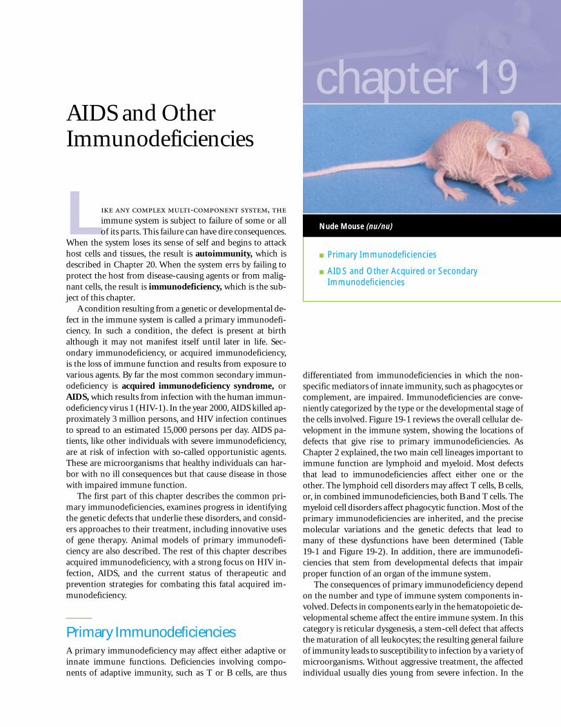
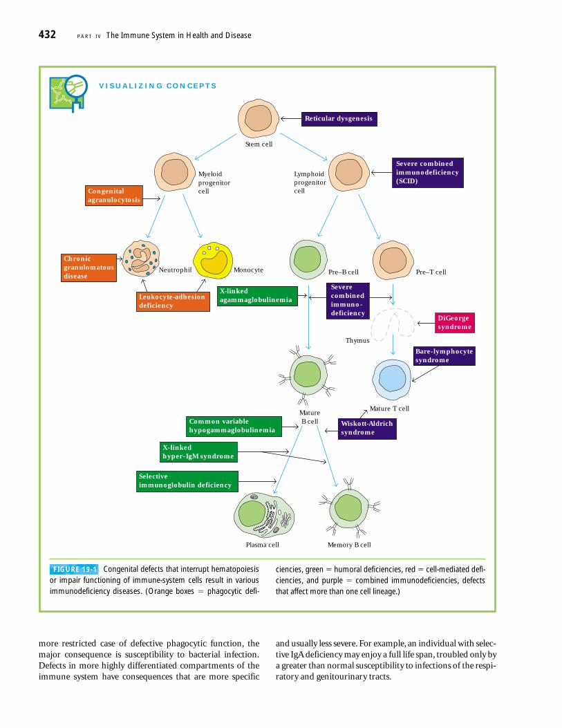
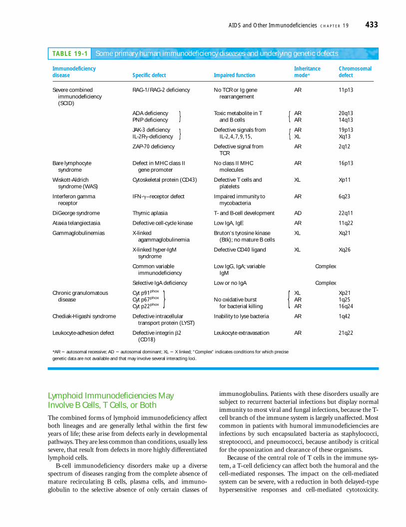
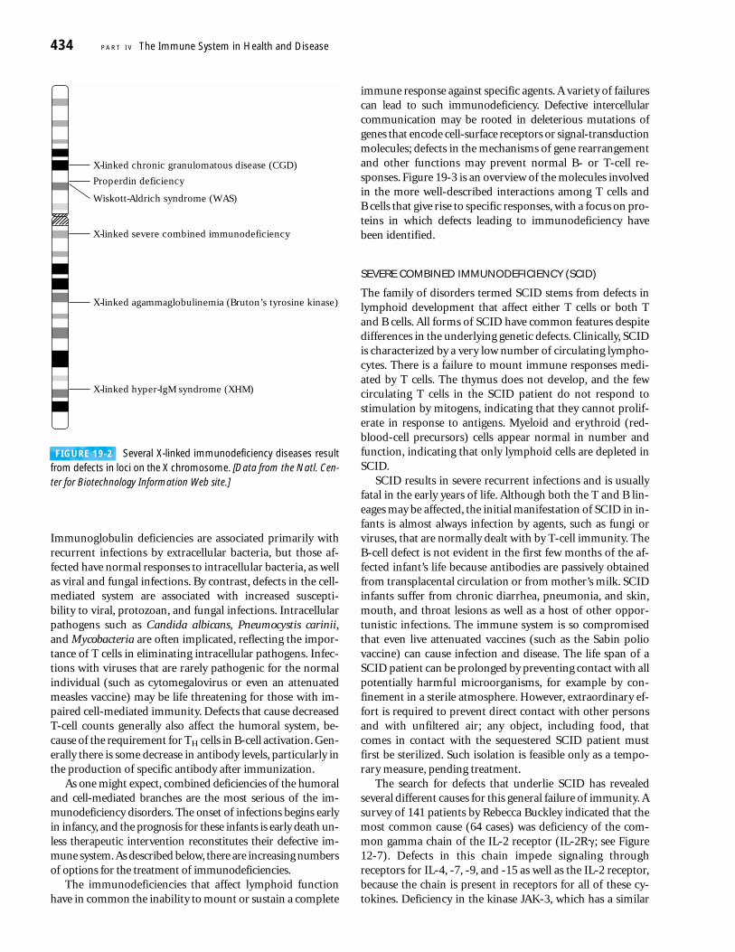

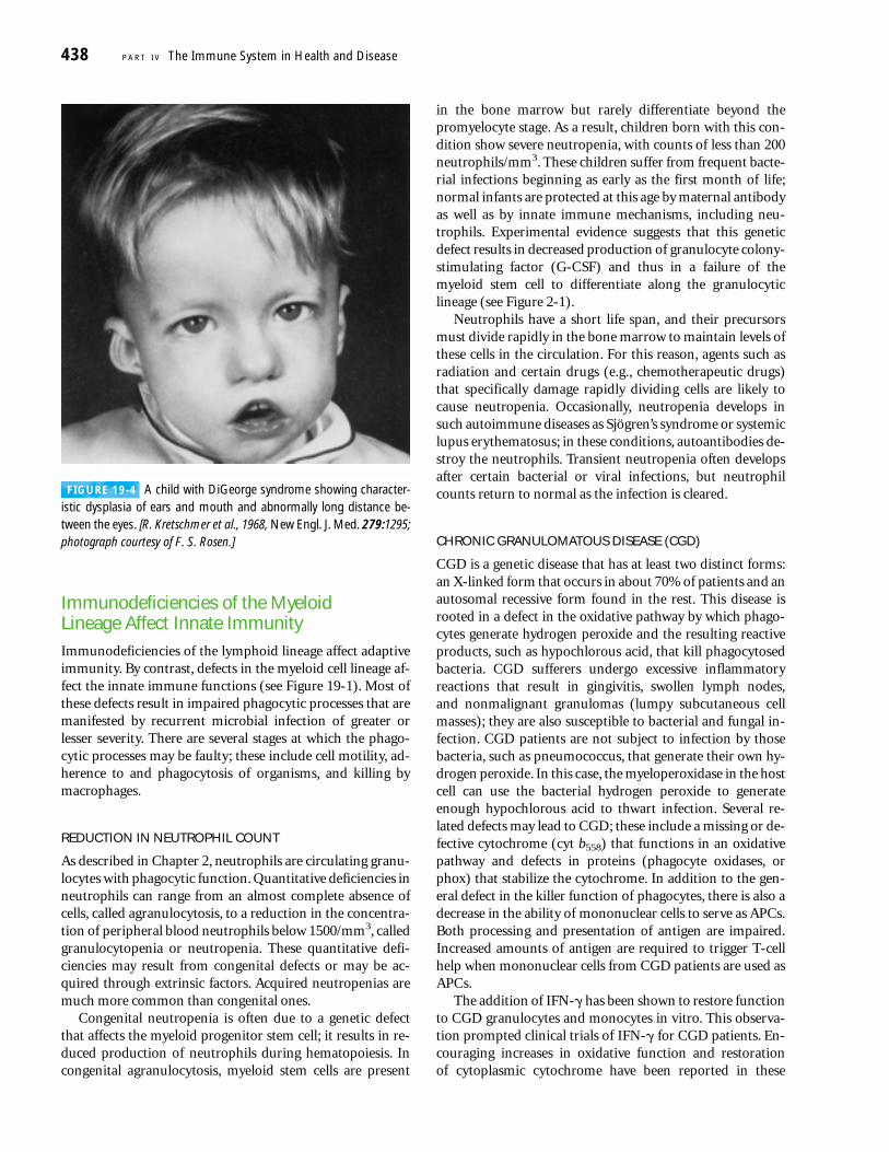
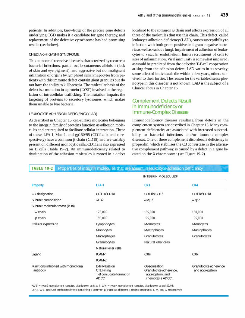
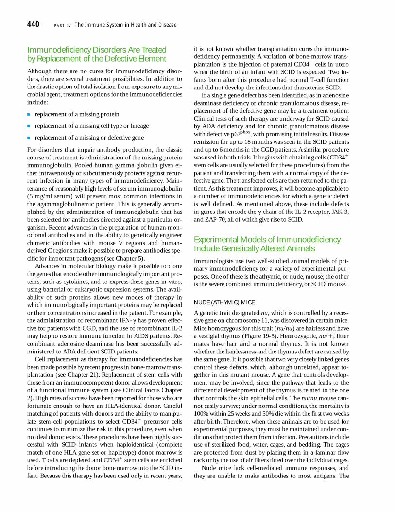
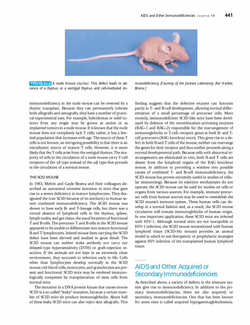
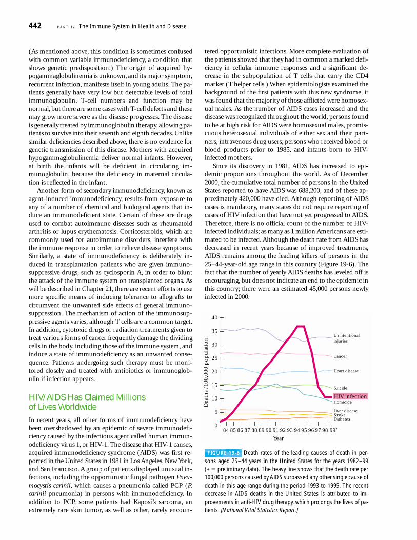
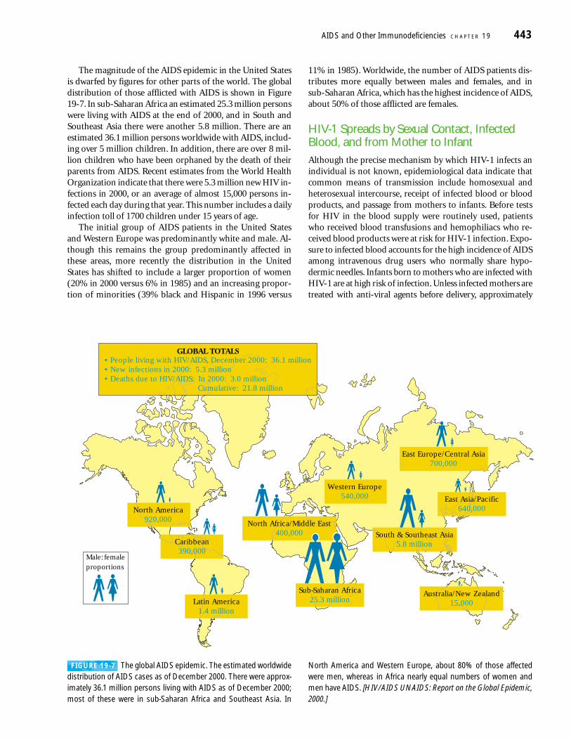
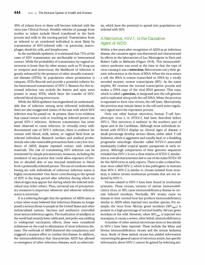
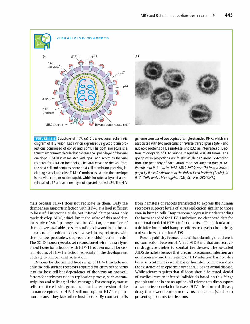
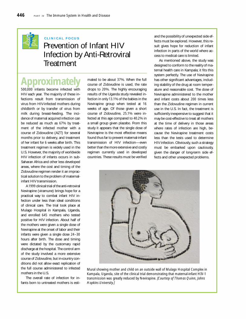
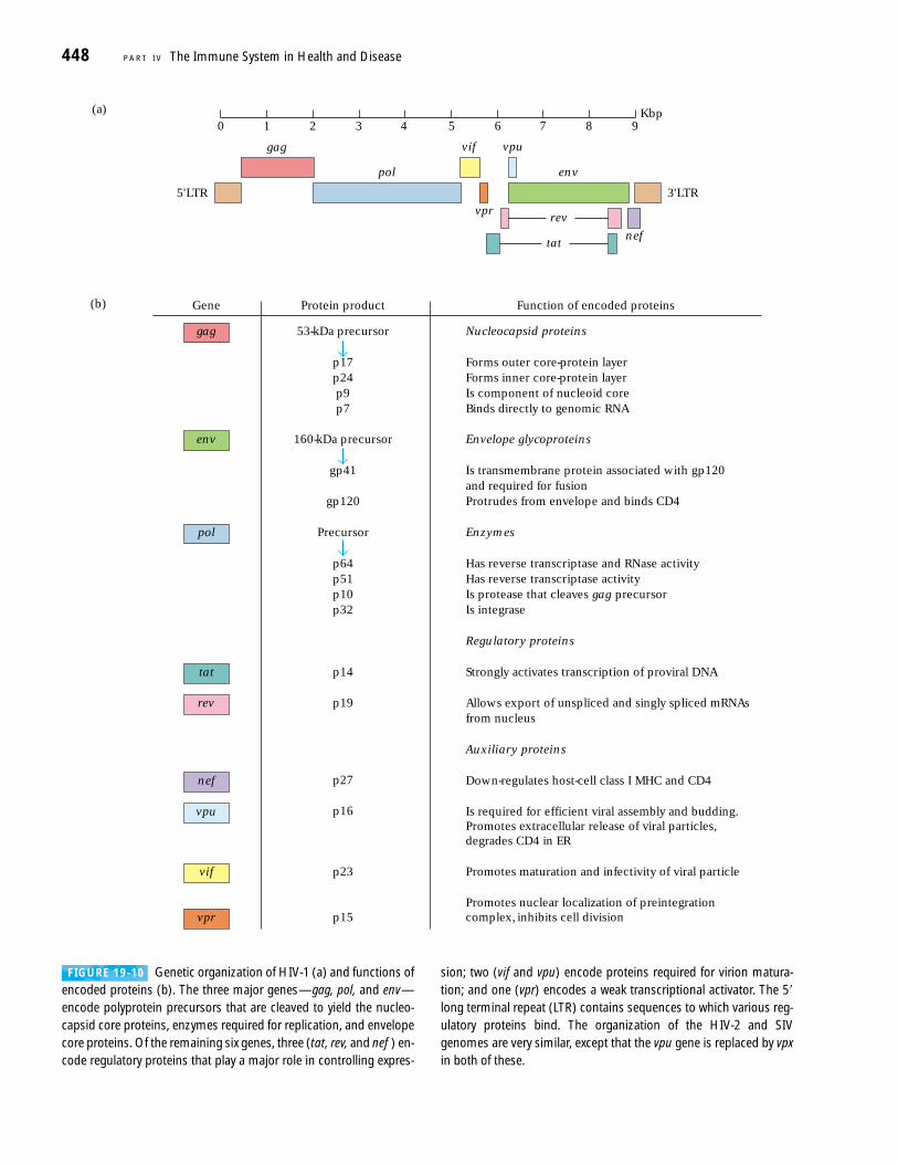
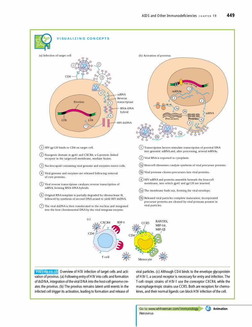
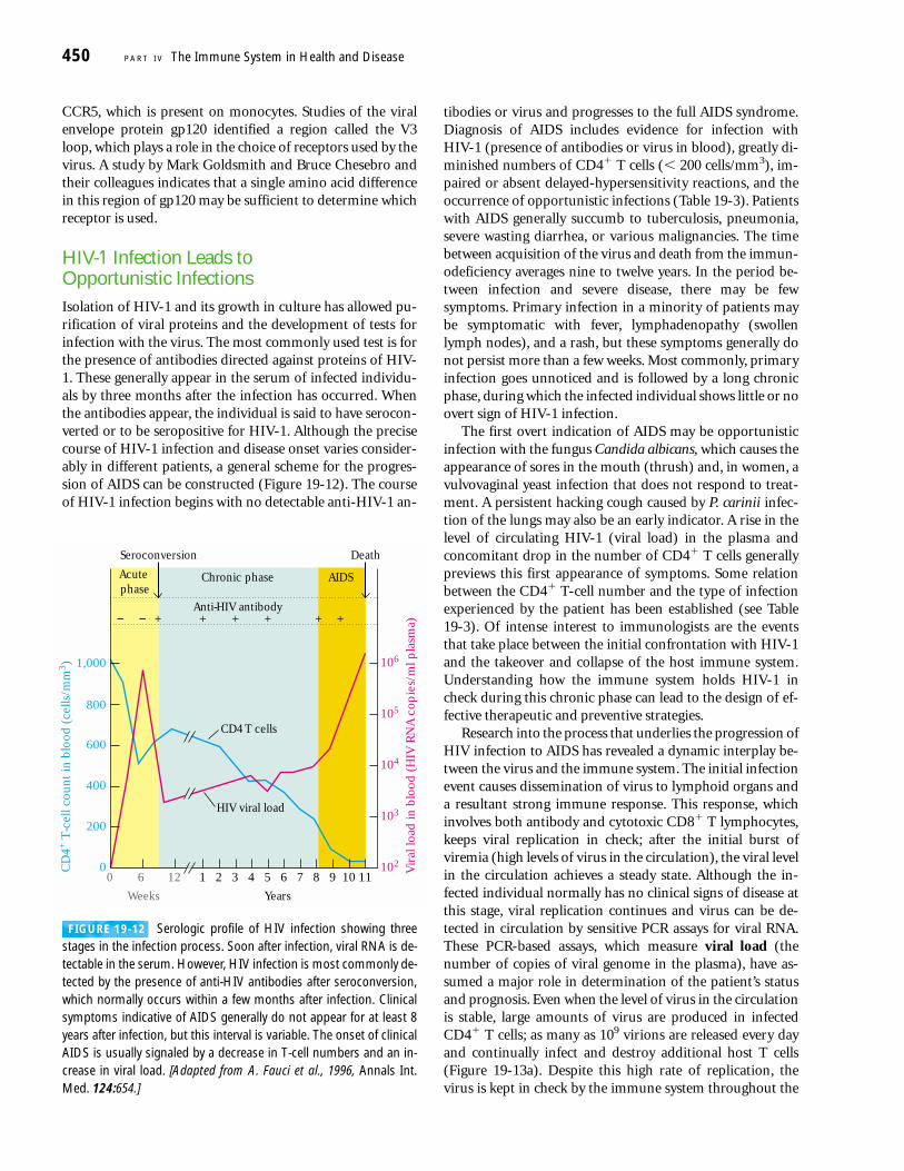
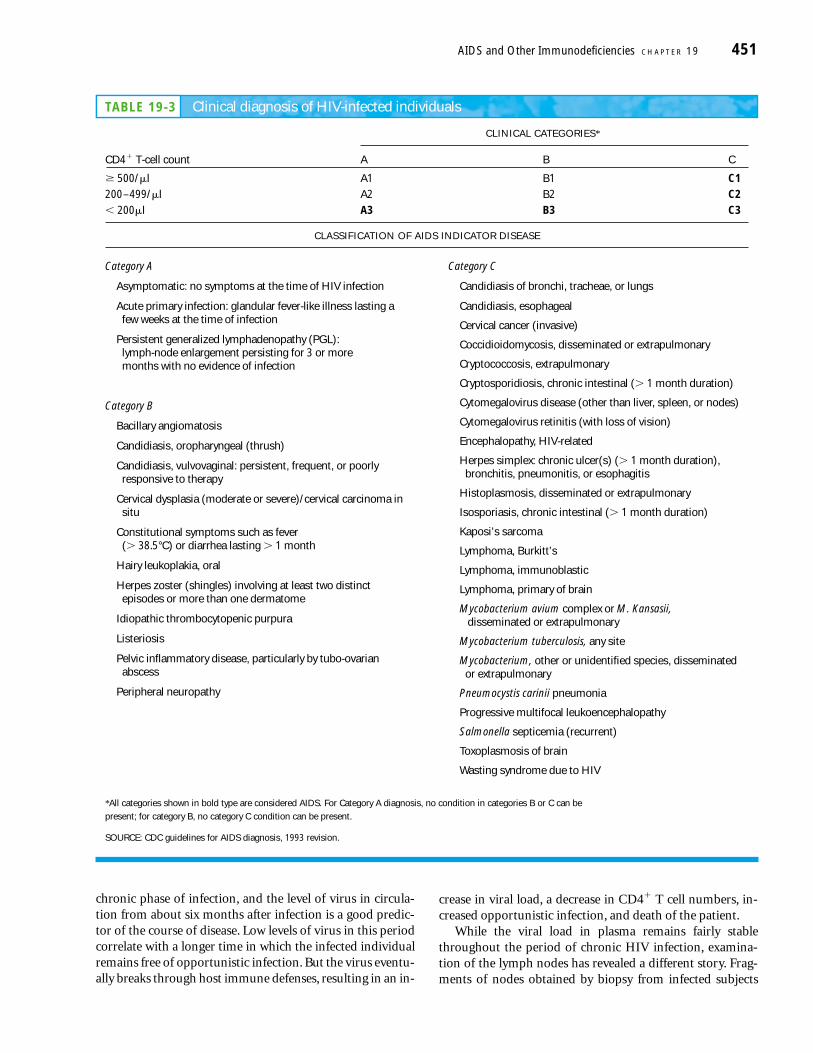
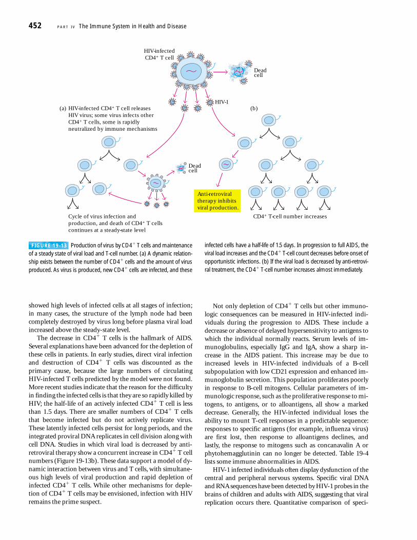
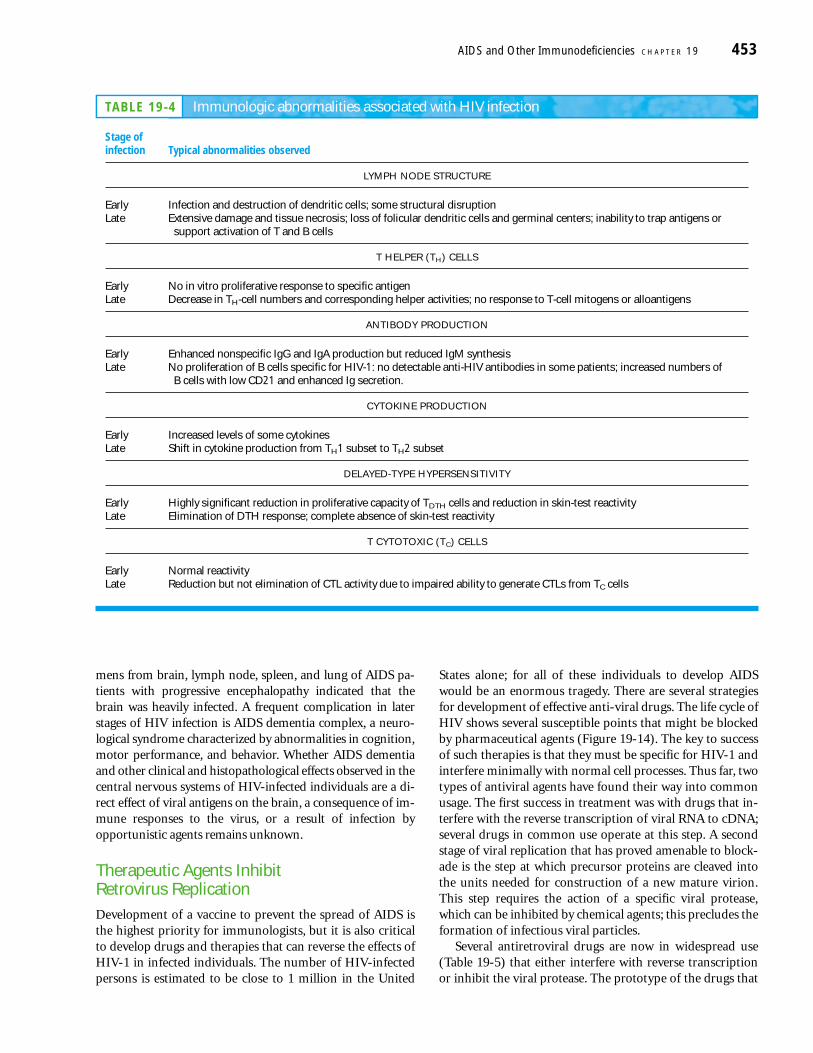
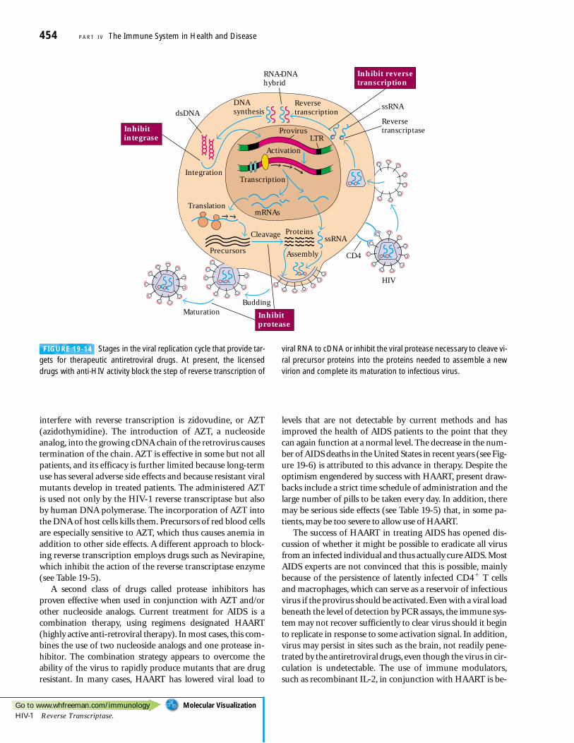
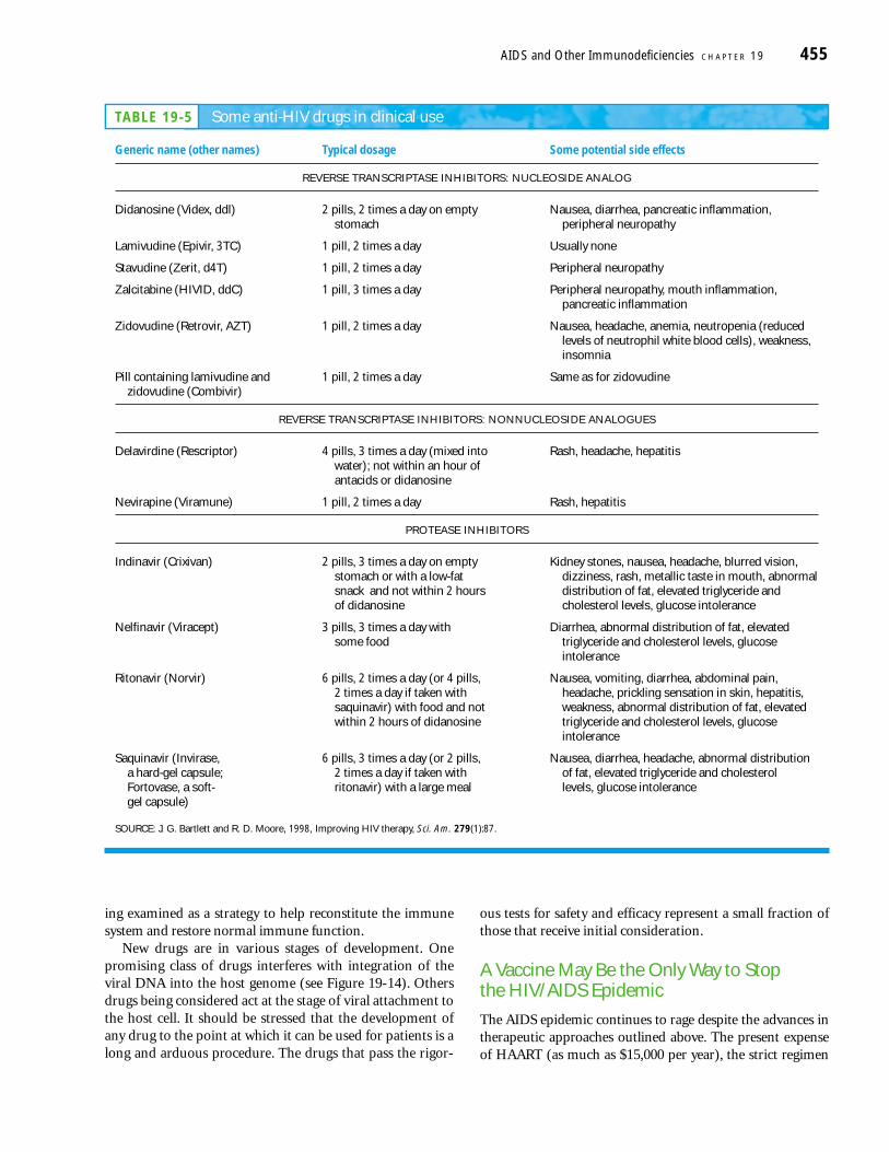

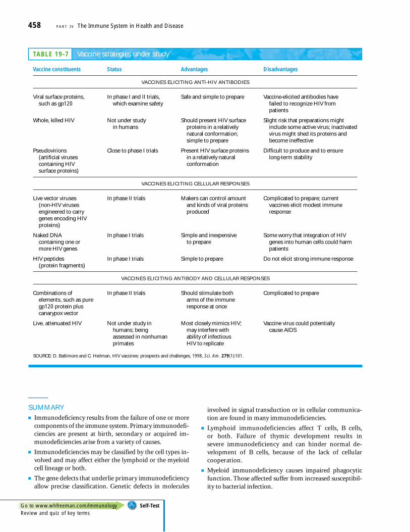
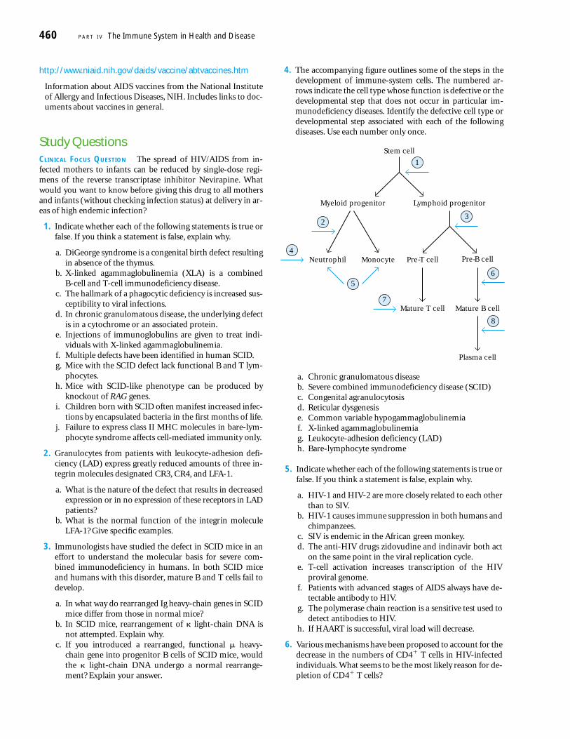


![IAC 11/17/21 Utilities[199] Ch 19, p.1 CHAPTER 19 ... - Iowa.gov](https://static.documents.pub/doc/80x56/6323f34ff021b67e740852a0/iac-111721-utilities199-ch-19-p1-chapter-19-iowagov.jpg)


