Endobacteria affect the metabolic profile of theirhost Gigaspora margarita, an arbuscularmycorrhizal fungusemi_2246 2083..2095
Alessandra Salvioli,1 Marco Chiapello,1
Joel Fontaine,2 Anissa Lounes Hadj-Sahraoui,2,3
Anne Grandmougin-Ferjani,2 Luisa Lanfranco1 andPaola Bonfante1,4*1Department of Plant Biology University of Torino,Torino, Italy.2Univ Lille Nord de France, F-59000 Lille, France.3ULCO, UCEIV, F-62228 Calais, France.4IPP-CNR, Torino, Italy.
Summary
The aim of this paper was to understand whether theendobacterium identified as Candidatus Glomerib-acter gigasporarum has an effect on the biology of itshost, the arbuscular mycorrhizal fungus Gigasporamargarita, through the study of the modificationsinduced on the fungal proteome and lipid profile. Theavailability of G. margarita cured spores (i.e. sporesthat do not contain bacteria), represented a crucialtool to enable the comparison between two fungalhomogeneous populations in the presence and theabsence of the bacterial components. Our resultsdemonstrate that the endobacterial presence leads toa modulation of fungal protein expression in all thedifferent conditions we tested (quiescent, germinat-ing and strigolactone-elicited germinating spores),and in particular after treatment with a strigolactoneanalogue. The fungal fatty acid profile resulted to bemodified both quantitatively and qualitatively in theabsence of endobacteria, being fatty acids less abun-dant in the cured spores. The results offer one of thefirst comparative metabolic studies of an AM fungusinvestigated under different physiological conditions,reveal that endobacteria have an important impacton the host fungal activity, influencing both proteinexpression and lipid profile, and suggest that the bac-terial absence is perceived by G. margarita as astimulus which activates stress-responsive proteins.
Introduction
The arbuscular mycorrhizal (AM) symbiosis is the mostwidespread mycorrhizal type, occurring in more than 80%of the land plants, and involving as symbiotic fungi theGlomeromycota, an ancient phylum that has coevolvedwith plants for at least 400 million years (Bonfante andGenre, 2008). The AM fungi contribute significantly to soilnutrient uptake in plants, increasing their productivity andconferring resistance to stresses. At the same time, asobligate biotrophs, they depend on the plant for carbohy-drates, being so far unculturable in the absence of theirhost. Their uniqueness is also mirrored by other biologicaltraits: they possess thousands of syncytial nuclei in theirsspores and hyphae (Parniske, 2008) and are consideredasexual microbes, even if genetically distinct AM fungianastomose and perform genetic crosses (Croll et al.,2009). Lastly, they are known to contain endobacteria intheir cytoplasm, which represent therefore the third com-ponent of mycorrhizal associations (Bonfante and Anca,2009).
As for insect endosymbionts, the presence of endobac-teria inside Glomeromycota cytoplasm has long beendocumented by electron microscopy, which has distin-guished two bacterial morphotypes. The first has beendetected inside AMF spores and hyphae colonizing plantroots sampled in the field. It is coccoid in shape, and hasbeen labelled ‘bacterium-like organism’ (BLO) for longtime, since its identity has been only recently solved asrelated to Mollicutes (Naumann et al., 2010). The otherbacterial type, rod-shaped and restricted to a single AMFfamily (Gigasporaceae), has been studied in more detail.Use of a combination of microscopy and molecular analy-sis of 16S rRNA has led to the description of these latterorganisms as bacteria related to Burkholderia (Bianciottoet al., 1996). They were placed in a new taxon named‘Candidatus Glomeribacter gigasporarum’ because of theirunculturability (Bianciotto et al., 2003). Isolate BEG34 ofGigaspora margarita and its endobacterium CandidatusG. gigasporarum are currently used as a model systemto investigate endobacteria–AM fungi interactions. Theseendocellular bacteria represent a stable and homoge-neous population inside the G. margarita cytoplasm andare vertically transmitted (Bianciotto et al., 2004). They
possess a Gram-negative cell wall, are rod-shaped withan approximate size of 0.8–1.2 ¥ 1.5–2.0 mm, and occursingly or in groups inside fungal vacuole-like structures,being surrounded by a fungal membrane.
Our knowledge on the interaction between AM fungi andtheir endobacteria is still limited and fragmentary, mainlybecause of the physiological features of both the partners(both of them are obligate endosymbionts). The recentachievement of a G. margarita sporal line, which is devoidof bacteria (‘cured’ spores), helped us to shed some lighton the effects of the endobacterium on its host (Luminiet al., 2007). Spores of the cured line have cells distinct invacuole morphology, cell wall organization, lipid bodiesand pigment granules. The absence of bacteria seems notto affect the symbiotic capacities of the fungal host, while itinfluences its presymbiotic growth to a great extent in termsof hyphal elongation and branching in response to a rootexudates treatment (Lumini et al., 2007). The active frac-tion of root plant exudates, which is responsible for AMhyphal branching in the vicinity of the host root, wasisolated and described as a molecule belonging to thestrigolactone family (Akiyama et al., 2005). In addition totheir effect in stimulating fungal presymbiotic development(Tamasloukht et al., 2003; Besserer et al., 2006; Bessereret al., 2008), strigolactones also seem to be perceived bythe endobacterium, which responds to this stimulus with anincrease in bacterial division mirrored by the upregulationof the FTz gene (Anca et al., 2009).
In this paper we investigate the impact of the endobac-terium Candidatus G. gigasporarum on the proteome andlipid profile of its fungal host G. margarita. The availabilityof G. margarita cured spores (Lumini et al., 2007) allowedus to compare fungal populations containing or not thebacterial symbiont.
Since recent findings suggested that the endobacteriamay modulate their life cycle according to the stagesof the fungal host development (Anca et al., 2009), theproteome profile was investigated considering differentphysiological conditions, i.e quiescent, germinatingspores and spores treated with a synthetic analogue ofnatural strigolactones.
Current knowledge on AM fungal proteome is limited toa few reports, and one in particular lists proteins detectedin Glomus intraradices mycelium (Recorbet et al., 2009).In this context, our findings show that endobacteria haveimportant impacts on the fungal proteome and lipid profile,and also represent one of the first comparative metabolicstudies of an AM fungus, investigating two fungal linesunder three physiological conditions.
Results
No evident phenotypic differences were found betweengerminating spores from the wild-type (wt) and cured lines
after 10 days of germination. In independent preliminarytests, a more intense branching was detected in the wtline after treatment with the strigolactone analogue GR 24after 15 days of incubation (data not shown). All theseresults are in agreement with what shown in Lumini andcolleagues (2007).
Protein profiles of wt and cured spores
Two-dimensional (2D) protein maps were obtained for thewt and cured fungus under three physiological conditions,i.e, quiescent, germinating and GR24-treated germinatingspores. On the whole, a total of 320 individual spots weredetected, that were reproducibly displayed within thewindow of pH 4–7 and molecular mass 5–200 kDa.Among the spots, 159 were present in each consideredcondition (quiescent, germinating or GR4-treated germi-nating spores). Gels from wt and cured spores were com-pared pairwise in the three conditions leading to thedetection of spots which were differentially expressed ina qualitative (presence versus absence) or quantitative(different level of expression) way. All the data are sche-matically presented in Fig. 1.
A total of 87 proteins were cut from the 2D gels and sentfor sequencing; 17 out of them were chosen among themost highly and constitutively present spots (that is, theywere common to all the considered conditions), whilethe other 70 corresponded to differentially expressedproteins. Out of these 87, 43 proteins were identified, 10constitutively and 33 differentially expressed. The resultsof protein identification are listed in Table 1.
Constitutively expressed proteins
Since data concerning G. margarita protein expressionare so far quite scanty, we selected some of the moreintense spots among the 159 that were common to all theconsidered conditions. A reliable result was obtained for10 out of 17 spots originally sent for sequencing.
Consistently with their constant detection in both wtand cured two-dimensional gel electrophoresis (2D-E)maps at high level of intensity (data not shown), the spotsfrom 1 to 10 were identified as proteins mainly involved insome central metabolic pathways (i.e. respiration, energyproduction, fatty acid (FA) and sterol ester biosynthesis).Protein identification by database search retrieved besthits belonging to a wide range of organisms, from plantsto bacteria. However, the constant observation of the cor-responding proteins in all the analysed maps stronglysupports the hypothesis that they belong to the fungalproteome.
Spots 1, 2, 3 and 4 share the same Mw but havedifferent pI; taken together, they account for a consider-able part of the total amount of protein detected on the
2D-E maps. They seem to be related to proteins withdifferent functions, and belonging to a wide range oforganisms.
Spot 5 was identified as an uncharacterized conservedprotein of unknown function, while spot 6 showed highestsimilarity with a synaptotagmin protein 2 from Caenorhab-ditis elegans. Synaptotagmins are globally known asmembrane proteins involved in vesicle-mediated trans-port and exocytosis. De novo search for spot 7 led to theidentification of an acyl-CoA desaturase; delta-9 FAdesaturases, found in various eukaryotes and bacteria,play essential roles in FA metabolism, being involved inthe CoA-bound desaturation of FAs.
For spot 8, the best hit was represented by a cyto-chrome C oxidase from the plant Cephalotaxus fortunei,the enzyme responsible for energy production via themitochondrial respiratory chain.
Mascot search for spot 9 led to the identification of aglutamine synthetase (GS); although a sequence of a GSfrom the AM fungus G. intraradices is already present inpublic databases (gi|161406807), the peptides retrievedfor spot 9 showed the best similarity with a GS from theectomycorrhizal fungus Suillus bovinus. In any case, theGS from G. intraradices (gi|161406807) shows a calcu-lated Mass/pI of 42.179/5.46, which is highly consistent
with what observed on our 2D-E map. This protein repre-sents a central enzyme of nitrogen metabolism sinceit allows assimilation of nitrogen and biosynthesis ofglutamine (Breuninger et al., 2004). It has been describedas crucial for nitrogen metabolism in germinating spores,and its mRNA has been reported not to be differentiallyexpressed in AM fungi exposed to root exudates anddifferent N sources (Breuninger et al., 2004; Gachomoet al., 2009). A possible functional regulation driven byphosphorylation has been reported for this enzyme inplants (Riedel et al., 2001). Spot 10 shared the highestsimilarity with the protein isocitrate lyase from Pichiaangusta. This protein represents a key enzyme of theglyoxylate cycle, which was suggested to play a centralrole in the flow of carbon during AM symbiosis (Bagoet al., 2002). A high expression of the ICL gene transcriptwas observed in the AM fungus G. intraradices germinat-ing spores and extraradical mycelium (Lammers et al.,2001).
Wild-type and cured spores: differentiallyexpressed proteins
A number of spots differentially expressed between wtand cured spores was identified under the three physi-ological conditions considered, showing both qualitative(presence versus absence) or quantitative (different levelof expression) differences (Fig. 1 and Table 2).
Quiescent spores. For the quiescent spores condition, 23spots were selected for sequencing, and good resultswere obtained for 12.
Spots 11 and 12, from the quiescent wt and cured sporemap, turned out to represent the same protein, a super-oxide dismutase (SOD) from G. margarita. This protein
Fig. 1. Schematic representation of theproteins detected in each 2-DE experiment;numbers outside the common area indicatequalitatively (not underlined) and quantitatively(underlined) differentially expressed proteins.
Table 2. Differentially expressed spots identified for each physiolo-gical condition by Mascot or de novo search.
represents one of the few described so far for G. marga-rita and the transcript of the corresponding gene wasalready demonstrated to be differentially expressedduring the fungal life cycle, with the highest mRNA level inthe symbiotic phase (Lanfranco et al., 2005). On the map,spots 11 and 12 shared the same mass, but possessedslightly different isoelectric points. The observation ofa shift in the isoelectric point is often due to post-translational modifications such as glycosylation or phos-phorylation. In silico analysis of the G. margarita SODsequence shows the presence of 5 and 10 predictedsites for phosphorylation and glycosylation respectively.Some SOD from animals are known to be glycosylated(Oda et al., 1994; Tang et al., 1994) or to possess putativeglycosylation sites (Cheng et al., 2006a,b). It has alsobeen shown that the cytoplasmic Listeria monocytogenesMnSOD is phosphorylated on serine and threonine resi-dues and less active when bacteria reach the stationaryphase (Archambaud et al., 2006).
The sequence obtained from spot 13, isolated fromquiescent wt spores, showed the highest similarity withflavin containing monoxygenases (FMOs) from the purplesea urchin Strongylocentrotus purpuratus. This proteinbelongs to a group of microsomal proteins involved in theprocess of non-nutritional foreign compounds metabolismknown as xenobiotics. Generally, FMO converts lipophilicnucleophile xenobiotics to more polar, readily excretedmetabolites, decreasing their pharmacological activity.The FMOs oxygenate nucleophilic O, N, S and Se atomsof a wide range of substrates, such as amines, amides,thiols and sulfides. Fungi are known to possess FMOs; forexample, an FMO from the yeast Schizosaccharomycespombe was identified, and its mechanism of action wasdescribed (Eswaramoorthy et al., 2006).
Spot number 20 was identified in the quiescent curedcondition as a subtilisin-kexin convertase. Subtilisin-likeproteases (SLPs) form a superfamily of enzymes that actto degrade protein substrates. In fungi, SLPs can playeither a general nutritive role, or may play specific roles incell metabolism; in addition, they have been shown to actas pathogenicity or virulence factors (Bryant et al., 2009).These proteins have been recently characterized in fungi,which interact with other eukaryotes, like the endophyticfungus Epichloë festucae and some entomopathogenicfungi (Bryant et al., 2009; Fang et al., 2009).
Germinating spores. The number of differentiallyexpressed proteins identified from germinating sporeswas very low: only 4 proteins out of the 20 selectedcould be reliably identified. Two proteins (Spots 24 and25), which can be related to a metabolically active con-dition (elongation factor and enolase), were identifiedin wt spores. By contrast spot 26, which was specificallydetected in the cured condition, showed the highest
similarity with a protein from Gibberella zeae, which rep-resents a putative stress-inducible heat shock proteinHSP30 (Seymour and Piper, 1999).
GR24-treated spores. The third considered conditionwas represented by spores germinating under treatmentwith the strigolactone analogue GR24. Strigolactones arecarotenoid-derived molecules which are naturally presentin plant root exudates; they are known to stimulate thegrowth and branching of AM germinating hyphae, as wellas the activity of fungal mitochondria. Cured sporesdisplay a reduced response to such a treatment, in termsof hyphal branching and growth (Lumini et al., 2007).
The GR24-treated condition detected the highestnumber of differentially expressed proteins (a total of 16out of 27 sent for sequencing). Some interesting spotsof putatively endobacterial origin were identified in thewt condition (see ‘Spots of potentially bacterial origin’paragraph below). On the fungal side, a protein that wasdetected in cured GR24-treated condition and describedas a hypothetical protein from G. zeae was identifiedas a putative peroxyredoxin (spot 40). This group ofproteins are possibly involved in redox-regulated anti-oxidant defence, a mechanism well described also infungi (Belozerskaya and Gessler, 2007). Similarly to whatobserved for germinating spores, two heat-shock proteinsseem to be more expressed in cured than wt spores afterstrigolactone analogue treatment (spots 38 and 42).
Cross-comparison between conditions
As a further step, a global statistical analysis was appliedto all the 2D maps obtained, in order to identify spotsspecifically expressed in one of the considered physi-ological conditions. Following this comparative analysis,spot 43 was identified to be specifically expressed inGR24-treated spores, in wt as well as in cured condition.This protein, which shares higher similarity with atyrosine-protein phosphatase from Drosophila melano-gaster, seems not to be expressed in quiescent and ger-minating spores.
Spot 35, which was originally identified as moreexpressed in wt versus cured spores in GR24-treatedcondition, and identified as an outer membrane porinprotein C (ompC), also revealed to be specificallyexpressed during strigolactone treatment, as it wasabsent in quiescent and germinating maps. Althoughthe Mascot search suggested a possible bacterial originfor this spot, the Blast analysis against the CandidatusGlomeribacter sequence database (S. Ghignone, I.A.Anca, A. Salvioli, L. Lanfranco and P. Bonfante, in prepa-ration) did not produce any significant hit, suggesting thatthis protein might not belong to the endobacterium. This
is consistent with its detection in GR24-treated curedspores, even though at a lower concentration. We canthus postulate that this protein belongs to the fungal pro-teome; indeed in eukaryotic cells beta-barrel proteinssimilar to bacterial ompC are known to be involved inmitochondrial outer membrane synthesis (Becker et al.,2009; Walther et al., 2009).
Spots of potentially bacterial origin
The database search for protein identification retrieved abacterial entry as best hit for the 39% of the identifiedproteins (17 spots out of 43). To check the possibility thatthese proteins belong indeed to the endobacterium, thecorresponding sequences were blasted against the provi-sional database constructed on the basis of the Candida-tus G. gigasporarum genome sequencing (S. Ghignone,I.A. Anca, A. Salvioli, L. Lanfranco and P. Bonfante, inpreparation). For six of the analysed proteins (here iden-tified in Table 1 as spot 18, 30, 31, 32, 33 and 36), a resultwith E value < e-50 was obtained, suggesting that in thesecases a protein belonging to the endobacterium proteomewas sequenced. Consistently with their nature, these sixputative endobacterial proteins were uniquely detected inmaps coming from the WT condition, and four out of themfrom the spores treated with GR24. Spot 18, 30 and 36show the highest similarity with proteins involved in thegeneral bacterial cell functioning (protein synthesis, mem-brane transport and structural component). Spots 32 and33 were identified as an ATP-dependent chaperoneprotein ClpB and a 60 kDa chaperonin groEL respectively.They are related to the chaperonin pathway. Similarly,
spot 31 was identified as a bacterial peroxyredoxin, athiol-specific antioxidant protein, which is considered toact as redox-regulated chaperone involved in bacterialantioxidant defence (Kumsta and Jakob, 2009). As dem-onstrated in early studies, the universal heat shock chap-eronin groEL is constitutively highly expressed inBuchnera and in other endosymbionts and host-restrictedorganisms (Aksoy, 2000). An increased investment inmechanisms for protein stabilization has been postulatedto have evolved as a compensation for accumulatedmutations that reduce protein stability (Wernegreen andMoran, 2000; Van Ham et al., 2003).
Lipid profile
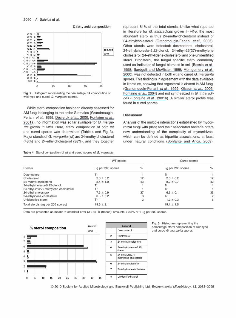
The FA composition of wt and cured spores of G. marga-rita was established following a chromatographic method-ology (Table 3 and Fig. 2), and the results are consistentwith what previously observed for this fungus (Bentivengaand Morton, 1996). The FA composition of wt sporesranged from C12:0 to C22:1. The predominant FA com-pounds were C16:0 (palmitic acid) and C18:1w9 (oleicacid); they constituted more than 69% of the total FA with35.8% of C18:1w9 and 33.7% of C16:0. In cured spores asignificant reduction of C16:0, C18:1w5, C20:3 and C20:4proportions was observed. The content of major FAsC16:0 and C18:1w9 decreased of three- and twofold,respectively, in comparison with wt spores values. Areduced content of other minor FAs: C16:1w9, C16:1w5,C18:1w7, C18:3 and C20:1 was also observed. Moreover,the total FA amount decreased more than twofold in curedspores.
Table 3. Fatty acid composition of wt and cured spores of G. margarita.
Total fatty acids (mg per 200 spores) 1723.6 � 141.5 784.8 � 212.6*
Data are presented as means � standard error (n = 3). The asterisk indicates significantly differences between spores type according tonon-parametric permutation test (P < 0.05). Tr (traces): amounts < 0.5% or 1 mg per 200 spores.
Endobacteria affect proteins and lipids in Gigaspora 2089
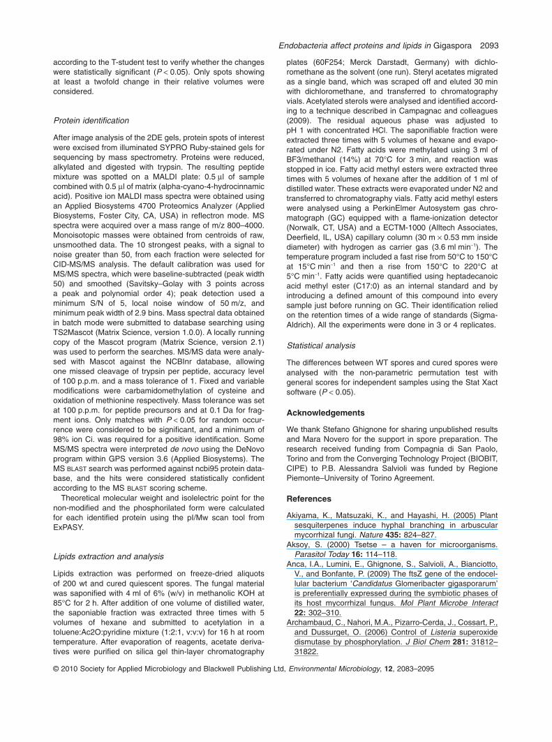
While sterol composition has been already assessed forAM fungi belonging to the order Glomales (Grandmougin-Ferjani et al., 1999; Declerck et al., 2000; Fontaine et al.,2001a), no information was so far available for G. marga-rita grown in vitro. Here, sterol composition of both wtand cured spores was determined (Table 4 and Fig. 3).Major sterols of G. margarita (wt) are 24-methylcholesterol(43%) and 24-ethylcholesterol (38%), and they together
represent 81% of the total sterols. Unlike what reportedin literature for G. intraradices grown in vitro, the mostabundant sterol is thus 24-methylcholesterol instead of24-ethylcholesterol (Grandmougin-Ferjani et al., 2005).Other sterols were detected: desmosterol, cholesterol,24-ethylcholesta-5.22-dienol, 24-ethyl-25(27)-methylenecholesterol, 24-ethylidene cholesterol and one unidentifiedsterol. Ergosterol, the fungal specific sterol commonlyused as indicator of fungal biomass in soil (Bossio et al.,1998; Bardgett and McAlister, 1999; Montgomery et al.,2000), was not detected in both wt and cured G. margaritaspores. This finding is in agreement with the data availablein literature, showing that ergosterol is absent in AM fungi(Grandmougin-Ferjani et al., 1999; Olsson et al., 2003;Fontaine et al., 2004) and not synthesized in G. intraradi-ces (Fontaine et al., 2001b). A similar sterol profile wasfound in cured spores.
Discussion
Analysis of the multiple interactions established by mycor-rhizal fungi with plant and their associated bacteria offersnew understanding of the complexity of mycorrhizas,which can be defined as tripartite associations, at leastunder natural conditions (Bonfante and Anca, 2009).
Fig. 2. Histogram representing the percentage FA composition ofwild-type and cured G. margarita spores.
Table 4. Sterol composition of wt and cured spores of G. margarita.
Details of these interactions are still unclear since thelimited availability of genome sequences for mycorrhizalfungi has only allowed a study of the impact of associatedbacteria on the transcriptome profile of Laccaria bicolor(Deveau et al., 2007). In this case, the helper bacterium P.fluorescens stimulated fungal growth and development aswell as altered fungal gene expression, leading to activa-tion of genes potentially involved in recognition processes,transcription regulation and synthesis of primary metabo-lism proteins. Unlike ectomycorrhizal fungi, the genomeof an AM fungus has not been completely analysedyet (http://mycor.nancy.inra.fr/IMGC/genomesequencing.html), making a transcriptomic investigation on AM fungal/bacterial interaction not currently feasible.
Information on the AM fungal proteome is quite scanty.The most exhaustive proteomic study is representedby the work of Recorbet and colleagues (2009), in whichthe authors isolated and identified 92 proteins from theextraradical mycelium of G. intraradices with the GeLC-MS/MS high-throughput technique.
Our results demonstrate that the endobacterial pres-ence leads to a modulation of fungal protein expression inall the different conditions we tested and, in particular,after treatment with a strigolactone analogue. GR24-treatment allowed in fact the detection of the highestnumber of differentially expressed proteins between wtand cured condition, in agreement with Lumini and col-leagues (2007), who showed that wt spores are moreresponsive to GR24 in terms of hyphal growth andbranching. The current view suggests that strigolactone-treated spores might be in a more active status (Bessereret al., 2006; Besserer et al., 2008); very recently Buckingand colleagues (2008) strengthened this vision, showingthat the expression of genes involved in primary metabolicpathways are induced by root exudates stimulation. Theglobal statistical analysis revealed that two proteins arespecific of this treatment. Interestingly, one of themwas identified as a tyrosine-protein phosphatase, whichbelongs to a group of enzymes that, together with tyrosinekinases, regulate the phosphorylation state of manyimportant signalling molecules. Such a protein could be agood candidate for the identification of components of thesignal transduction cascades involved in the perception ofthe branching factor.
The second observation is that five out of these GR24-stimulated proteins resulted to be bacterial proteins.Under this condition, an increase of bacterial divisionswas in fact observed (Anca et al., 2009), which could leadto an easier detection of bacterial proteins. Thus, thedetection of bacterial protein in the GR24-treated wtspores map can be related to the fact that bacteria aremore abundant in this condition, and/or can indicate thatsuch proteins are part of the molecular response of theendobacterium to the strigolactone stimulus.
By contrast, cured line-specific proteins were relativelymore limited (11) and four out of them were identified asstress-responsive proteins. Even if the cured line keepsits symbiotic capabilities, the germinating myceliumresulted to be less efficient in developing and contactingthe host roots in axenic cultures (Lumini et al., 2007). Itseems therefore that the G. margarita perceives the bac-terium absence as an indirect or direct stimulus, whichactivates stress-responsive proteins.
The impact of the endobacterium on the metabolism ofits host is also demonstrated by the strong differences inthe FA profile. Lipids are crucial molecules for signallingand functioning of AM symbiosis (Bucher, 2010). In fact,AM fungi store predominantly the organic carbon acquiredfrom the plant root as lipids. In addition, it was suggestedthat fungal FA metabolism may play a major role in theobligate biotrophism of AM fungi (Trepanier et al., 2005).
The FA profile here obtained for the wt spores is in goodagreement with literature data for G. margarita, showing apredominance of the FA C18:1w9 and a lower presenceof C16:1w5, which is, by contrast, highly represented inother AM fungi (for example, in G. intraradices, this FArepresents the 50-70% of the total neutral lipids content)(Graham et al., 1995; Bentivenga and Morton, 1996;Olsson and Johansen, 2000). The comparison of FA com-position from wt and cured spores highlighted importantdifferences, showing that the total FA content decreasesin the absence of the endobacteria and that the specificprofile is affected in the cured spores. However, anddifferently from the protein profile, bacteria-specific FAswere not detected in the wt spores. The relatively lownumber of bacteria per spores and/or some similaritieswith fungal FA could explain the absence of bacterialmarkers in the FA profile. Several b-Proteobacteria are infact characterized by C16:0 and C18:1w7 (Krejci andKroppenstedt, 2006), which are also the major FA in fungi.
As a second point, our biochemical analyses are ingood agreement with morphological observations of thecured G. margarita spores, which revealed a decrease ofsize and number of lipid masses (Lumini et al., 2007). Thelower total FA content in the cured line could also point tothe absence of the fungal membrane, which regularlysurrounds the endobacteria as detected at ultrastructurallevel (Bonfante et al., 1994).
It is known that fungal storage lipids mainly consist ofneutral lipid FAs (Olsson and Johansen, 2000), of whichtriacylglycerols are the dominant type in spores andvesicles. Triacylglycerols are specifically enriched in16-carbon FA (Grandmougin-Ferjani et al., 2005). Ourresults show that FA C16:0 (palmitic acid) resulted to beless abundant in the cured spores. Since palmitic acidis the direct product of the fatty acid synthase (FAS)complex, prior to any subsequent modification (elonga-tion, desaturation, etc.), the data may suggest that the
Endobacteria affect proteins and lipids in Gigaspora 2091
cured spores are less efficient in FA biosynthesis and/orstorage. Moreover, we cannot completely rule out that theoxidative stress highlighted by the stress-responsive pro-teins detected in cured spores has an impact on metabolicenzymes and on FA profile. Indeed, the reduction ofthe polyinsatured FA C18:3 could suggest an inductionof lipid peroxydation in cured spores. Since lipids storedinside the spores are used to sustain germination andpre-symbiotic growth (Bago et al., 1999; Fontaine et al.,2001b), a decreased FA availability in spores could beproposed to explain the reduced pre-symbiotic growthobserved in the cured fungus (Lumini et al., 2007).
Differently from the FA, sterol biosynthesis was notaffected by endobacteria, since the peculiar profile foundfor wt G. margarita, was maintained also in the cured line.
In conclusion, the comparison between the proteic andlipidic profiles of G. margarita spores containing endobac-teria versus its cured line has offered a first snapshot ofthe biological impact of the Candidatus G. gigasporarumon its fungal host: its absence does not affect the expres-sion of crucial enzymes for the fungal metabolism like GSor isocitrate lyase. By contrast, heat shock proteins areunambiguously upregulated, suggesting that the fungushas to face a stress situation. In the mean time, FA profilechanges, and the synthesis of palmitic acid decreases;this fact can be morphologically mirrored by thedecreased lipid storage observed in cured spores and,on the physiological side, can offer an explanation to thereduced pre-symbiotic growth. This metabolic snapshotseems therefore to put forward a model in which G. mar-garita and Candidatus G. gigasporarum live together in awell-established balance; as a consequence, the absenceof the endobacterium is mirrored by changes in physi-ological and molecular fungal features.
Experimental procedures
Fungal material
Spores of G. margarita Becker and Hall (BEG 34; depositedat the European Bank of Glomeromycota) containing Candi-datus G. gigasporarum endobacteria were used for all experi-ments, together with the cured spores, which were obtainedas described in Lumini and colleagues (2007). Spores arereferred as wt (wild-type) spores, while the spores withoutbacteria are identified as ‘cured’. The cured status was rou-tinely checked with PCR by using specific primers (Luminiet al., 2007). To allow them to germinate, the spores wereplaced in 60 mm of diameter Petri dishes with 1 ml of sterilewater, and let in the dark at 28–30°C for ten days.
Wild-type and cured germinating spores were also treatedwith a chemically synthesized strigolactone, a moleculecalled GR24 that is analogue to the natural strigolactonescontained in roots exudates (Buee et al., 2000; Akiyamaet al., 2005). This molecule was kindly provided by PeterKarlowsky (Göttingen University, Germany), and was added
to the germination water to the final concentration of0.01 p.p.m.
Protein extraction
The protein extraction was performed on 400 quiescent, ger-minating and GR24-treated wt and cured spores for eachexperiment, according to the procedure described by Bestel-Corre and colleagues (2002). The pellet was dried and resus-pended in 800 ml of solubilization buffer containing 9 M urea,2% Triton X-100. Lipids and nucleic acids were removedby supercentrifuging at 25 krpm during 30 min (BeckmanCoulter mod. Avanti j-301), and protein content of the super-natants was quantified by the method of Bradford, usingbovine serum albumin (BSA) as a standard.
2D-Electrophoresis
The first dimension was performed with 17 cm IPG strips, pH4–7 (Bio-Rad), followed by the second dimension on a 10%SDS-polyacrylamide gels (0.1 cm ¥ 19 cm ¥ 23 cm). Electro-phoresis was run for 5 h at 10°C under constant mA (24 foreach gel). Gels were then fixed in 10% methanol, 7% aceticacid solution for 30 min, and stained with the SYPRO Rubyfluorescent dye (Molecular Probes, Invitrogen, Carlsbad, CA,USA) according to manufacturer’s instructions, and visual-ized under UV illumination at 365 nm using the MolecularImager VersaDoc MP 4000 Imaging System (Bio-Rad). Threereplicates were considered for each fungal sample.
2D-PAGE analysis
Digital images of the gels were analysed using PDQuest2-DE Analysis Software (Bio-Rad). Protein spots were con-sidered only when their intensity was at least 20 times thebackground intensity. The similarity index between two gelswas calculated as the ratio between the common spots andthe total detected spots. The correlation coefficient was cal-culated by the PDQuest software. In order to verify whetherthe same protein content was separated on the gels, a two-step statistical analysis was performed. First, normality testswere performed (Kolmogorov–Smirnov test and Shapiro–Wilk test). Then, as the samples were not normally distrib-uted, the Wilcoxon–Mann–Whitney test was used to compareboth the technical replicates and the biological replicates.
To identify spots of interest, gels from wild-type spores andcured spores were compared pairwise, in the three treat-ments (quiescent, germinating or GR4-treated spores).’
The PDQuest software allowed us to perform both aqualitative (presence versus absence) and a quantitativeanalysis was done. For the quantitative analysis, spots wereconsidered differentially expressed if they were at least twiceas intense as in the comparative gel.
Finally, a cross-comparison among the maps obtainedin the different conditions (i.e. quiescent, germinating andGR24-treated spores) was done using PDQuest 2-DE Analy-sis Software (Bio-Rad) as already described, and consideringtwo replicate gels for each condition. Relative spot volumesof the replicate gels were compared and were analysed
according to the T-student test to verify whether the changeswere statistically significant (P < 0.05). Only spots showingat least a twofold change in their relative volumes wereconsidered.
Protein identification
After image analysis of the 2DE gels, protein spots of interestwere excised from illuminated SYPRO Ruby-stained gels forsequencing by mass spectrometry. Proteins were reduced,alkylated and digested with trypsin. The resulting peptidemixture was spotted on a MALDI plate: 0.5 ml of samplecombined with 0.5 ml of matrix (alpha-cyano-4-hydrocinnamicacid). Positive ion MALDI mass spectra were obtained usingan Applied Biosystems 4700 Proteomics Analyzer (AppliedBiosystems, Foster City, CA, USA) in reflectron mode. MSspectra were acquired over a mass range of m/z 800–4000.Monoisotopic masses were obtained from centroids of raw,unsmoothed data. The 10 strongest peaks, with a signal tonoise greater than 50, from each fraction were selected forCID-MS/MS analysis. The default calibration was used forMS/MS spectra, which were baseline-subtracted (peak width50) and smoothed (Savitsky–Golay with 3 points acrossa peak and polynomial order 4); peak detection used aminimum S/N of 5, local noise window of 50 m/z, andminimum peak width of 2.9 bins. Mass spectral data obtainedin batch mode were submitted to database searching usingTS2Mascot (Matrix Science, version 1.0.0). A locally runningcopy of the Mascot program (Matrix Science, version 2.1)was used to perform the searches. MS/MS data were analy-sed with Mascot against the NCBInr database, allowingone missed cleavage of trypsin per peptide, accuracy levelof 100 p.p.m. and a mass tolerance of 1. Fixed and variablemodifications were carbamidomethylation of cysteine andoxidation of methionine respectively. Mass tolerance was setat 100 p.p.m. for peptide precursors and at 0.1 Da for frag-ment ions. Only matches with P < 0.05 for random occur-rence were considered to be significant, and a minimum of98% ion Ci. was required for a positive identification. SomeMS/MS spectra were interpreted de novo using the DeNovoprogram within GPS version 3.6 (Applied Biosystems). TheMS BLAST search was performed against ncbi95 protein data-base, and the hits were considered statistically confidentaccording to the MS BLAST scoring scheme.
Theoretical molecular weight and isolelectric point for thenon-modified and the phosphorilated form were calculatedfor each identified protein using the pI/Mw scan tool fromExPASY.
Lipids extraction and analysis
Lipids extraction was performed on freeze-dried aliquotsof 200 wt and cured quiescent spores. The fungal materialwas saponified with 4 ml of 6% (w/v) in methanolic KOH at85°C for 2 h. After addition of one volume of distilled water,the saponiable fraction was extracted three times with 5volumes of hexane and submitted to acetylation in atoluene:Ac2O:pyridine mixture (1:2:1, v:v:v) for 16 h at roomtemperature. After evaporation of reagents, acetate deriva-tives were purified on silica gel thin-layer chromatography
plates (60F254; Merck Darstadt, Germany) with dichlo-romethane as the solvent (one run). Steryl acetates migratedas a single band, which was scraped off and eluted 30 minwith dichloromethane, and transferred to chromatographyvials. Acetylated sterols were analysed and identified accord-ing to a technique described in Campagnac and colleagues(2009). The residual aqueous phase was adjusted topH 1 with concentrated HCl. The saponifiable fraction wereextracted three times with 5 volumes of hexane and evapo-rated under N2. Fatty acids were methylated using 3 ml ofBF3/methanol (14%) at 70°C for 3 min, and reaction wasstopped in ice. Fatty acid methyl esters were extracted threetimes with 5 volumes of hexane after the addition of 1 ml ofdistilled water. These extracts were evaporated under N2 andtransferred to chromatography vials. Fatty acid methyl esterswere analysed using a PerkinElmer Autosystem gas chro-matograph (GC) equipped with a flame-ionization detector(Norwalk, CT, USA) and a ECTM-1000 (Alltech Associates,Deerfield, IL, USA) capillary column (30 m ¥ 0.53 mm insidediameter) with hydrogen as carrier gas (3.6 ml min-1). Thetemperature program included a fast rise from 50°C to 150°Cat 15°C min-1 and then a rise from 150°C to 220°C at5°C min-1. Fatty acids were quantified using heptadecanoicacid methyl ester (C17:0) as an internal standard and byintroducing a defined amount of this compound into everysample just before running on GC. Their identification reliedon the retention times of a wide range of standards (Sigma-Aldrich). All the experiments were done in 3 or 4 replicates.
Statistical analysis
The differences between WT spores and cured spores wereanalysed with the non-parametric permutation test withgeneral scores for independent samples using the Stat Xactsoftware (P < 0.05).
Acknowledgements
We thank Stefano Ghignone for sharing unpublished resultsand Mara Novero for the support in spore preparation. Theresearch received funding from Compagnia di San Paolo,Torino and from the Converging Technology Project (BIOBIT,CIPE) to P.B. Alessandra Salvioli was funded by RegionePiemonte–University of Torino Agreement.
References
Akiyama, K., Matsuzaki, K., and Hayashi, H. (2005) Plantsesquiterpenes induce hyphal branching in arbuscularmycorrhizal fungi. Nature 435: 824–827.
Aksoy, S. (2000) Tsetse – a haven for microorganisms.Parasitol Today 16: 114–118.
Anca, I.A., Lumini, E., Ghignone, S., Salvioli, A., Bianciotto,V., and Bonfante, P. (2009) The ftsZ gene of the endocel-lular bacterium ‘Candidatus Glomeribacter gigasporarum’is preferentially expressed during the symbiotic phases ofits host mycorrhizal fungus. Mol Plant Microbe Interact22: 302–310.
Archambaud, C., Nahori, M.A., Pizarro-Cerda, J., Cossart, P.,and Dussurget, O. (2006) Control of Listeria superoxidedismutase by phosphorylation. J Biol Chem 281: 31812–31822.
Endobacteria affect proteins and lipids in Gigaspora 2093
Bago, B., Pfeffer, P.E., Douds, D.D., Jr, Brouillette, J.,Becard, G., and Shachar-Hill, Y. (1999) Carbon metabolismin spores of the arbuscular mycorrhizal fungus Glomusintraradices as revealed by nuclear magnetic resonancespectroscopy. Plant Physiol 121: 263–272.
Bago, B., Pfeffer, P.E., Zipfel, W., Lammers, P., and Shachar-Hill, Y. (2002) Tracking metabolism and imaging transportin arbuscular mycorrhizal fungi. Metabolism and transportin AM fungi. Plant Soil 244: 189–197.
Bardgett, R.D., and McAlister, E. (1999) The measurementof soil fungal : bacterial biomass ratios as an indicator ofecosystem self-regulation in temperate meadow grass-lands. Biol Fertil Soils 29: 282–290.
Becker, T., Gebert, M., Pfanner, N., and van der Laan, M.(2009) Biogenesis of mitochondrial membrane proteins.Curr Opin Cell Biol 21: 484–493.
Belozerskaya, T.A., and Gessler, N.N. (2007) Reactiveoxygen species and the strategy of antioxidant defense infungi: a review. Appl Biochem Microbiol 43: 506–515.
Bentivenga, S.P., and Morton, J.B. (1996) Congruence offatty acid methyl ester profiles and morphological charac-ters of arbuscular mycorrhizal fungi in Gigasporaceae.Proc Natl Acad Sci USA 93: 5659–5662.
Besserer, A., Puech-Pages, V., Kiefer, P., Gomez-Roldan, V.,Jauneau, A., Roy, S., et al. (2006) Strigolactones stimulatearbuscular mycorrhizal fungi by activating mitochondria.Plos Biol 4: 1239–1247.
Besserer, A., Becard, G., Jauneau, A., Roux, C., andSejalon-Delmas, N. (2008) GR24, a synthetic analog ofstrigolactones, stimulates the mitosis and growth of thearbuscular mycorrhizal fungus Gigaspora rosea by boost-ing its energy metabolism. Plant Physiol 148: 402–413.
Bestel-Corre, G., Dumas-Gaudot, E., Poinsot, V., Dieu, M.,Dierick, J.F., van Tuinen, D., et al. (2002) Proteome analy-sis and identification of symbiosis-related proteins fromMedicago truncatula Gaertn. by two-dimensional electro-phoresis and mass spectrometry. Electrophoresis 23: 122–137.
Bianciotto, V., Minerdi, D., Perotto, S., and Bonfante, P.(1996) Cellular interactions between arbuscular mycor-rhizal fungi and rhizosphere bacteria. Protoplasma 193:123–131.
Bianciotto, V., Lumini, E., Bonfante, P., and Vandamme, P.(2003) ‘Candidatus Glomeribacter gigasporarum’ gen.nov., sp nov., an endosymbiont of arbuscular mycorrhizalfungi. Int J Syst Evol Microbiol 53: 121–124.
Bianciotto, V., Genre, A., Jargeat, P., Lumini, E., Becard, G.,and Bonfante, P. (2004) Vertical transmission of endobac-teria in the arbuscular mycorrhizal fungus Gigasporamargarita through generation of vegetative spores. ApplEnviron Microbiol 70: 3600–3608.
Bonfante, P., and Anca, I.A. (2009) Plants, mycorrhizal fungi,and bacteria: a network of interactions. Annu Rev Microbiol63: 363–383.
Bonfante, P., and Genre, A. (2008) Plants and arbuscularmycorrhizal fungi: an evolutionary-developmental perspec-tive. Trends Plant Sci 13: 492–498.
Bonfante, P., Balestrini, R., and Mendgen, K. (1994) Storageand secretion processes in the spore of Gigasporamargarita Becker and Hall as revealed by high-pressurefreezing and freeze-substitution. New Phytol 128: 93–101.
Bossio, D.A., Scow, K.M., Gunapala, N., and Graham,K.J. (1998) Determinants of soil microbial communities:effects of agricultural management, season, and soil typeon phospholipid fatty acid profiles. Microb Ecol 36: 1–12.
Breuninger, M., Trujillo, C.G., Serrano, E., Fischer, R., andRequena, N. (2004) Different nitrogen sources modulateactivity but not expression of glutamine synthetase inarbuscular mycorrhizal fungi. Fungal Genet Biol 41: 542–552.
Bryant, M.K., Schardl, C.L., Hesse, U., and Scott, B. (2009)Evolution of a subtilisin-like protease gene family in thegrass endophytic fungus Epichloe festucae. BMC Evol Biol9: Advanced online publication, doi: 10.1186/1471-2148-9-168.
Bucher, M. (2010) A novel lipid signal in the arbuscularmycorrhizal symbiosis within eyesight? New Phytol 185:593–595.
Bucking, H., Abubaker, J., Govindarajulu, M., Tala, M.,Pfeffer, P.E., Nagahashi, G., et al. (2008) Root exudatesstimulate the uptake and metabolism of organic carbon ingerminating spores of Glomus intraradices. New Phytol180: 684–695.
Buee, M., Rossignol, M., Jauneau, A., Ranjeva, R., andBecard, G. (2000) The pre-symbiotic growth of arbus-cular mycorrhizal fungi is induced by a branching factorpartially purified from plant root exudates. Mol PlantMicrobe Interact 13: 693–698.
Campagnac, E., Fontaine, J., Sahraoui, A., Laruelle, F.,Durand, R., and Grandmougin-Ferjani, A. (2009) Fenpropi-morph slows down the sterol pathway and the develop-ment of the arbuscular mycorrhizal fungus Glomusintraradices. Mycorrhiza 19: 365–374.
Cheng, W.T., Tung, Y.H., Chiou, T.T., and Chen, J.C. (2006a)Cloning and characterisation of mitochondrial manganesesuperoxide dismutase (mtMnSOD) from the giant freshwa-ter prawn Macrobrachium rosenbergii. Fish ShellfishImmunol 21: 453–466.
Cheng, W.T., Tung, Y.H., Liu, C.H., and Chen, J.C. (2006b)Molecular cloning and characterisation of copper/zincsuperoxide dismutase (Cu,Zn-SOD) from the giant fresh-water prawn Macrobrachium rosenbergii. Fish ShellfishImmunol 21: 102–112.
Croll, D., Giovannetti, M., Koch, A.M., Sbrana, C., Ehinger,M., Lammers, P.J., and Sanders, I.R. (2009) Nonselfvegetative fusion and genetic exchange in the arbuscularmycorrhizal fungus Glomus intraradices. New Phytol 181:924–937.
Declerck, S., Cranenbrouck, S., Dalpe, Y., Seguin, S.,Grandmougin-Ferjani, A., Fontaine, J., and Sancholle, M.(2000) Glomus proliferum sp nov.: a description based onmorphological, biochemical, molecular and monoxeniccultivation data. Mycologia 92: 1178–1187.
Deveau, A., Palin, B., Delaruelle, C., Peter, M., Kohler, A.,Pierrat, J.C., et al. (2007) The mycorrhiza helperPseudomonas fluorescens BBc6R8 has a specific primingeffect on the growth, morphology and gene expression ofthe ectomycorrhizal fungus Laccaria bicolor S238N. NewPhytol 175: 743–755.
Eswaramoorthy, S., Bonanno, J.B., Burley, S.K., andSwaminathan, S. (2006) Mechanism of action of a flavin-containing monooxygenase. PNAS 103: 9832–9837.
Fang, W.G., Feng, J., Fan, Y.H., Zhang, Y.J., Bidochka, M.J.,Leger, R.J.S., and Pei, Y. (2009) Expressing a fusionprotein with protease and chitinase activities increasesthe virulence of the insect pathogen Beauveria bassiana.J Invertebr Pathol 102: 155–159.
Fontaine, J., Grandmougin-Ferjani, A., Hartmann, M.A., andSancholle, M. (2001a) Sterol biosynthesis by the arbus-cular mycorrhizal fungus Glomus intraradices. Lipids 36:1357–1363.
Fontaine, J., Grandmougin-Ferjani, A., and Sancholle, M.(2001b) Lipid metabolism of the endomycorrhizal fungus:Glomus intraradices. C R Acad Sci III 324: 847–853.
Fontaine, J., Grandmougin-Ferjani, A., Glorian, V., andDurand, R. (2004) 24-Methyl/methylene sterols increase inmonoxenic roots after colonization by arbuscular mycor-rhizal fungi. New Phytol 163: 159–167.
Gachomo, E., Allen, J.W., Pfeffer, P.E., Govindarajulu, M.,Douds, D.D., Jin, H.R., et al. (2009) Germinating sporesof Glomus intraradices can use internal and exogenousnitrogen sources for de novo biosynthesis of amino acids.New Phytol 184: 399–411.
Graham, J.H., Hodge, N.C., and Morton, J.B. (1995) Fatty-acid methyl-ester profiles for characterization of glomaleanfungi and their endomycorrhizae. Appl Environ Microbiol61: 58–64.
Grandmougin-Ferjani, A., Dalpe, Y., Hartmann, M.A., Laruelle,F., and Sancholle, M. (1999) Sterol distribution in arbuscularmycorrhizal fungi. Phytochemistry 50: 1027–1031.
Grandmougin-Ferjani, A., Fontaine, J., and Durand, R.(2005) Carbon metabolism, lipid composition and metabo-lism in arbuscular mycorrhizal fungi. In In Vitro Cultureof Mycorrhizas. Declerck, S., Strullu, D. G. and Fortin, J.(eds). Berlin, Germany: Springer, pp. 159–180.
Krejci, E., and Kroppenstedt, R.M. (2006) Differentiation ofspecies combined into the Burkholderia cepacia complexand related taxa on the basis of their fatty acid patterns.J Clin Microbiol 44: 1159–1164.
Kumsta, C., and Jakob, U. (2009) Redox-regulated chaper-ones. Biochemistry 48: 4666–4676.
Lammers, P.J., Jun, J., Abubaker, J., Arreola, R., Gopalan,A., Bago, B., et al. (2001) The glyoxylate cycle in an arbus-cular mycorrhizal fungus. Carbon flux and gene expres-sion. Plant Physiol 127: 1287–1298.
Lanfranco, L., Novero, M., and Bonfante, P. (2005) The myc-orrhizal fungus Gigaspora margarita possesses a CuZnsuperoxide dismutase that is up-regulated during symbio-sis with legume hosts. Plant Physiol 137: 1319–1330.
Lumini, E., Bianciotto, V., Jargeat, P., Novero, M., Salvioli, A.,Faccio, A., et al. (2007) Presymbiotic growth and sporalmorphology are affected in the arbuscular mycorrhizalfungus Gigaspora margarita cured of its endobacteria. CellMicrobiol 9: 1716–1729.
Montgomery, H.J., Monreal, C.M., Young, J.C., and Seifert,K.A. (2000) Determination of soil fungal biomass from soilergosterol analyses. Soil Biol Biochem 32: 1207–1217.
Naumann, M., Schüssler, A., and Bonfante, P. (2010) Theobligate endobacteria of arbuscular mycorrhizal fungi are
ancient heritable components related to the Mollicutes.ISME J (in press): doi: 10.1038/ismej.2010.21
Oda, A., Bannai, C., Yamaoka, T., Katori, T., Matsushima, T.,and Yamashita, K. (1994) Inactivation of Cn,Zn-Superoxidedismutase by in-vitro glycosylation and in erythrocytes ofdiabetic-patients. Horm Metab Res 26: 1–4.
Olsson, P.A., and Johansen, A. (2000) Lipid and fatty acidcomposition of hyphae and spores of arbuscular mycor-rhizal fungi at different growth stages. Mycol Res 104:429–434.
Olsson, P.A., Larsson, L., Bago, B., Wallander, H., and vanAarle, I.M. (2003) Ergosterol and fatty acids for biomassestimation of mycorrhizal fungi. New Phytol 159: 7–10.
Parniske, M. (2008) Arbuscular mycorrhiza: the motherof plant root endosymbioses. Nat Rev Microbiol 6: 763–775.
Recorbet, G., Rogniaux, H., Gianinazzi-Pearson, V., andDumas-Gaudot, E. (2009) Fungal proteins in the extra-radical phase of arbuscular mycorrhiza: a shotgun pro-teomic picture. New Phytol 181: 248–260.
Riedel, J., Tischner, R., and Mack, G. (2001) The chloroplas-tic glutamine synthetase (GS-2) of tobacco is phosphory-lated and associated with 14-3-3 proteins inside thechloroplast. Planta 213: 396–401.
Seymour, I.J., and Piper, P.W. (1999) Stress induction ofHSP30, the plasma membrane heat shock protein geneof Saccharomyces cerevisiae, appears not to use knownstress-regulated transcription factors. Microbiology-Sgm145: 231–239.
Tamasloukht, M., Sejalon-Delmas, N., Kluever, A., Jauneau,A., Roux, C., Becard, G., and Franken, P. (2003) Rootfactors induce mitochondrial-related gene expression andfungal respiration during the developmental switch fromasymbiosis to presymbiosis in the arbuscular mycorrhizalfungus Gigaspora rosea. Plant Physiol 131: 1468–1478.
Tang, L., Ou, X., Henkleduhrsen, K., and Selkirk, M.E. (1994)Extracellular and cytoplasmic Cuzn superoxide dismutasesfrom Brugia lymphatic filarial nematode parasites. InfectImmun 62: 961–967.
Trepanier, M., Becard, G., Moutoglis, P., Willemot, C., Gagne,S., Avis, T.J., and Rioux, J.A. (2005) Dependence ofarbuscular-mycorrhizal fungi on their plant host forpalmitic acid synthesis. Appl Environ Microbiol 71: 5341–5347.
Van Ham, R.C.H., Kamerbeek, J., Palacios, C., Rausell, C.,Abascal, F., Bastolla, U., et al. (2003) Reductive genomeevolution in Buchnera aphidicola. Proc Natl Acad Sci USA100: 581–586.
Walther, D.M., Papic, D., Bos, M.P., Tommassen, J., andRapaport, D. (2009) Signals in bacterial beta-barrelproteins are functional in eukaryotic cells for targeting toand assembly in mitochondria. Proc Natl Acad Sci USA106: 2531–2536.
Wernegreen, J.J., and Moran, N.A. (2000) Decay of mutual-istic potential in aphid endosymbionts through silencing ofbiosynthetic loci: Buchnera of Diuraphis. Proc R Soc LondB Biol Sci 267: 1423–1431.
Endobacteria affect proteins and lipids in Gigaspora 2095