147 0003–004X/98/0102–0147$05.00 American Mineralogist, Volume 83, pages 147–158, 1998 Aqueous Pb sorption by hydroxylapatite: Applications of atomic force microscopy to dissolution, nucleation, and growth studies STEVEN K. LOWER, 1, *PATRICIA A. MAURICE, 1 SAMUEL J. TRAINA, 2 AND ERNEST H. CARLSON 1 1 Department of Geology, Kent State University, Kent, Ohio 44242, U.S.A. 2 School of Natural Resources, Ohio State University, Columbus, Ohio 43210, U.S.A. ABSTRACT A combination of atomic force microscopy, scanning election microscopy, transmission electron microscopy, energy dispersive spectroscopy, electron diffraction, and X-ray dif- fraction were used to study reactions of 0.5–500 mg/L aqueous Pb with Ca 5 (PO 4 ) 3 OH, hydroxylapatite (HAP), at pH 6 and 22 8C. Following 2 h reaction time, concentrations of Pb aq ([Pb aq ]) decreased from 500 mg/L to ,100 mg/L, and from 0.5–100 mg/L to ,15 mg/L. This loss of Pb aq from solution (i.e., sorption) resulted partially from simultaneous dissolution of HAP and precipitation of Pb 5 (PO 4 ) 3 OH, hydroxypyromorphite (HPY), or another solid Pb phase. The initial saturation state with respect to HPY (defined as the ratio of the ion activity product to equilibrium solubility product) influenced strongly precipitation processes. At a high degree of saturation (initial [Pb aq ] . 100 mg/L), small nuclei or aggregates of poorly crystalline HPY precipitated homogeneously in solution. At intermediate saturation (initial [Pb aq ] ;10–100 mg/L), large, euhedral needles of HPY precipitated homogeneously in solution. At a low degree of saturation (initial [Pb aq ] , 10 mg/L), a needle-like Pb-con- taining phase grew heterogeneously on HAP. These results agree well with concepts de- rived from nucleation and growth theories and demonstrate that initial saturation state influences strongly the sorption process. INTRODUCTION Pb is one of the most ubiquitous and hazardous envi- ronmental pollutants. As levels of Pb in the environment continue to grow, it is becoming increasingly important to develop methodologies for in-situ remediation of Pb- contaminated sites. The release rate of Pb from different sources depends upon Pb mineralogy, with slow rates of Pb dissolution resulting in decreased Pb bioavailability (Davis et al. 1993). Similarly, the slow rate of dissolution of Pb minerals in an organism’s gastrointestinal tract is believed to decrease Pb bioavailability (Davis et al. 1992; Ruby et al. 1992). If these premises are correct, the bio- availability of Pb should be reduced by trapping it in minerals with low solubilities. Nriagu (1972, 1973a, 1973b) suggested the application of phosphate as an in-situ method to control hazardous quantities of Pb. The relative solubilities of Pb com- pounds indicate that lead phosphates are more stable un- der ambient environmental conditions than lead oxides, hydroxides, carbonates, and sulfates (Nriagu 1984; Ruby et al. 1994). Suzuki et al. (1981, 1982, 1984) have shown that Ca 5 (PO 4 ) 3 OH, hydroxylapatite (HAP), immobilizes aqueous Pb (Pb aq ). * Present address: Department of Geological Sciences, Virgin- ia Polytechnic Institute and State University, Blacksburg, Virgin- ia 24061, U.S.A. Absorption of Pb on the surface of HAP, followed by cation substitution with Ca was suggested as the mecha- nism on the basis of macroscopic solution data (Suzuki et al. 1984; Takeuchi et al. 1988; Takeuchi and Arai 1990). In contrast, Ma et al. (1993, 1994a, 1994b, 1995) and Xu and Schwartz (1994) proposed that the dissolution of HAP provides phosphate for the subsequent precipi- tation of Pb 5 (PO 4 ) 3 OH, hydroxypyromorphite (HPY) from solutions containing Pb aq on the basis of macro- scopic data with some direct microscopic, spectroscopic, and diffraction evidence. Considerable research has focused on metal adsorption to mineral surfaces (e.g., Benjamin and Leckie 1981). As described by Sposito (1986), sorption is the loss of a chemical species from an aqueous solution to a contigu- ous solid phase. Two of the principle mechanisms of sorption include adsorption, the two-dimensional accu- mulation of matter at the mineral-water interface; and precipitation, the three-dimensional growth of a solid phase. Precipitation reactions are not understood as well (e.g., Scheidegger and Sparks 1996) but may be initiated through homogeneous or heterogeneous nucleation, may involve coprecipitation, and may include surface precip- itation (Sposito 1986). The reaction mechanism cannot be determined using macroscopic data alone (Sposito 1986) but instead must be determined using a combination of macroscopic and surface-sensitive approaches (e.g., Junta and Hochella 1994; O’Day et al. 1994).
Transcript
1470003–004X/98/0102–0147$05.00
American Mineralogist, Volume 83, pages 147–158, 1998
Aqueous Pb sorption by hydroxylapatite: Applications of atomic force microscopy todissolution, nucleation, and growth studies
STEVEN K. LOWER,1,* PATRICIA A. MAURICE,1 SAMUEL J. TRAINA,2 AND ERNEST H. CARLSON1
1Department of Geology, Kent State University, Kent, Ohio 44242, U.S.A.2School of Natural Resources, Ohio State University, Columbus, Ohio 43210, U.S.A.
ABSTRACT
A combination of atomic force microscopy, scanning election microscopy, transmissionelectron microscopy, energy dispersive spectroscopy, electron diffraction, and X-ray dif-fraction were used to study reactions of 0.5–500 mg/L aqueous Pb with Ca5(PO4)3OH,hydroxylapatite (HAP), at pH 6 and 22 8C. Following 2 h reaction time, concentrations ofPbaq ([Pbaq]) decreased from 500 mg/L to ,100 mg/L, and from 0.5–100 mg/L to ,15mg/L. This loss of Pbaq from solution (i.e., sorption) resulted partially from simultaneousdissolution of HAP and precipitation of Pb5(PO4)3OH, hydroxypyromorphite (HPY), oranother solid Pb phase.
The initial saturation state with respect to HPY (defined as the ratio of the ion activityproduct to equilibrium solubility product) influenced strongly precipitation processes. Ata high degree of saturation (initial [Pbaq] . 100 mg/L), small nuclei or aggregates of poorlycrystalline HPY precipitated homogeneously in solution. At intermediate saturation (initial[Pbaq] ;10–100 mg/L), large, euhedral needles of HPY precipitated homogeneously insolution. At a low degree of saturation (initial [Pbaq] , 10 mg/L), a needle-like Pb-con-taining phase grew heterogeneously on HAP. These results agree well with concepts de-rived from nucleation and growth theories and demonstrate that initial saturation stateinfluences strongly the sorption process.
INTRODUCTION
Pb is one of the most ubiquitous and hazardous envi-ronmental pollutants. As levels of Pb in the environmentcontinue to grow, it is becoming increasingly importantto develop methodologies for in-situ remediation of Pb-contaminated sites. The release rate of Pb from differentsources depends upon Pb mineralogy, with slow rates ofPb dissolution resulting in decreased Pb bioavailability(Davis et al. 1993). Similarly, the slow rate of dissolutionof Pb minerals in an organism’s gastrointestinal tract isbelieved to decrease Pb bioavailability (Davis et al. 1992;Ruby et al. 1992). If these premises are correct, the bio-availability of Pb should be reduced by trapping it inminerals with low solubilities.
Nriagu (1972, 1973a, 1973b) suggested the applicationof phosphate as an in-situ method to control hazardousquantities of Pb. The relative solubilities of Pb com-pounds indicate that lead phosphates are more stable un-der ambient environmental conditions than lead oxides,hydroxides, carbonates, and sulfates (Nriagu 1984; Rubyet al. 1994). Suzuki et al. (1981, 1982, 1984) have shownthat Ca5(PO4)3OH, hydroxylapatite (HAP), immobilizesaqueous Pb (Pbaq).
* Present address: Department of Geological Sciences, Virgin-ia Polytechnic Institute and State University, Blacksburg, Virgin-ia 24061, U.S.A.
Absorption of Pb on the surface of HAP, followed bycation substitution with Ca was suggested as the mecha-nism on the basis of macroscopic solution data (Suzukiet al. 1984; Takeuchi et al. 1988; Takeuchi and Arai1990). In contrast, Ma et al. (1993, 1994a, 1994b, 1995)and Xu and Schwartz (1994) proposed that the dissolutionof HAP provides phosphate for the subsequent precipi-tation of Pb5(PO4)3OH, hydroxypyromorphite (HPY)from solutions containing Pbaq on the basis of macro-scopic data with some direct microscopic, spectroscopic,and diffraction evidence.
Considerable research has focused on metal adsorptionto mineral surfaces (e.g., Benjamin and Leckie 1981). Asdescribed by Sposito (1986), sorption is the loss of achemical species from an aqueous solution to a contigu-ous solid phase. Two of the principle mechanisms ofsorption include adsorption, the two-dimensional accu-mulation of matter at the mineral-water interface; andprecipitation, the three-dimensional growth of a solidphase. Precipitation reactions are not understood as well(e.g., Scheidegger and Sparks 1996) but may be initiatedthrough homogeneous or heterogeneous nucleation, mayinvolve coprecipitation, and may include surface precip-itation (Sposito 1986). The reaction mechanism cannot bedetermined using macroscopic data alone (Sposito 1986)but instead must be determined using a combination ofmacroscopic and surface-sensitive approaches (e.g., Juntaand Hochella 1994; O’Day et al. 1994).
148 LOWER ET AL.: Pb SORPTION BY HYDROXYLAPATITE
FIGURE 1. (a) Schematic illustration of the role of saturationon the competing processes of nucleation (increasing DGrxn) andgrowth (decreasing DGrxn) (modified after Berner 1981). As thedegree of saturation is increased, there is a decrease in both thefree energy barrier to nucleation and the size of the stable nu-cleus. (b) Predicted morphology of precipitates at various solu-tion saturation states which, for HPY, are strongly dependentupon the initial aqueous Pb concentration ([Pbaq]i). (DGrxn 5Gibbs energy, V 5 saturation state, E 5 activation energy, n 5size of critical nucleus, subscripts refer to [Pbaq]i in mg/L).
This study combines ex-situ tapping-mode atomicforce microscopy (TMAFM), scanning electron micros-copy (SEM), transmission electron microscopy (TEM),energy-dispersive spectroscopy (EDS), electron diffrac-tion (ED), and X-ray diffraction (XRD) to characterizethe reactants and products involved in Pbaq sorption byHAP at various initial Pbaq concentrations ([Pbaq]i). Ourresearch built upon previous studies by Ma et al. (1993,1994a), by using the same types of reactants and reactionconditions. These techniques allowed us to differentiatethe sorption of Pbaq by homogeneous vs. heterogeneousprecipitation; however, we were unable to determinewhether Pbaq also adsorbed to the HAP surface. The com-bined approach allowed comparison of newly developedAFM techniques with more classical SEM and TEM tech-niques to evaluate ex-situ AFM applications to studiesinvolving environmental particles.
BACKGROUND
Two competing processes, nucleation and crystalgrowth, occur during the precipitation or crystallizationof a solid phase from solution (Berner 1981). When anucleus forms in solution an interface is created. Thiscreation of a solid-liquid interface has a positive contri-bution to the net free energy of crystallization (DGrxn)(Drever 1988), given by the following equation:
DGrxn 5 DGsurf 2 DGbulk,
where DGsurf is the surface contribution and DGbulk is thebulk contribution.
Homogeneous nucleation, which precedes growth, canoccur spontaneously in solution when saturation exceedsa critical value (Nielsen 1964). The essential requirementfor this type of precipitation is the formation of a stableembryonic aggregate of ions or molecules. Such nucleiform as the result of collisions among ions or moleculesin solution, although such collisions do not always yieldstable nuclei. As a nucleus grows from solution, it en-counters a free energy barrier to further growth. This in-terfacial free energy (DGsurf ) dominates DGrxn until thesystem reaches a maximum value (Fig. 1a). At this point,the energy released from the formation of bonds in thebulk structure (DGbulk) dominates, and growth processesoccur spontaneously. Hence the nucleus, which is nowconsidered a crystal, grows by a decrease in the free en-ergy of crystallization (Nielsen 1964; Berner 1981). Nu-cleation and growth continue until a sufficient quantity ofdissolved constituents are removed from solution, therebyrelieving the supersaturation of the system. Consequently,the processes of nucleation and growth compete for dis-solved components.
The saturation state of a system (defined as the ratioof the ion activity product to the equilibrium solubilityproduct) exerts a strong influence on the competing nu-cleation and growth processes, as shown schematically inFigure 1a. As the degree of saturation (V) is increased,there is a decrease in both the free energy barrier to nu-cleation (E) and in the size of the stable nucleus (n). The
competition of nucleation and growth processes at differ-ent saturation states determines crystal size and mor-phology (Fig. 1b). At large values of saturation the energybarrier to nucleation is relatively low, and smaller nucleiare stable. Consequently, many small, poorly crystallinenuclei form rapidly in solution thereby consuming mostof the dissolved ions, leaving little for growth (Berner1981). At lesser values of saturation, the energy barrierto nucleation is greater, and large nuclei are stable. Hence,only a few nuclei form in solution and most ions areconsumed by growth processes resulting in fewer butlarger euhedral crystals, which show high crystallinity(Berner 1981). At extremely small values of saturation,the energy barrier to nucleation and the size required fora stable nucleus are too great. Under such conditions,nuclei form in solution but are unstable and disaggregrate.Hence homogeneous nucleation does not occur. In suchcases, nucleation can only take place when the presenceof another solid phase reduces the energy barrier to nu-cleation (Steefel and van Cappellen 1990). This process,called heterogeneous nucleation, is the growth of a solidphase on the surface of another mineral. The suitabilityof a substrate for heterogeneous nucleation mainly de-pends on the degree of similarity between the structuresof the two phases (Schwertmann and Cornell 1991).
MATERIALS
Hydroxylapatite
We used synthetic HAP from Bio-Rad Laboratories(Hercules, California, U.S.A.), which was grown by treat-ing brushite (CaHPO4·2H2O) with alkali under slow stir-ring (Tiselius et al. 1956). This material was used as a
149LOWER ET AL.: Pb SORPTION BY HYDROXYLAPATITE
FIGURE 2. SEM of hydroxylapatite (HAP) reactant used inbatch sorption experiments showing plate-like particles of vari-ous sizes (image size 5 570 3 410 mm).
FIGURE 3. X-ray diffractograms of: (a) Holly Springs, Geor-gia HAP; (b) synthetic HAP reactant used in this study (reactionsof HAP with , 10 mg/L [Pbaq]i produced similar diffractograms);(c) product from reaction of HAP with 10 mg/L [Pbaq]i (the majorhydroxypyromorphite (HPY) peaks are labeled ‘‘P’’); (d) productfrom reaction of HAP with 100 mg/L [Pbaq]i (the major HAPpeaks are labeled ‘‘A’’); (e) product from reaction of HAP with500 mg/L [Pbaq]i; and (f ) synthetic HPY. At [Pbaq]i 10 mg/L, HPYwas detected in the reaction product.
reactant directly as received from the manufacturer, withno grinding or washing procedure.
HAP consists of plate-like crystalline particles ,200mm on edge (Fig. 2). EDS showed only Ca and P. Thelarge difference in refractive indices of HAP and brushiteallowed us to determine that no observable residualbrushite remained. However the HAP crystals were toosmall to obtain an optic axis figure with a polarizing mi-croscope. Therefore we are uncertain whether it wasmonoclinic (P21/b) (Elliott 1971; Elliott et al. 1973) orhexagonal (P63/m) (Kay et al. 1964; Hughes et al. 1989,1990).
Although HAP was the only phase detected by XRD,the peaks were broader and less intense than those pro-duced by natural HAP obtained from a talc-schist in Hol-ly Springs, Georgia (specimen no. R9498 provided by theNational Museum of Natural History/Smithsonian Insti-tution) (Figs. 3a and 3b). Because both HAPs wereground to the same size, this may indicate poor crystal-linity of the synthetic HAP. The synthetic material wasprecipitated at temperatures ,100 8C and atmosphericpressure (Tiselius et al. 1956), unlike most natural apatite,which forms at much higher temperatures and pressures(Nash 1984). If the synthetic HAP is indeed poorly crys-talline, it may react differently than other apatite phases.For example, amorphous materials have been shown tobe more reactive than crystalline materials of the samecomposition (Stumm and Morgan 1981; Parks 1990).
After annealing the HAP at temperatures .800 8C, theXRD pattern showed that an additional phase, whitlockite[Ca3(PO4)2], is present. This result indicates Ca-deficien-cies in the HAP structure because Ca-deficient HAP isless stable thermally than stoichiometric HAP, which isnot affected by such temperatures (Posner et al. 1984).Such results agree well with analyses of Tiselius et al.(1956), who observed that the Ca to P ratio of a solutionin apparent equilibrium with the HAP was less than thatexpected for stoichiometric HAP, for which Ca/P equals1.667.
The structural and chemical variations described aboveare common to most HAPs whether they are synthetic ornatural (Posner et al. 1984). Such variations complicatekinetic studies that rely solely on macroscopic observa-tions of solution chemistry.
HydroxypyromorphiteHPY is known to precipitate when HAP is reacted with
solutions of Pbaq (e.g., Ma et al. 1993). Therefore, a spec-imen of synthetic HPY (sample provided by V. Laperche,Ohio State University) was characterized structurally andchemically. This material was synthesized according tothe methods of Narasaraju et al. (1972) by reacting leadnitrate with diammonium hydrogen phosphate at pH 12and 37 8C. HPY was the only phase detected by XRD(Fig. 3f ). Only Pb and P were detected by EDS.
METHODS
Batch experimentsBatch sorption experiments were conducted in high-
density polyethylene bottles (Nalgene). Pb solutions weremade from Pb(NO3)2 stock solution (Spex), using Milli-Q H2O. Reagent grade sodium hydroxide (Fisher Scien-tific), nitric acid (Aldrich), or both were used to adjustthe pH.
Sorption experiments were conducted by reacting 0.1g of HAP with 200 mL solutions containing 0, 0.5, 1, 10,100, or 500 mg/L [Pbaq]i. The HAP was added to H2Oand stirred until the solution pH stabilized at an average
150 LOWER ET AL.: Pb SORPTION BY HYDROXYLAPATITE
FIGURE 4. HAP reacted with 100 mg/L [Pbaq]i. Comparisonof TMAFM height (top) and amplitude (bottom) images (scanarea of each image is 4.50 mm on a side; light areas in heightimage correspond to high regions, the highest of which is ;400nm). These images were collected simultaneously and show twodistinct particle morphologies: needles and clusters of roundedmaterial. Complementary SEM/EDS, TEM/EDS, and XRD re-sults, indicate the needles are HPY and the rounded clusters areHAP. The height image shows accurate vertical dimensions whilethe amplitude image provides sharper visual details. Pores in thepolycarbonate membrane substrate can be seen in the lower rightcorner of each image. Tip-sample convolution occurred when theneedles were oriented perpendicular to the pc membrane (seearrow).
value of seven. Pbaq was then added to the system andthe pH was immediately adjusted to six. The reaction ves-sel was covered with aluminum foil and placed on a shak-er table at ambient temperature (;22 8C) for 2 h.
Subsequent to reaction, samples were split for variousinstrumental analyses. Following removal of small vol-umes of sample for AFM, SEM, and TEM analyses, aportion of the remaining suspension was filtered through0.1 mm polycarbonate membranes and immediately ana-lyzed for Pbaq and phosphate. The remainder of the so-lution was centrifuged for XRD analysis.
Each experiment was repeated to assess the reproduc-ibility of the instrumental and chemical techniques. AFM,SEM, TEM, EDS, ED, and XRD analyses were per-formed on two to four replicates of each experiment.Chemical analyses were performed on three replicatesfrom each experiment.
Atomic force microscopy
We used a Digital Instruments Nanoscope III AFMwith a Nikon binocular optical microscope attachmentand tapping-mode capability. The principles of AFM asapplied to geologic materials have been discussed in de-tail (e.g., Hochella 1990; Eggleston 1994). Several re-searchers describe the application of AFM to environ-mental particles and discuss suggested imagingparameters and identification of artifacts (e.g., Nagy andBlum 1994; Maurice and Lower 1998).
TMAFM imaging of particles requires use of a substratematerial. We chose 0.1 mm polycarbonate membranes assubstrates. Because detailed measurements of particle di-mensions were not conducted, an atomically flat substratewas unnecessary. Because particles much smaller than 0.1mm in diameter were retained on the membranes, loss offine particulates is not a concern in imaging.
For TMAFM analysis, small volumes of reacted solution(;0.5–1.0 mL) were syringe filtered onto the membranes.To reduce the potential for precipitation or dissolution upondrying, air was immediately forced from a clean syringethrough each membrane, removing the remaining solutionfrom the filter housing. The membranes were then placedsample-side-up on tissues to wick away remaining moistureand quickly transferred to a clean container with a protectivecover. Upon drying, the membranes were attached gently tometal AFM stubs using double-sided tape and Parafilm. Ob-servations with the binocular microscope attachment of theAFM insured that the Parafilm did not remove observableamounts of material from the membranes. A comparisonwith TEM images demonstrated that these preparation tech-niques were non-destructive.
Samples were imaged in air shortly after preparation be-cause adhesion of particles to the membrane decreases withtime. The main benefit of TMAFM for particulate studiesis a reduction in lateral frictional forces upon scanning, rel-ative to the commonly used contact mode of imaging(Zhong et al. 1993), thereby reducing the probability ofplucking particles off the substrate surface during imaging.
Relatively sharp etched silicon tips were used to reduce tip-sample convolution (Barron et al. 1997).
Data were collected simultaneously in the traditionalheight mode and amplitude mode (Fig. 4). Height images
151LOWER ET AL.: Pb SORPTION BY HYDROXYLAPATITE
TABLE 1. Changes in [Pbaq] upon 2 h reaction with HAP
[Pbaq] initial(mg/L)
[Pbaq] final (mg/L)
This workMa et al.
(1993, 1994a)
50010010510.50
72 8006.73.2
—0.60.40.4
89 4005.6
—0.7
——1.3
Note: All analyses had a relative standard deviation ,5%.
provide a direct representation of sample microtopogra-phy, and are true to scale in x, y, and z directions. Am-plitude images display the rate of change in surface mi-crotopography. For very rough surfaces, including theparticles imaged here, the amplitude images were perhapsthe most important part of the imaging procedure becausethey provided easily interpretable representations of sur-faces that were often much clearer than the height images.However, height (z) data are meaningless in amplitudeimages so that these data could not be used for three-dimensional measurements. With the exception of Figure4a, all figures presented herein are in amplitude mode.Hence height scales are not presented.
Other microscopy proceduresSEM analysis was conducted on a JEOL 6300-V SEM
and a Princeton Gamma Tech ISIABT SEM (model SX-40A) equipped with a microanalyzer EDS. TEM analysiswas conducted on a Philips CM200 with a Philips MSEDS and ED capabilities.
After a sample was analyzed by TMAFM, it was coat-ed with carbon or gold and analyzed with SEM and EDS.Samples for TEM analysis were prepared by allowing afew drops of reacted solution to dry on a copper grid.Larger particles did not adhere well to the grid. Hence,TEM analyses may have been biased toward smaller sizeparticles. Each sample was analyzed at numerous loca-tions to provide a representative sampling scheme. Byanalyzing the same samples with SEM, TEM, and AFM,in some instances we could use EDS capabilities of theother microscopes to help interpret AFM images that lackspectroscopic data.
X-ray diffractionXRD analysis was performed using an automated Ri-
gaku u-u goniometer equipped with a monochromator. Alldiffraction patterns were obtained using CuKa radiationat 45 kV and 35 mA. Measurements were made from 2u5 3 to 708 using a scan-speed of 0.5 to 1.08 2u per min.Samples were prepared by centrifuging reacted solutionsat 2400 rpm for a period of 1 h to concentrate particles.0.1 mm in size (Jackson 1969). The centrifuged solutionwas decanted and the slurry of concentrated particles wasthen placed on ceramic tile and allowed to air dry. Side-loaded mounts were prepared to insure a random orien-tation of grains (Morris et al. 1984). Sometimes there wasan insufficient quantity of particles remaining because toomany were lost to other preparation techniques. For thesesamples, a back-loaded mount was prepared (Moore andReynolds 1989).
Chemical analysisAn inductively coupled plasma emission spectrometer
was used to analyze dissolved [Pbaq] . 500 mg/L, and anatomic absorption spectrophotometer and graphite fur-nace was used to analyze dissolved [Pbaq] , 500 mg/L.Dissolved phosphate was measured colorimetrically(Murphy and Riley 1962).
Although the final [Pbaq] was determined for each ex-periment, phosphate analysis was conducted only on thereaction blank and showed that HAP dissolution did in-deed occur within 2 h. Previous studies (e.g., Ma et al.1993; Xu and Schwartz 1994) of similar Pb-HAP systemsconcentrated on identifying changes in Pbaq, Caaq, andphosphate concentrations over time. We chose not to re-peat these detailed macroscopic measurements; insteadPbaq and phosphate were analyzed as necessary to checkour results against the previous determinations. The pre-cision of all chemical analyses was within 5% relativestandard deviation.
RESULTS
Macroscopic measurements
Final [Pbaq] for each experiment (Table 1) agree wellwith results of Ma et al. (1993, 1994a). These macro-scopic observations showed rapid loss of Pbaq by adsorp-tion to the surface of HAP or the precipitation of a solidPb phase. For example, an [Pbaq]i of 100 mg/L was re-duced to less than 10 mg/L in only 2 h and to ;1 mg/Lin 12 h, after which concentration remained approximate-ly constant.
0 mg/L [Pbaq]i
Reaction of HAP with H2O at pH 6, in the absence ofadded Pbaq served as a blank. TMAFM of the reactionblank showed clusters or hillocks of material on the sur-faces of HAP particles, with no obvious crystallographi-cally controlled dissolution features (Fig. 5). No etch pitswere observed at achievable resolution ($100 nm). TheHAP surface did not possess features resembling needlesor elongate rods, which were commonly found whenHAP was reacted with Pbaq. Nor did SEM and TEM anal-yses reveal etch pits, instead rounded edges and smallerparticle sizes relative to unreacted HAP were seen (Lower1997). The absence of etch pits and the presence ofrounded edges and rounded hillocks on the surface sug-gests bulk-transport controlled dissolution (Berner 1978).XRD and EDS results were identical to those of unreactedHAP (see Fig. 3b), confirming that the HAP was not con-verted to another Ca-phosphate phase.
152 LOWER ET AL.: Pb SORPTION BY HYDROXYLAPATITE
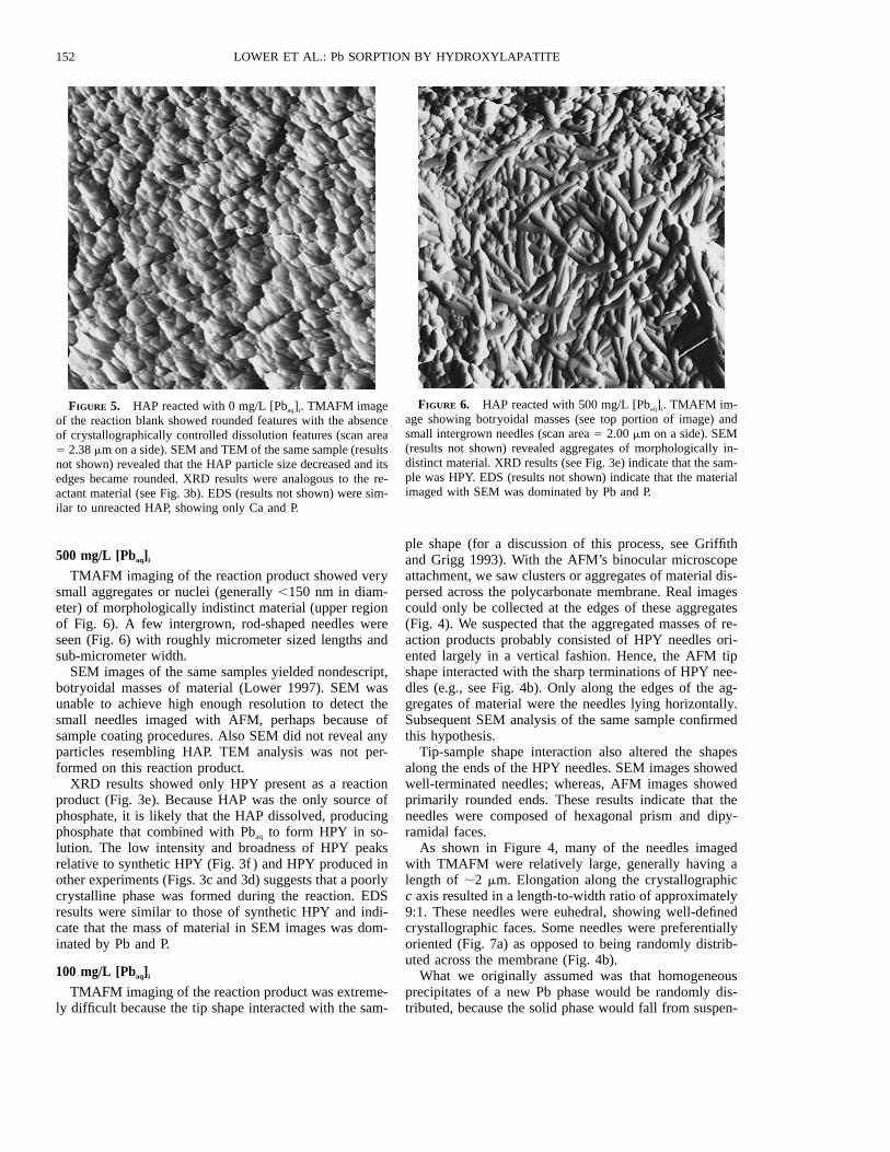
FIGURE 5. HAP reacted with 0 mg/L [Pbaq]i. TMAFM imageof the reaction blank showed rounded features with the absenceof crystallographically controlled dissolution features (scan area5 2.38 mm on a side). SEM and TEM of the same sample (resultsnot shown) revealed that the HAP particle size decreased and itsedges became rounded. XRD results were analogous to the re-actant material (see Fig. 3b). EDS (results not shown) were sim-ilar to unreacted HAP, showing only Ca and P.
FIGURE 6. HAP reacted with 500 mg/L [Pbaq]i. TMAFM im-age showing botryoidal masses (see top portion of image) andsmall intergrown needles (scan area 5 2.00 mm on a side). SEM(results not shown) revealed aggregates of morphologically in-distinct material. XRD results (see Fig. 3e) indicate that the sam-ple was HPY. EDS (results not shown) indicate that the materialimaged with SEM was dominated by Pb and P.
500 mg/L [Pbaq]i
TMAFM imaging of the reaction product showed verysmall aggregates or nuclei (generally ,150 nm in diam-eter) of morphologically indistinct material (upper regionof Fig. 6). A few intergrown, rod-shaped needles wereseen (Fig. 6) with roughly micrometer sized lengths andsub-micrometer width.
SEM images of the same samples yielded nondescript,botryoidal masses of material (Lower 1997). SEM wasunable to achieve high enough resolution to detect thesmall needles imaged with AFM, perhaps because ofsample coating procedures. Also SEM did not reveal anyparticles resembling HAP. TEM analysis was not per-formed on this reaction product.
XRD results showed only HPY present as a reactionproduct (Fig. 3e). Because HAP was the only source ofphosphate, it is likely that the HAP dissolved, producingphosphate that combined with Pbaq to form HPY in so-lution. The low intensity and broadness of HPY peaksrelative to synthetic HPY (Fig. 3f ) and HPY produced inother experiments (Figs. 3c and 3d) suggests that a poorlycrystalline phase was formed during the reaction. EDSresults were similar to those of synthetic HPY and indi-cate that the mass of material in SEM images was dom-inated by Pb and P.
100 mg/L [Pbaq]i
TMAFM imaging of the reaction product was extreme-ly difficult because the tip shape interacted with the sam-
ple shape (for a discussion of this process, see Griffithand Grigg 1993). With the AFM’s binocular microscopeattachment, we saw clusters or aggregates of material dis-persed across the polycarbonate membrane. Real imagescould only be collected at the edges of these aggregates(Fig. 4). We suspected that the aggregated masses of re-action products probably consisted of HPY needles ori-ented largely in a vertical fashion. Hence, the AFM tipshape interacted with the sharp terminations of HPY nee-dles (e.g., see Fig. 4b). Only along the edges of the ag-gregates of material were the needles lying horizontally.Subsequent SEM analysis of the same sample confirmedthis hypothesis.
Tip-sample shape interaction also altered the shapesalong the ends of the HPY needles. SEM images showedwell-terminated needles; whereas, AFM images showedprimarily rounded ends. These results indicate that theneedles were composed of hexagonal prism and dipy-ramidal faces.
As shown in Figure 4, many of the needles imagedwith TMAFM were relatively large, generally having alength of ;2 mm. Elongation along the crystallographicc axis resulted in a length-to-width ratio of approximately9:1. These needles were euhedral, showing well-definedcrystallographic faces. Some needles were preferentiallyoriented (Fig. 7a) as opposed to being randomly distrib-uted across the membrane (Fig. 4b).
What we originally assumed was that homogeneousprecipitates of a new Pb phase would be randomly dis-tributed, because the solid phase would fall from suspen-
153LOWER ET AL.: Pb SORPTION BY HYDROXYLAPATITE
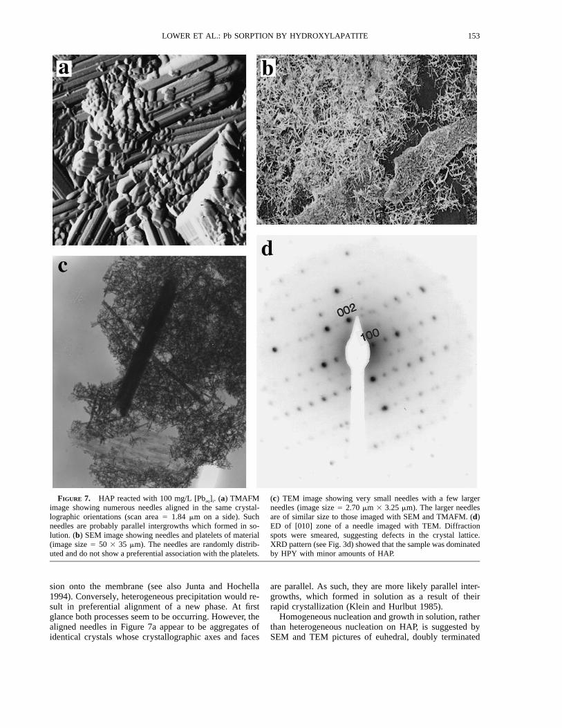
FIGURE 7. HAP reacted with 100 mg/L [Pbaq]i. (a) TMAFMimage showing numerous needles aligned in the same crystal-lographic orientations (scan area 5 1.84 mm on a side). Suchneedles are probably parallel intergrowths which formed in so-lution. (b) SEM image showing needles and platelets of material(image size 5 50 3 35 mm). The needles are randomly distrib-uted and do not show a preferential association with the platelets.
(c) TEM image showing very small needles with a few largerneedles (image size 5 2.70 mm 3 3.25 mm). The larger needlesare of similar size to those imaged with SEM and TMAFM. (d)ED of [010] zone of a needle imaged with TEM. Diffractionspots were smeared, suggesting defects in the crystal lattice.XRD pattern (see Fig. 3d) showed that the sample was dominatedby HPY with minor amounts of HAP.
sion onto the membrane (see also Junta and Hochella1994). Conversely, heterogeneous precipitation would re-sult in preferential alignment of a new phase. At firstglance both processes seem to be occurring. However, thealigned needles in Figure 7a appear to be aggregates ofidentical crystals whose crystallographic axes and faces
are parallel. As such, they are more likely parallel inter-growths, which formed in solution as a result of theirrapid crystallization (Klein and Hurlbut 1985).
Homogeneous nucleation and growth in solution, ratherthan heterogeneous nucleation on HAP, is suggested bySEM and TEM pictures of euhedral, doubly terminated
154 LOWER ET AL.: Pb SORPTION BY HYDROXYLAPATITE
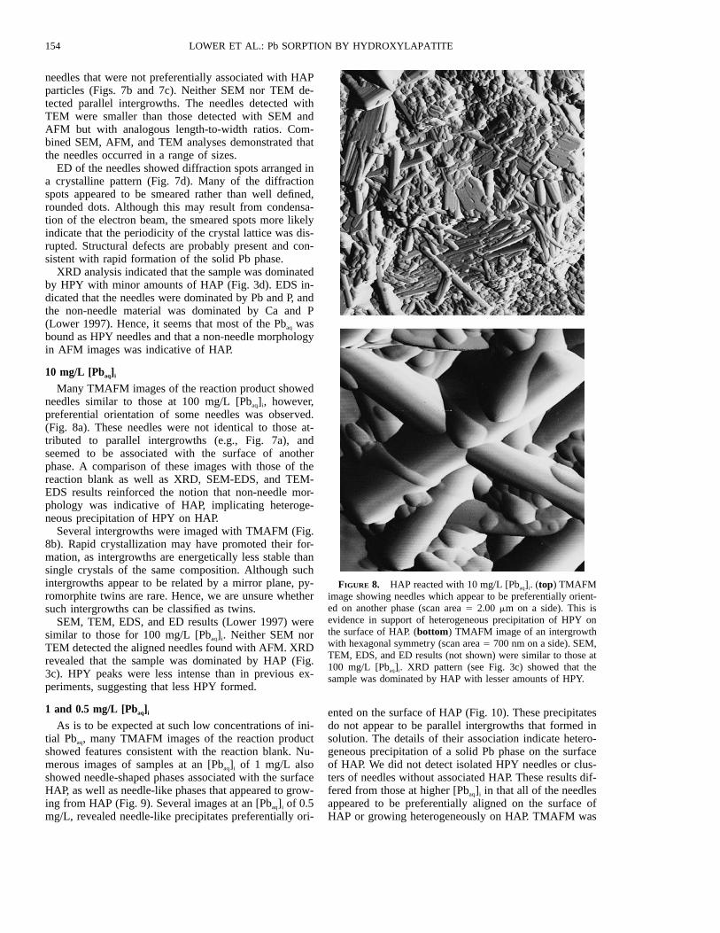
FIGURE 8. HAP reacted with 10 mg/L [Pbaq]i. (top) TMAFMimage showing needles which appear to be preferentially orient-ed on another phase (scan area 5 2.00 mm on a side). This isevidence in support of heterogeneous precipitation of HPY onthe surface of HAP. (bottom) TMAFM image of an intergrowthwith hexagonal symmetry (scan area 5 700 nm on a side). SEM,TEM, EDS, and ED results (not shown) were similar to those at100 mg/L [Pbaq]i. XRD pattern (see Fig. 3c) showed that thesample was dominated by HAP with lesser amounts of HPY.
needles that were not preferentially associated with HAPparticles (Figs. 7b and 7c). Neither SEM nor TEM de-tected parallel intergrowths. The needles detected withTEM were smaller than those detected with SEM andAFM but with analogous length-to-width ratios. Com-bined SEM, AFM, and TEM analyses demonstrated thatthe needles occurred in a range of sizes.
ED of the needles showed diffraction spots arranged ina crystalline pattern (Fig. 7d). Many of the diffractionspots appeared to be smeared rather than well defined,rounded dots. Although this may result from condensa-tion of the electron beam, the smeared spots more likelyindicate that the periodicity of the crystal lattice was dis-rupted. Structural defects are probably present and con-sistent with rapid formation of the solid Pb phase.
XRD analysis indicated that the sample was dominatedby HPY with minor amounts of HAP (Fig. 3d). EDS in-dicated that the needles were dominated by Pb and P, andthe non-needle material was dominated by Ca and P(Lower 1997). Hence, it seems that most of the Pbaq wasbound as HPY needles and that a non-needle morphologyin AFM images was indicative of HAP.
10 mg/L [Pbaq]i
Many TMAFM images of the reaction product showedneedles similar to those at 100 mg/L [Pbaq]i, however,preferential orientation of some needles was observed.(Fig. 8a). These needles were not identical to those at-tributed to parallel intergrowths (e.g., Fig. 7a), andseemed to be associated with the surface of anotherphase. A comparison of these images with those of thereaction blank as well as XRD, SEM-EDS, and TEM-EDS results reinforced the notion that non-needle mor-phology was indicative of HAP, implicating heteroge-neous precipitation of HPY on HAP.
Several intergrowths were imaged with TMAFM (Fig.8b). Rapid crystallization may have promoted their for-mation, as intergrowths are energetically less stable thansingle crystals of the same composition. Although suchintergrowths appear to be related by a mirror plane, py-romorphite twins are rare. Hence, we are unsure whethersuch intergrowths can be classified as twins.
SEM, TEM, EDS, and ED results (Lower 1997) weresimilar to those for 100 mg/L [Pbaq]i. Neither SEM norTEM detected the aligned needles found with AFM. XRDrevealed that the sample was dominated by HAP (Fig.3c). HPY peaks were less intense than in previous ex-periments, suggesting that less HPY formed.
1 and 0.5 mg/L [Pbaq]i
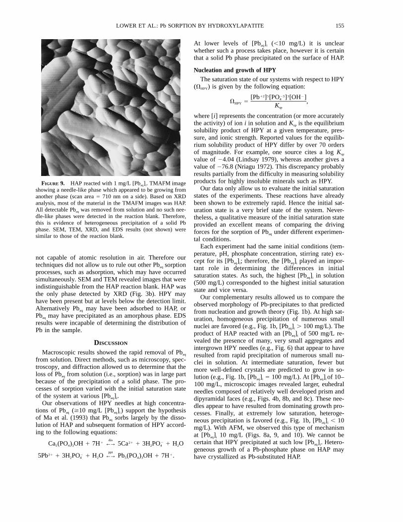
As is to be expected at such low concentrations of ini-tial Pbaq, many TMAFM images of the reaction productshowed features consistent with the reaction blank. Nu-merous images of samples at an [Pbaq]i of 1 mg/L alsoshowed needle-shaped phases associated with the surfaceHAP, as well as needle-like phases that appeared to grow-ing from HAP (Fig. 9). Several images at an [Pbaq]i of 0.5mg/L, revealed needle-like precipitates preferentially ori-
ented on the surface of HAP (Fig. 10). These precipitatesdo not appear to be parallel intergrowths that formed insolution. The details of their association indicate hetero-geneous precipitation of a solid Pb phase on the surfaceof HAP. We did not detect isolated HPY needles or clus-ters of needles without associated HAP. These results dif-fered from those at higher [Pbaq]i in that all of the needlesappeared to be preferentially aligned on the surface ofHAP or growing heterogeneously on HAP. TMAFM was
155LOWER ET AL.: Pb SORPTION BY HYDROXYLAPATITE
FIGURE 9. HAP reacted with 1 mg/L [Pbaq]i. TMAFM imageshowing a needle-like phase which appeared to be growing fromanother phase (scan area 5 710 nm on a side). Based on XRDanalysis, most of the material in the TMAFM images was HAP.All detectable Pbaq was removed from solution and no such nee-dle-like phases were detected in the reaction blank. Therefore,this is evidence of heterogeneous precipitation of a solid Pbphase. SEM, TEM, XRD, and EDS results (not shown) weresimilar to those of the reaction blank.
not capable of atomic resolution in air. Therefore ourtechniques did not allow us to rule out other Pbaq sorptionprocesses, such as adsorption, which may have occurredsimultaneously. SEM and TEM revealed images that wereindistinguishable from the HAP reaction blank. HAP wasthe only phase detected by XRD (Fig. 3b). HPY mayhave been present but at levels below the detection limit.Alternatively Pbaq may have been adsorbed to HAP, orPbaq may have precipitated as an amorphous phase. EDSresults were incapable of determining the distribution ofPb in the sample.
DISCUSSION
Macroscopic results showed the rapid removal of Pbaq
from solution. Direct methods, such as microscopy, spec-troscopy, and diffraction allowed us to determine that theloss of Pbaq from solution (i.e., sorption) was in large partbecause of the precipitation of a solid phase. The pro-cesses of sorption varied with the initial saturation stateof the system at various [Pbaq]i.
Our observations of HPY needles at high concentra-tions of Pbaq ($10 mg/L [Pbaq]i) support the hypothesisof Ma et al. (1993) that Pbaq sorbs largely by the disso-lution of HAP and subsequent formation of HPY accord-ing to the following equations:
dis1 21 2Ca (PO ) OH 1 7H ←→ 5Ca 1 3H PO 1 H O5 4 3 2 4 2
ppt21 2 15Pb 1 3H PO 1 H O ←→ Pb (PO ) OH 1 7H .2 4 2 5 4 3
At lower levels of [Pbaq]i (,10 mg/L) it is unclearwhether such a process takes place, however it is certainthat a solid Pb phase precipitated on the surface of HAP.
Nucleation and growth of HPYThe saturation state of our systems with respect to HPY
(VHPY) is given by the following equation:12 5 23 3 2[Pb ] [PO ] [OH ]4V 5 ,HPY Ksp
where [i] represents the concentration (or more accuratelythe activity) of ion i in solution and Ksp is the equilibriumsolubility product of HPY at a given temperature, pres-sure, and ionic strength. Reported values for the equilib-rium solubility product of HPY differ by over 70 ordersof magnitude. For example, one source cites a log Ksp
value of 24.04 (Lindsay 1979), whereas another gives avalue of 276.8 (Nriagu 1972). This discrepancy probablyresults partially from the difficulty in measuring solubilityproducts for highly insoluble minerals such as HPY.
Our data only allow us to evaluate the initial saturationstates of the experiments. These reactions have alreadybeen shown to be extremely rapid. Hence the initial sat-uration state is a very brief state of the system. Never-theless, a qualitative measure of the initial saturation stateprovided an excellent means of comparing the drivingforces for the sorption of Pbaq under different experimen-tal conditions.
Each experiment had the same initial conditions (tem-perature, pH, phosphate concentration, stirring rate) ex-cept for its [Pbaq]i; therefore, the [Pbaq]i played an impor-tant role in determining the differences in initialsaturation states. As such, the highest [Pbaq]i in solution(500 mg/L) corresponded to the highest initial saturationstate and vice versa.
Our complementary results allowed us to compare theobserved morphology of Pb-precipitates to that predictedfrom nucleation and growth theory (Fig. 1b). At high sat-uration, homogeneous precipitation of numerous smallnuclei are favored (e.g., Fig. 1b, [Pbaq]i . 100 mg/L). Theproduct of HAP reacted with an [Pbaq]i of 500 mg/L re-vealed the presence of many, very small aggregates andintergrown HPY needles (e.g., Fig. 6) that appear to haveresulted from rapid precipitation of numerous small nu-clei in solution. At intermediate saturation, fewer butmore well-defined crystals are predicted to grow in so-lution (e.g., Fig. 1b, [Pbaq]i 5 100 mg/L). At [Pbaq]i of 10–100 mg/L, microscopic images revealed larger, euhedralneedles composed of relatively well developed prism anddipyramidal faces (e.g., Figs. 4b, 8b, and 8c). These nee-dles appear to have resulted from dominating growth pro-cesses. Finally, at extremely low saturation, heteroge-neous precipitation is favored (e.g., Fig. 1b, [Pbaq]i , 10mg/L). With AFM, we observed this type of mechanismat [Pbaq]i 10 mg/L (Figs. 8a, 9, and 10). We cannot becertain that HPY precipitated at such low [Pbaq]i. Hetero-geneous growth of a Pb-phosphate phase on HAP mayhave crystallized as Pb-substituted HAP.
156 LOWER ET AL.: Pb SORPTION BY HYDROXYLAPATITE
FIGURE 10. HAP reacted with 0.5 mg/L [Pbaq]i. TMAFM im-age showing a needle-like phase preferentially oriented on thesurface of another phase (scan area 5 1.60 mm on a side). Cou-pled with other results, this is evidence of heterogeneous nucle-ation of a solid Pb phase on the surface of HAP. SEM, TEM,XRD, and EDS results (not shown) were similar to those of thereaction blank.
FIGURE 11. HAP reacted with 100 mg/L [Pbaq]i and allowedto age for several days. SEM image of needles of HPY (imagesize 5 23 3 16 mm). The needles of HPY grew significantlylarger because of Ostwald ripening (compare with Fig. 8b); yet,some small needles remained as rosettes on the surface of theselarger needles.
Sample aging effectsThe experiment with an [Pbaq]i of 100 mg/L was al-
lowed to react for another five days, filtered and analyzedwith TMAFM and SEM. TMAFM was unsuccessful be-cause of tip-sample shape interaction as discussed above.SEM results showed much larger needles than those im-aged after 2 h (e.g., length .25 mm and width .5 mm),and many had small rosettes on their surfaces (Fig. 11).EDS showed that these needles were dominated by Pband P.
The needles of HPY appear to increase in size as theyaged in solution, perhaps because of Ostwald ripening(Steefel and van Cappellen 1990). The small needles ofHPY that precipitated after 2 h (e.g., Fig. 7b) had a largersurface area per unit volume relative to the aged needles,and therefore they may have been unstable. Apparently,the small HPY crystals were a precursor phase that un-derwent a transformation to a more stable morphology.Ostwald ripening generally occurs by the gradual increasein particle size, so that we cannot at present explain thebimodal particle size distribution (large needles vs. smallrosettes). Although the smaller rosettes may have precip-itated upon drying, this is unlikely given the drying pro-cedure that we used.
In most cases HAP removes all detectable Pbaq fromsolution in a matter of minutes to hours. Hence the systemis in apparent macroscopic equilibrium with respect toPbaq. However these results indicate that although the sys-tem may have reached a macroscopic equilibrium, it hadnot achieved microscopic equilibrium. This result may
have important implications for use of apatites to reme-diate Pb contaminated soils, in that the alternating wetand dry cycles of soil environments may influence thekinetics of the overall process.
Comparison of AFM, SEM, and TEMThis study benefited greatly from the complementary
information provided by each form of microscopy. Ingeneral, we found that it was rewarding to apply all threetechniques to the same sample to gain a better under-standing of reaction processes. AFM provided higher res-olution of surfaces and morphological details that pro-moted the comparison of microscopic images tomacroscopic results in greater detail, especially at thelowest [Pbaq]i. SEM provided better images of larger nee-dles (i.e., needles with lengths .2 mm), and TEM pro-vided the best images of extremely small needles (i.e.,needles with lengths ,1 mm). Spectroscopic and diffrac-tion capabilities of SEM and TEM facilitated identifica-tion and allowed determination of phase association inheterogeneous samples.
A major disadvantage of AFM was tip-sample shape in-teraction that commonly precluded imaging and sometimesdistorted morphology (i.e., rounding of the ends of needles).This study demonstrated the importance of applying otherforms of microscopy to validate AFM observations. Anotherdisadvantage of AFM was that its analysis time was muchlonger than those for similar samples with SEM and TEM.Disadvantages of SEM and TEM lie in the destructive na-ture of these techniques. For example, samples are coatedwith carbon or gold. Furthermore, the electron beam canconvert HAP to other Ca-phosphate phases during imaging(Huaxia and Marquis 1991).
Finally, AFM can be operated in ambient environmentalconditions. Hence, the validation of AFM in air allows sucha technique to be applied to a solution environment to ob-
157LOWER ET AL.: Pb SORPTION BY HYDROXYLAPATITE
serve reactions in real time. It is perhaps this final point thatsets AFM apart from the other forms of microscopy.
ACKNOWLEDGMENTS
This research benefited greatly from discussions with many colleagues,including: V. Laperche (Ohio State University), J. Hughes (Miami Uni-versity), and M.F. Hochella Jr. and his research group at Virginia Poly-technic Institute and State University. M. Manecki (Kent State University)and R. Klouda (Liquid Crystal Institute) provided assistance with SEM;H. Colijn (Ohio State University) provided assistance with TEM and ED.Funding for S.K.L. was provided by the Water Resources Research Insti-tute at Kent State University. S.K.L. also thanks J. Lower and T. Lowerfor their continued support and encouragement. Finally, we thank J. Ra-kovan (Virginia Polytechnic Institute and State University) and an anon-ymous reviewer for their helpful comments.
REFERENCES CITED
Barron, V., Galvez, N., Hochella, M.F. Jr., and Torrent, J. (1997) Epitaxialovergrowth of goethite on hematite synthesized in phosphate media: Ascanning force and transmission electron microscopy study. AmericanMineralogist, 83, 1089–1098.
Benjamin, M.M. and Leckie, J.O. (1981) Multi-site adsorption of Cd, Co,Zn, and Pb on amorphous iron oxyhydroxide. Journal of Colloid andInterface Science, 79, 209–221.
Berner, R.A. (1978) Rate control of mineral dissolution under earth surfaceconditions. American Journal of Science, 278, 1235–1252.
(1981) Kinetics of weathering and diagenesis. In MineralogicalSociety of America Reviews in Mineralogy, 8, 111–134.
Davis, A., Ruby, M.V., and Bergstrom, P.D. (1992) Mineralogic controlson arsenic and lead bioavailability in soils from the Butte mining dis-trict, Montana, USA. Environmental Science and Technology, 26, 461–468.
Davis, A., Drexler, J.W., Ruby, M.V., and Nicholson, A. (1993) Micro-mineralogy of mine wastes in relation to lead bioavailability, Butte,Montana. Environmental Science and Technology, 27, 1415–1425.
Drever, J.I. (1988) The geochemistry of natural waters (2nd edition), 437p. Prentice Hall, Englewood Cliffs, New Jersey.
Eggleston, C. (1994) High resolution scanning probe microscopy: Tip-surface interaction, artifacts, and applications in mineralogy and geo-chemistry. In A.E. Blum and K. Nagy, Eds., Scanning probe micros-copy of clay minerals, p. 1–90. Clay Minerals Society, Boulder,Colorado.
Elliott, J.C. (1971) Monoclinic space group of hydroxyapatite. NaturePhysical Science, 230, 72.
Griffith, J.E. and Grigg, D.A. (1993) Dimensional metrology with scan-ning probe microscopes. Journal of Applied Physics, 74, R83–R109.
Hochella, M.F. Jr. (1990) Atomic structure, microtopography, composition,and reactivity of mineral surfaces. In Mineralogical Society of AmericaReviews in Mineralogy, 23, 87–132.
Huaxia, J. and Marquis, P.M. (1991) Modification of hydroxyapatite dur-ing transmission electron microscopy. Journal of Materials Science Let-ters, 10, 132–134.
Hughes, J.M., Cameron, M., and Crowley, D.K. (1989) Structural varia-tions in natural F, OH, and Cl apatites. American Mineralogist, 74, 870–876.
(1990) Crystal structures of natural ternary apatites: Solid solutionin the Ca5(PO4)3X (X 5 F, OH, Cl) system. American Mineralogist, 75,295–304.
Jackson, M.L. (1969) Soil chemical analysis-Advanced course (2nd edi-tion), 845 p. Department of Soil Science, University of Madison,Wisconsin.
Junta, J.L. and Hochella, M.F. Jr. (1994) Manganese (II) oxidation at min-eral surfaces: A microscopic and spectroscopic study. Geochimica etCosmochimica Acta, 58, 4985–4999.
Kay, M.I., Young, R.A., and Posner, A.S. (1964) Crystal structure of hy-droxyapatite. Nature, 204, 1050–1052.
Klein, C. and Hurlbut, C.S. Jr. (1985) Manual of mineralogy (20th edi-tion), 596 p. Wiley, New York.
Lindsay, W.L. (1979) Chemical equilibria in soils, 449 p. Wiley, NewYork.
Lower, S.K. (1997) Pb sorption to hydroxylapatite: A microscopic, spec-troscopic, and diffraction study, 170 p. M.S. thesis, Kent State Univer-sity, Kent, Ohio.
Ma, Q.Y., Traina, S.J., Logan, T.J., and Ryan, J.A. (1993) In situ leadimmobilization by apatite. Environmental Science and Technology, 27,1803–1810.
Ma, Q.Y., Logan, T.J., Traina, S.J., and Ryan, J.A. (1994a) Effects ofNO3
2, Cl2, F2, SO422, and CO3
22 on Pb21 immobilization by hydroxy-apatite. Environmental Science and Technology, 28, 408–418.
Ma, Q.Y., Traina, S.J., Logan, T.J., and Ryan, J.A. (1994b) Effects ofaqueous Al, Cd, Cu, Fe(II), Ni, and Zn on Pb immobilization by hy-droxyapatite. Environmental Science and Technology, 28, 1219–1228.
Ma, Q.Y., Logan, T.J., and Traina, S.J. (1995) Lead immobilization fromaqueous solutions and contaminated soils using phosphate rocks. En-vironmental Science and Technology, 29, 1118–1126.
Maurice, P.A. and Lower, S.K. (1998) Using atomic force microscopy tostudy soil mineral reactions. Advances in Agronomy, in press.
Moore, D.M. and Reynolds, R.C. Jr. (1989) X-ray diffraction and identi-fication and analysis of clay minerals, 332 p. Oxford University Press,New York.
Morris, M.C., McMurdie, H.F., Evans, E.H., Pavetzkin, B., Parker, H.S.,Pyrros, N.P., and Hubbard, C.R. (1984) Standard X-ray diffraction pow-der patterns-section 20, 145 p. National Bureau of Standards, Washing-ton, D.C.
Murphy, J. and Riley, J.P. (1962) A modified single solution method forthe determination of phosphate in natural waters. Analytica ChimicaActa, 27, 31–36.
Nagy, K. and Blum, A.E. (1994) Scanning probe microscopy of clay min-erals, 239 p. Clay Minerals Society, Boulder, Colorado.
Narasaraju, T.S.B., Singh, R.P., and Rao, V.L.N. (1972) A new method ofpreparation of solid solutions of calcium and lead hydroxylapatite. Jour-nal of Inorganic and Nuclear Chemistry, 34, 2072–2074.
Nash, W.P. (1984) Phosphate minerals in terrestrial igneous and metamor-phic rocks. In J.O. Nriagu and P.B. Moore, Eds., Phosphate minerals,p. 215–241. Springer, Berlin.
Nielsen, A.E. (1964) The kinetics of precipitation, 151 p. MacMillan, NewYork.
Nriagu, J.O. (1972) Lead orthophosphates: I. Solubility and hydrolysis ofsecondary lead orthophosphate. Inorganic Chemistry, 11, 2499–2503.
(1973a) Lead orthophosphates: II. Stability of chloropyromorphiteat 258 C. Geochimica et Cosmochimica Acta, 37, 367–377.
(1973b) Lead orthophosphates: III. Stability of fluoropyromorphiteand bromopyromorphite at 258 C. Geochimica et Cosmochimica Acta,37, 1735–1743.
(1984) Formation and stability of base metal phosphates in soilsand sediments. In J.O. Nriagu and P.B. Moore, Eds., Phosphate min-erals, p. 319–329. Springer, Berlin.
O’Day, P.A., Brown, G.E., and Parks, G.A. (1994) X-ray absorption spec-troscopy of cobalt(II) multinuclear surface complexes and surface pre-cipitates on kaolinite. Journal of Colloid and Interface Science, 165,269–289.
Parks, G.A. (1990) Surface energy and adsorption at mineral-water inter-faces: An introduction. In Mineralogical Society of America Reviewsin Mineralogy, 23, 133–175.
Posner, A.S., Blumenthal, N.C., and Betts, F. (1984) Chemistry and struc-ture of precipitated hydroxyapatites. In J.O. Nriagu and P.B. Moore,Eds., Phosphate minerals, p. 330–350. Springer, Berlin.
Ruby, M.V., Davis, A., Kempton, J.H., Drexler, J.W., and Bergstron, P.D.(1992) Lead bioavailability: Dissolution kinetics under simulated gastricconditions. Environmental Science and Technology, 26, 1242.
Ruby, M.V., Davis, A., and Nicholson, A. (1994) In situ formation of leadphosphates in soils as a method to immobilize lead. Environmental Sci-ence and Technology, 28, 646–654.
Scheidegger, A.M. and Sparks, D.L. (1996) Kinetics of formation anddissolution of nickel surface precipitates on pyrophyllite. Chemical Ge-ology, 132, 157–164.
158 LOWER ET AL.: Pb SORPTION BY HYDROXYLAPATITE
Schwertmann, U. and Cornell, R.M. (1991) Iron oxides in the laboratory,132 p. Weinheim, New York.
Sposito, G. (1986) Distinguishing adsorption from surface precipitation.In J.A. Davis and K.F. Hayes, Eds., Geochemical processes at mineralsurfaces, p. 217–228. American Chemical Society, Washington.
Steefel, C.I. and Van Cappellen, P. (1990) A new kinetic approach tomodeling water-rock interaction: The role of nucleation, precursors, andOstwald ripening. Geochimica et Cosmochimica Acta, 54, 2657–2677.
Stumm, W. and Morgan, J.J. (1981) Aquatic chemistry (2nd edition), 796p. Wiley, New York.
Suzuki, T., Hatsushika, T., and Hayakawa, Y. (1981) Synthetic hydroxy-apatites employed as inorganic cation exchangers. Journal of the Chem-ical Society Faraday Transactions I, 77, 1059–1062.
Suzuki, T., Hatsushika, T., Miyake, M. (1982) Synthetic hydroxyapatitesas inorganic cation exchangers: 2. Journal of the Chemical Society Far-aday Transactions I, 78, 3605–3611.
Suzuki, T., Ishigaki, K., Miyake, M. (1984) Synthetic hydroxyapatites asinorganic cation exchangers: 3. Exchange characteristics of lead ions
(Pb21). Journal of the Chemical Society Faraday Transactions I, 80,3157–3165.
Takeuchi, Y. and Arai, H. (1990) Removal of coexisting Pb21, Cu21 andCd21 ions from water by addition of hydroxyapatite powder, 3. pH andsample conditioning effects. Journal of Chemical Engineering of Japan,23, 75–80.
Takeuchi, Y., Suzuki, T., and Arai, H.J. (1988) A study of equilibrium andmass transfer in processes for removal of heavy-metal ions by hydrox-yapatite. Journal of Chemical Engineering of Japan, 21, 98–100.
Tiselius, A., Hjerten, S., and Levin, O. (1956) Protein chromatography oncalcium phosphate columns. Archives of Biochemistry and Biophysics,65, 132–155.
Xu, Y. and Schwartz, F.W. (1994) Lead immobilization by hydroxyapatitein aqueous solutions. Journal of Contaminant Hydrology, 15, 187–206.
Zhong, Q., Inniss, D., Kjoller, K., and Elings, V.B. (1993) Fractured poly-mer/silica fiber surface studied by tapping mode atomic force micros-copy. Surface Science Letters, 290, L688–L692.
MANUSCRIPT RECEIVED APRIL 14, 1997MANUSCRIPT ACCEPTED SEPTEMBER 5, 1997