Hamer et al. BMC Microbiology 2014, 14:223http://www.biomedcentral.com/1471-2180/14/223
RESEARCH ARTICLE Open Access
Replication of Brucella abortus and Brucellamelitensis in fibroblasts does not requireAtg5-dependent macroautophagyIsabelle Hamer1*, Emeline Goffin1,3, Xavier De Bolle2, Jean-Jacques Letesson2 and Michel Jadot1
Abstract
Background: Several intracellular bacterial pathogens have evolved subtle strategies to subvert vesicular traffickingpathways of their host cells to avoid killing and to replicate inside the cells. Brucellae are Gram-negative facultativeintracellular bacteria that are responsible for brucellosis, a worldwide extended chronic zoonosis. Following invasion,Brucella abortus is found in a vacuole that interacts first with various endosomal compartments and then withendoplasmic reticulum sub-compartments. Brucella establishes its replication niche in ER-derived vesicles. In thepast, it has been proposed that B. abortus passed through the macroautophagy pathway before reaching its nicheof replication. However, recent experiments provided evidence that the classical macroautophagy pathway was notinvolved in the intracellular trafficking and the replication of B. abortus in bone marrow-derived macrophages andin HeLa cells. In contrast, another study showed that macroautophagy favoured the survival and the replication ofBrucella melitensis in infected RAW264.7 macrophages. This raises the possibility that B. abortus and B. melitensisfollowed different intracellular pathways before replicating. In the present work, we have addressed this issue bycomparing the replication rate of B. abortus and B. melitensis in embryonic fibroblasts derived from wild-type andAtg5−/− mice, Atg5 being a core component of the canonical macroautophagic pathway.
Results: Our results indicate that both B. abortus S2308 and B. melitensis 16M strains are able to invade andreplicate in Atg5-deficient fibroblasts, suggesting that the canonical Atg5-dependent macroautophagic pathway isdispensable for Brucella replication. The number of viable bacteria was even slightly higher in Atg5−/− fibroblaststhan in wild-type fibroblasts. This increase could be due to a more efficient uptake or to a better survival rate ofbacteria before the beginning of the replication in Atg5-deficient cells as compared to wild-type cells. Moreover,our data show that the infection with B. abortus or with B. melitensis does not stimulate neither the conversion ofLC3-I to LC3-II nor the membrane recruitment of LC3 onto the BCV.
Conclusion: Our study suggests that like Brucella abortus, Brucella melitensis does not subvert the canonicalmacroautophagy to reach its replicative niche or to stimulate its replication.
BackgroundMany intracellular bacteria have developed strategiesto hijack the intracellular trafficking machinery of thehost cell in order to escape lysosomal degradation ensuringtheir survival and their replication [1]. For example, Myco-bacterium tuberculosis blocks the maturation of phago-somes into the degradative phagolysosomes by producing
* Correspondence: [email protected] Unit in Molecular Physiology (URPhyM), NAmur Research Institutefor LIfe Sciences (NARILIS), University of Namur, Namur, BelgiumFull list of author information is available at the end of the article
lipids that mimic the phosphoinositides and inhibit thefusion between phagosomes and lysosomes [2]. Somebacteria, including Coxiella burnetii, Legionella pneu-mophila and Staphylococcus aureus can survive andreplicate for some time in autophagosome-like vacuolesby delaying [3,4] or by blocking [5] their maturation intoautophagolysosomes.After its uptake by HeLa cells, Brucella abortus is recov-
ered in a vacuole (BCV) that transiently interacts withearly and late endosomes and perhaps lysosomes, succes-sively acquiring markers of endosomal compartments such
Ltd. This is an Open Access article distributed under the terms of the Creativeommons.org/licenses/by/4.0), which permits unrestricted use, distribution, andiginal work is properly credited. The Creative Commons Public Domaing/publicdomain/zero/1.0/) applies to the data made available in this article,
Hamer et al. BMC Microbiology 2014, 14:223 Page 2 of 9http://www.biomedcentral.com/1471-2180/14/223
as EEA1 (Early Endosome Antigen 1), Rab5, Rab7 andLAMP-1 (Lysosomal-associated membrane protein 1)[6]. During these different steps of maturation, the BCVbecomes acidic allowing the expression of genes encodingthe VirB type IV secretion system (T4SS) [6]. Brucellaavoids lysosomal degradation by blocking the phagosome-lysosome fusion probably by a mechanism dependenton lipid rafts and perhaps on cyclic ß-1,2-glucans [7–9].Afterwards, the BCV interacts in a sustained way withsubdomains of the endoplasmic reticulum, called ERES(endoplasmic reticulum exit sites) and at around 12 h p.i.,Brucella abortus starts to replicate in ER-derived vesicleslabelled with ER specific markers, such as sec61ß and cal-nexin [6,10,11]. Later on, from 48 h p.i., Starr et al. [12]demonstrated that these replicative BCV (rBCV) could beconverted into LAMP-1 and Rab7-positive compartments(called autophagic BCV or aBCV) that would be involvedin the completion of the intracellular Brucella lifecycleand could promote its cell-to-cell spreading [12].Earlier studies had already revealed that some bacteria
resided in autophagosome-like vacuoles characterized bymultilamellar membranes after 24 h of infection and thatBrucella replication was increased when macroautophagywas activated by serum starvation, suggesting that B. abor-tus transits through the autophagic pathway before reach-ing its replicative compartment [11,13]. Since then, manyproteins implicated in the regulation of macroautophagy(Atg proteins) have been discovered [14,15]. The initiationof autophagosome formation requires the ULK complexand the class III phosphatidylinositol 3-P kinase (PI3K)complex. The nucleation of the isolation membrane re-quires the recruitment of additional Atg proteins andautophagy-specific PtdIns(3)P effectors [14,15]. Theexpansion of the isolation membrane relies on twoubiquitylation-like reactions. The first one drives theconjugation of Atg12 to Atg5 in the presence of Atg7and Atg10. Atg5-Atg12 conjugates are non-covalentlyassociated to Atg16L (Atg16-like) forming multimericcomplexes of approximately 800 kDa [16–18]. The sec-ond reaction conjugates the cytosolic soluble LC3-I(microtubule-associated protein 1 light chain 3) to a phos-phatidylethanolamine (PE) in the presence of Atg4, Atg3and Atg7 producing the membrane-associated LC3-IIform [19–21]. The Atg5-Atg12 conjugates are essentialfor the maturation of the isolation membrane into autop-hagosome and targeting of LC3 to the membrane [18].Recently, using epithelial cells and macrophages defi-
cient in one of the regulatory proteins of the conventionalmacroautophagic pathway, Starr et al. [12] have found thatcore proteins of this canonical macroautophagy machinerysuch as ULK-1, Beclin1, Atg5, Atg7, LC3B were not neces-sary for the intracellular trafficking of B. abortus betweenthe endocytic compartments and the ER-derived vesiclesand for its replication [12]. Nevertheless, the conversion
of rBCV to aBCV at a later stage of infection, i.e. 48 h and72 h p.i., seems to be dependent on ULK-1, Beclin1,Atg14L and hVps34 but independent on Atg5, Atg7,Atg16L1 and Atg4B [12]. On the other hand, Guo et al.[22] have observed that infection by B. melitensis inducedmacroautophagy that in turn favoured its replication inRAW264.7 macrophages [22]. This later study raises thepossibility that in contrast to B. abortus, B. melitensiscould subvert macroautophagy to replicate in host cells.In our present work, we addressed this issue using embry-onic fibroblasts from wild-type and Atg5-knockout miceinfected or not with B. abortus and B. melitensis.
ResultsRelative abundance of LC3-I and LC3-II in infected mouseembryonic fibroblastsAs it has been shown that B. melitensis stimulated macro-autophagy in macrophages to favour its replication [22],we sought to determine whether this also occurred in in-fected MEFs. First, we established clones stably transfectedwith GFP-LC3 to monitor the formation of autophagicvacuoles by fluorescence microscopy. As expected [19], inbasal conditions, the fluorescent staining in GFP-LC3 ex-pressing cells was faint and diffuse while under starvationconditions, it was more punctuate, due to the recruitmentof LC3 onto autophagosomal membranes (Additional file 1).In contrast, when the same cells were infected with B. abor-tus or with B. melitensis, the GFP-LC3 staining remaineddiffuse and colocalisation between GFP-LC3 and TexasRed-labelled bacteria was only very occasionally detected.Then, we examined the relative abundance of LC3-I andLC3-II by Western blotting. Preliminary experimentsshowed that in WT MEFs, LC3-II was detected even inbasal conditions (Figure 1A). After 2 h of starvation inEBSS, the abundance of both LC3-I and LC3-II decreased,probably due to an acceleration of the autophagic flowsince LC3-II is degraded when autophagosomes fuse withlysosomes. In contrast, the LC3-II/LC3-I ratio increasedin the presence of bafilomycin, a vacuolar H+-ATPase in-hibitor known to block autophagosome/lysosome fusion.As expected, in Atg5−/− MEFs, LC3-II was never detectedwhatever the cell culture conditions because the presenceof Atg5 is absolutely required for the LC3 recruitmentonto autophagosome membrane [19]. In WT MEFs in-fected with B. abortus or with B. melitensis, the relativeabundance of LC3-I and LC3-II at 18 h p.i. did not changewhen compared to non-infected MEFs (Figure 1B).
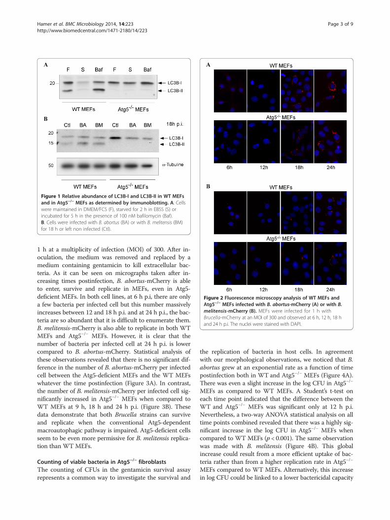
Replication of B. abortus- and B. melitensis-mCherry inAtg5−/− fibroblastsWe studied the contribution of the macroautophagic path-way on the replication of Brucellae using Atg5-deficientMEFs. First, we infected cells with B. abortus-mCherry(Figure 2A) or with B. melitensis-mCherry (Figure 2B) for
Figure 2 Fluorescence microscopy analysis of WT MEFs andAtg5−/− MEFs infected with B. abortus-mCherry (A) or with B.melitensis-mCherry (B). MEFs were infected for 1 h withBrucella-mCherry at an MOI of 300 and observed at 6 h, 12 h, 18 hand 24 h p.i. The nuclei were stained with DAPI.
Figure 1 Relative abundance of LC3B-I and LC3B-II in WT MEFsand in Atg5−/− MEFs as determined by immunoblotting. A. Cellswere maintained in DMEM/FCS (F), starved for 2 h in EBSS (S) orincubated for 5 h in the presence of 100 nM bafilomycin (Baf).B. Cells were infected with B. abortus (BA) or with B. melitensis (BM)for 18 h or left non infected (Ctl).
Hamer et al. BMC Microbiology 2014, 14:223 Page 3 of 9http://www.biomedcentral.com/1471-2180/14/223
1 h at a multiplicity of infection (MOI) of 300. After in-oculation, the medium was removed and replaced by amedium containing gentamicin to kill extracellular bac-teria. As it can be seen on micrographs taken after in-creasing times postinfection, B. abortus-mCherry is ableto enter, survive and replicate in MEFs, even in Atg5-deficient MEFs. In both cell lines, at 6 h p.i, there are onlya few bacteria per infected cell but this number massivelyincreases between 12 and 18 h p.i. and at 24 h p.i., the bac-teria are so abundant that it is difficult to enumerate them.B. melitensis-mCherry is also able to replicate in both WTMEFs and Atg5−/− MEFs. However, it is clear that thenumber of bacteria per infected cell at 24 h p.i. is lowercompared to B. abortus-mCherry. Statistical analysis ofthese observations revealed that there is no significant dif-ference in the number of B. abortus-mCherry per infectedcell between the Atg5-deficient MEFs and the WT MEFswhatever the time postinfection (Figure 3A). In contrast,the number of B. melitensis-mCherry per infected cell sig-nificantly increased in Atg5−/− MEFs when compared toWT MEFs at 9 h, 18 h and 24 h p.i. (Figure 3B). Thesedata demonstrate that both Brucella strains can surviveand replicate when the conventional Atg5-dependentmacroautophagic pathway is impaired. Atg5-deficient cellsseem to be even more permissive for B. melitensis replica-tion than WTMEFs.
Counting of viable bacteria in Atg5−/− fibroblastsThe counting of CFUs in the gentamicin survival assayrepresents a common way to investigate the survival and
the replication of bacteria in host cells. In agreementwith our morphological observations, we noticed that B.abortus grew at an exponential rate as a function of timepostinfection both in WT and Atg5−/− MEFs (Figure 4A).There was even a slight increase in the log CFU in Atg5−/−
MEFs as compared to WT MEFs. A Student’s t-test oneach time point indicated that the difference between theWT and Atg5−/− MEFs was significant only at 12 h p.i.Nevertheless, a two-way ANOVA statistical analysis on alltime points combined revealed that there was a highly sig-nificant increase in the log CFU in Atg5−/− MEFs whencompared to WT MEFs (p < 0.001). The same observationwas made with B. melitensis (Figure 4B). This globalincrease could result from a more efficient uptake of bac-teria rather than from a higher replication rate in Atg5−/−
MEFs compared to WT MEFs. Alternatively, this increasein log CFU could be linked to a lower bactericidal capacity
0
25
50
75
100
125
150
175
200
225
0 5 10 15 20 25 30
Nu
mb
er o
f b
acte
ria/
infe
cted
cel
l N
um
ber
of
bac
teri
a/in
fect
ed c
ell
Time postinfection (h)
WT MEFs
Atg5-/- MEFs
0
5
10
15
20
25
30
35
40
45
50
55
0 5 10 15 20 25 30
Time postinfection (h)
WT MEFs
Atg5-/- MEFs
#
##
##
A
B
Figure 3 Quantification of the infection of WT MEFs and Atg5−/−
MEFs with B. abortus-mCherry (A) or with B. melitensis-mCherry(B). MEFs were infected for 1 h with Brucella-mCherry at an MOI of300. Cells were observed by fluorescence microscopy at 6 h, 9 h,12 h, 18 h and 24 h p.i. Values represent the number of bacteria perinfected cell as means ± SEM with n ≥ 50, where n is the number ofobserved infected cells. Statistical significance was calculated usingthe Mann–Whitney Rank Sum Test. # and ## indicate a significantdifference with p <0.05 and p <0.01, respectively.
0
1
2
3
4
5
6
0 5 10 15 20 25 30
log
CF
U
Time postinfection (h)
WT MEFs
Atg5-/- MEFs
***
0
1
2
3
4
5
6
0 5 10 15 20 25 30
log
CF
U
Time postinfection (h)
WT MEFs
Atg5-/- MEFs
10 15 20
Time postinfection (h)
WT
Atg5
***
***
A
B
Figure 4 Intracellular growth of Brucella in WT and Atg5 −/−
MEFs. MEFs were infected for 1 h with B. abortus S2308 (A) or withB. melitensis 16M (B) at an MOI of 300. Log CFUs were obtainedfrom cell lysates of infected WT MEFs and Atg5−/− MEFs at the indicatedtime after infection. Results represent means ± SD measured fromat least three independent experiments made in triplicates. Statisticalsignificance was calculated using the Holm-Sidak multiple comparisonstest following a two-way ANOVA. p < 0.001 for both B. abortus andB. melitensis. *** indicates a highly significant difference using aStudent’s t-test.
Hamer et al. BMC Microbiology 2014, 14:223 Page 4 of 9http://www.biomedcentral.com/1471-2180/14/223
of Atg5-deficient cells compared to WT cells at earlystages of infection.
Intracellular replication of B. abortus and B. melitensis inthe presence of 3-methyladeninePrevious studies have shown that incubation of cells inthe presence of 3-methyladenine (3MA), an inhibitor ofclass III PI3K often used to block macroautophagy [23],impaired the replication of B. abortus [13] and B. meli-tensis [22] in HeLa cells and in RAW264.7 macrophages,respectively. These data are in contradiction with our re-sults showing that both bacterial strains are able to repli-cate in Atg5-deficient MEFs. Therefore, we sought todetermine the putative impact of 3MA on the replicationof Brucellae in WT MEFs. First, we assessed the numberof B. abortus-mCherry per infected cell in WT MEFspreincubated for 2 h in the presence or absence of 10 mM3MA. As shown in Figure 5A, this treatment had no
significant impact on the number of bacteria per infectedWT MEF. Similar results were obtained with WT MEFsinfected with B. melitensis-mCherry (Figure 5B). However,in this case, we observed a significant decrease (p < 0.01)in the number of bacteria per infected cell but only at24 h p.i. Next, we examined the impact of a pre-treatmentwith 3MA on Brucella replication in host cells usingthe gentamicin survival assay. Our results show that apre-incubation of WT MEFs with 3MA does not im-pair the replication of both B. abortus and B. melitensis(Figure 6 A-B).
DiscussionAfter internalisation, B. abortus is found inside individualvacuoles that interact transiently with endosomes and per-haps lysosomes [6]. Then, Brucella evades the endocyticpathway and reaches its replicative niche, an ER-derived
A
B
0
50
100
150
200
250
6 12 19 24
Nu
mb
er o
f b
acte
ria/
infe
cted
cel
l
Time postinfection (h)
WT MEFs
WT MEFs with 3MA
NS
NS
NS
#
0
5
10
15
20
25
30
35
6 12 19 24
Nu
mb
er o
f b
acte
ria/
infe
cted
cel
l
Time postinfection (h)
WT MEFs
WT MEFs with 3MA
NS
NS
##
#
Figure 5 Impact of 3MA on the infection of WT MEFs with B. abortus-mCherry (A) or with B. melitensis-mCherry (B). The number ofbacteria per infected cell was measured on at least 57 infected cells coming from two independent experiments. Values represent means ± SEM.Statistical significance was calculated using the Mann–Whitney Rank Sum Test. # and ## indicate a significant difference with p <0.05 and p <0.01,respectively. NS stands for non significant difference.
Hamer et al. BMC Microbiology 2014, 14:223 Page 5 of 9http://www.biomedcentral.com/1471-2180/14/223
compartment, by a still unknown mechanism. It is alsounclear whether Brucella transits through the autophagicpathway before its replication. Based on the appearance ofB. abortus in multilamellar structures looking like autop-hagosomes and on the decrease of its replication rate afterautophagy inhibition with 3MA, Pizarro-Cerda et al. [11]proposed that this bacterium passed through the autoph-agy pathway before reaching its niche of replication [13].In agreement with this assumption, Guo et al. (2012) no-ticed that inoculation of macrophages with B. melitensisstimulated autophagy and that a pre-treatment with 3MAreduced its growth rate [22]. In contrast, using macro-phages derived from KO mice or HeLa cells incubated inthe presence of siRNA targeting the autophagic machin-ery, Starr et al. [12] showed that B. abortus does not use
the conventional macroautophagic pathway either for itsintracellular trafficking between the endocytic compart-ments and the ER derived-vesicles or for its replication[12]. In our study, we sought to compare the fate of B.abortus and B. melitensis in Atg5-deficient MEFs, i.e. incells that are unable to set up the conventional pathway ofmacroautophagy even under starvation conditions.Our results show that both Brucella strains are able to
invade and replicate in Atg5−/− MEFs, indicating that Atg5is dispensable for the intracellular survival and replicationnot only of B. abortus but also of B. melitensis. We ob-served even a slight but significant increase in the log CFUin Atg5−/− MEFs infected with B. abortus or with B. meli-tensis when compared to WT MEFs, all time points com-bined. The counting of fluorescent bacteria per infected
0
1
2
3
4
5
6
7
0 5 10 15 20 25 30
log
CF
U
Time postinfection (h)
WT MEFs
WT MEFs with 3MA
10 15 20 25
Time postinfection (h)
WT MEFs
WT MEFs with 3
0
1
2
3
4
5
6
7
0 5 10 15 20 25 30
log
CF
U
Time postinfection (h)
WT MEFs
WT MEFs with 3MA
A
B
Figure 6 Impact of 3MA on the infection of WT MEFs with B.abortus S2308 (A) or with B. melitensis 16M (B). Results representlog CFUs (means ± SD) measured at various times postinfection in atleast three independent experiments made in triplicates.
Hamer et al. BMC Microbiology 2014, 14:223 Page 6 of 9http://www.biomedcentral.com/1471-2180/14/223
cell, which takes into account living bacteria but alsodead bacteria and bacteria that are no longer able toreplicate, indicates that for B. abortus, there is no differ-ence between the two cell lines even at short times post-infection (Figure 3A) whereas for B. melitensis, there isa significant increase in the Atg5−/− MEFs at 9, 18 h and24 h. p.i., as compared to WT MEFs (Figure 3B). There-fore, for B. abortus, the higher CFUs in Atg5−/− MEFs vsWT MEFs could be explained by an increase in the per-centage of infected cells among the cell population or by ahigher survival rate during the early times after infectionrather than by a higher replication rate. In contrast, for B.melitensis, the increase in the log CFU in Atg5-deficientcells could also result from a slight increase in the replica-tion rate.Next, our data reveal that there is no conversion of
LC3-I to LC3-II in WT MEFs upon Brucella invasionand that neither B. abortus nor B. melitensis is detectedin autophagic compartments stained with GFP-LC3,even under starvation conditions. This is consistent withthe results of Starr et al. [12], which also showed thatthe siRNA-mediated silencing of LC3B in HeLa cells didnot impair the maturation of the BCV into a replicative
niche in cells infected with B. abortus. In contrast, Guoet al. [22] proposed that B. melitensis infection inducedautophagy because they observed an accumulation ofGFP-LC3-positive autophagic vacuoles and a conversionof LC3-I to LC3-II in infected RAW264.7 macrophages,compared to control cells. Moreover, these authorsshowed that a treatment with the autophagy inhibitor3MA attenuated the replication efficiency of B. meliten-sis. It is not clearly indicated how long they incubatedcells with this compound but it has been demonstratedthat under nutrient-rich conditions, a prolonged treat-ment (up to 9 h) with 3MA could promote rather thaninhibit the autophagy flux [24]. In contrast to Guo et al.,[22], we did not observe a significant decrease in theCFU and in the number of Brucella per infected cells(except for B. melitensis at 24 h p.i.) in WT MEFs pre-treated with 3MA. This discrepancy could be explainedeither by the incubation conditions or by a cell-type spe-cificity. The subversion of the autophagic pathway by B.melitensis could occur in RAW264.7 macrophages butnot in MEFs.Given the multifactorial effects of 3MA on cell metab-
olism [25], cells derived from Atg5 KO mice represent amore reliable tool to study the role of autophagy in dif-ferent biological situations [18]. Based on our resultswith Atg5−/− MEFs, it is obvious that B. melitensis 16Mas well as B. abortus are able to replicate in cells deficientin the canonical macroautophagy pathway. However, wecannot rule out the involvement of autophagosomesformed by an Atg5 and Atg7-independent alternativemacroautophagy. Indeed, it has been demonstrated thatthe incubation of Atg5−/− MEF with etoposide, a proapop-totic molecule, induced autophagosome formation with-out conversion of LC3-I to LC3-II [26]. Likewise, Starret al. [12] have shown that the conversion of rBCVs intoaBCV that occurs at a very late stage after infection withB. abortus does not require several core autophagic pro-teins, of which Atg5 and LC3B [12]. These findings dem-onstrate that autophagic vacuoles can be formed in Atg5-deficient cells. However, these alternative macroautophagypathways, independent of Atg5 and LC3, are inhibited by3MA [12,26]. Thus, if Brucella subverts an alternativemacroautophagy pathway to reach its replicative niche inmouse embryonic fibroblasts, it should proceed by an-other mechanism because in our conditions of incubation,the replication efficiency is not impaired in WT MEFstreated with 3MA.Finally, it has been demonstrated that the intracellular
trafficking of B. abortus and B. melitensis could be differ-ent in some human trophoblastic cell lines [27]. There-fore, it could be interesting to study the involvement ofthe conventional and the alternative macroautophagypathways in other cell types, such as trophoblasts andperitoneal or bone marrow-derived macrophages.
Hamer et al. BMC Microbiology 2014, 14:223 Page 7 of 9http://www.biomedcentral.com/1471-2180/14/223
ConclusionCollectively, our data indicate on one hand that cell in-vasion with B. abortus and B. melitensis does not inducemacroautophagy in WT MEFs and on the other hand,that both Brucella strains can replicate in Atg5-deficientMEFs.
MethodsBacteria strainsBrucella abortus S2308 and Brucella melitensis 16M areCO2-independent virulent smooth strains. Brucella-mCherry strains constitutively express the fluorescentmCherry protein due to the intregration of a plasmidcontaining the coding sequence of mCherry and akanamycin resistance marker [28]. Before each infection,bacteria stored at −80°C were plated onto 2YT Agar (1.6%bacto-peptone, 1% yeast extract, 0.5% NaCl and 1.3%Agar) Petri dishes. For Brucella-mCherry, kanamycin(10 μg/mL) was added in this culture medium to maintainselection. After approximately 72 hours of incubation at37°C, a dozen or so isolated colonies were taken andcultured overnight at 37°C under agitation in 5 mL of2YT liquid medium (1% tryptone, 0.6% bacto-peptone,1% yeast extract and 0.5% NaCl) without antibiotics.
Host cellsWe used mouse embryonic fibroblasts from wild type (WTMEFs) and from Atg5 knockout mice (Atg5−/− MEFs) [29]available at the Riken BRC Cell Bank. Cells were culturedin Dulbecco’s modified Eagle medium (DMEM, Lonza)supplemented with 10% vol/vol fetal calf serum (FCS,Sigma). After counting in a Burker chamber, MEFs wereseeded at a density of 50,000 cells/well in 12-well platescontaining coverslips for the microscopy experiments andin 24-well plates in triplicates for the counting of CFUs.After seeding, cells were incubated overnight at 37°C under5% of CO2 before bacteria inoculation. When indicated,10 mM 3-methyladenine (Sigma, directly prepared in DMEMmedium) was added to the cell monolayers to inhibitautophagy prior to infection. To investigate the pres-ence of Brucella in LC3B-positive autophagosomes, weestablished stable clones of MEFs expressing GFP-LC3WT (plasmid pEX-GFP-hLC3WT, Addgene). Starvation-induced autophagy was obtained by a 2 h-incubation inEBSS medium (Earle’s Balanced Salt solution) after threewashes with PBS to remove serum.
Cell infection with BrucellaGrowth of bacteria was assessed by measuring the op-tical density (OD) at a wavelength of 600 nm consider-ing that an OD = 1 corresponds to 1×10⁹ bacteria/mL.Then, bacteria were sedimented by centrifugation at 900 gfor 10 min to discard 2YT medium and resuspended inthe same volume of DMEM+ 10% FCS. After dilution of
the bacterial suspension in an appropriate volume ofDMEM+ FCS to get an MOI (multiplicity of infection) of300, the culture medium present in 12-well plates contain-ing MEFs was withdrawn and replaced by the bacterialsuspension. The Petri dishes were centrifuged for 10 minat 400 g at 4°C to favour the adhesion of bacteria to thecell surface and then placed in a 5% CO2 incubator at37°C (this is the time zero postinfection). The passagefrom 4°C to 37°C aims at synchronizing the entry of bac-teria into the cells. After one hour of infection, wells werewashed thrice with sterile phosphate-buffered saline (PBS,136.9 mM NaCl, 2.7 mM KCl, 10.1 mM Na2HPO4 and1.8 mM KH2PO4) and further incubated for one hour withDMEM + FCS containing 50 μg of gentamicin per mL tokill extracellular bacteria. Afterwards, the medium waschanged and replaced by the medium containing 10 μgof gentamicin per mL until the end of the postinfectionperiod [28].For the counting of viable intracellular bacteria using
colony forming units (CFUs), after infection with Bru-cella, cells were washed thrice with PBS then lysed for10 min at room temperature in 800 μl of PBS containing0.1% Triton X-100 under manual agitation. Lysates werediluted from 10 to 1,000 times in PBS and plated onPetri dishes containing 2YT Agar. Petri dishes were incu-bated for three to four days at 37°C before the counting ofcolony forming units.
Fluorescence microscopyTo count the number of Brucella per infected cell, we in-fected MEFs with Brucella-mCherry. At various timepoints p.i., cells were washed twice with filtered dPBS (PBSsupplemented with 0.88 mM CaCl2 and 0.5 mM MgCl2),fixed for 20 min at room temperature in 4% paraformalde-hyde in cold PBS, then washed thrice with dPBS. Nucleiwere stained with 4’-6-diamidino-2-phenylindole (DAPI)prepared in PBS containing 0.1% Triton X-100 and washedthree times with PBS. Coverslips were mounted in Mowiolon glass plates. Fluorescence was observed using a Nikoni80 fluorescence microscope. In an attempt to detectBrucella in compartments stained with LC3, we infectedcells expressing GFP-LC3 with B. abortus S2308 or withB. melitensis 16M that do not express mCherry. After fix-ation, membrane permeabilisation with Triton X-100 (0.1%in dPBS) and blocking of unspecific sites with bovine serumalbumine (2% in dPBS), bacteria were detected with a mono-clonal antibody raised against the lipopolysaccharides ofBrucella (A76-12G12) [30] and a goat anti-mouse TexasRed-conjugated secondary antibody. Fluorescence was ob-served using a Leica TCD confocal fluorescence microscope.
Western blottingMEFs were washed three times with PBS and then incu-bated for 10 min in cold lysis buffer (10 mM Tris–HCl
Hamer et al. BMC Microbiology 2014, 14:223 Page 8 of 9http://www.biomedcentral.com/1471-2180/14/223
pH 7.4, 150 mM NaCl, 0.5% Triton X-100 and a protease-inhibitor cocktail (Roche)). After 10 min of rotation on awheel, cell lysates were centrifuged for 15 min at 13,000RPM at 4°C to sediment cell debris. Protein concentrationof these clear lysates was determined using the BCA(Bicinchoninic acid) protein assay (Pierce). Fifteen micro-grams of proteins were separated by SDS-PAGE 12% andthen, transferred onto polyvinyl difluoride (PVDF) mem-branes. Membranes were blocked for 1 h in PBS containing0.1% Tween 20 and 2% of blocking agent (GE Healthcare),then incubated for 2 h with a primary monoclonal anti-LC3B antibody (NanoTools, Germany) and a secondaryanti-mouse antibody conjugated to horseradish peroxidase(HRP). The activity of HRP was revealed by enhancedchemiluminescence (Perkin-Elmer).
Statistical analysisError bars indicate standard deviation (SD) or standarderror of the mean (SEM) as indicated in the legend. Statisticalsignificance was determined using SigmaPlot 11 software.Whenever possible, we have performed unpaired Student’st-tests. When the normality test (Shapiro-Wilk) or theequal variance test failed, we carried out a Mann–Whitneyrank sum test. A two-way ANOVA followed by a pairwisemultiple comparison procedure (Holm-Sidak method)was also carried out. Statistical significant differenceswere accepted for p < 0.05.
Ethics statementNo live animal was used in this work.
Additional file
Additional file 1: GFP-LC3 labelling in WT MEFs infected or notwith B. abortus or B. melitensis. WT MEFs stably expressing GFP-LC3were maintained under normal conditions (left) or under starved conditions(right). NI, BA and BM correspond to non infected cells, cells infected withB. abortus and cells infected with B. melitensis, respectively. MEFs were fixedat 10 h p.i. Bacteria were detected with a monoclonal anti-LPS antibody andan anti-mouse IgG Texas Red-conjugated secondary antibody. Nucleiwere stained with DAPI. Cells were observed by confocal fluorescencemicroscopy.
Competing interestsThe authors declare that they have no competing interests.
Authors’ contributionsIH, MJ, XDB, JJL conceived the study. IH and EG carried out the experiments.IH wrote the manuscript and all the authors read and approved the finalmanuscript.
AcknowledgmentsWe acknowledge Dr. Noboru Mizushima (Tokyo Medical and DentalUniversity) for providing WT and Atg5−/− MEFs. This work was supported bythe Actions de Recherches Concertées-Communauté Française de Belgique(Grant number Convention N°08/13-015) and the University of Namur. We
thank Thierry Arnould and Martine Raes (URBC, University of Namur) forfruitful discussions and access to the confocal microscopy.
Author details1Research Unit in Molecular Physiology (URPhyM), NAmur Research Institutefor LIfe Sciences (NARILIS), University of Namur, Namur, Belgium. 2ResearchUnit in Biology of Microorganisms (URBM), NAmur Research Institute for LIfeSciences (NARILIS), University of Namur, Namur, Belgium. 3Present address:Faculty of Veterinary Medicine-Department of infectious and parasiticdiseases, Laboratory of Immunology and Vaccinology, University of Liège,Liège, Belgium.
Received: 17 April 2014 Accepted: 13 August 2014Published: 2 September 2014
References1. Cemma M, Brumell JH: Interactions of pathogenic bacteria with
autophagy systems. Curr Biol 2012, 22(13):R540–R545.2. Vergne I, Fratti RA, Hill PJ, Chua J, Belisle J, Deretic V: Mycobacterium
3. Amer AO, Swanson MS: Autophagy is an immediate macrophageresponse to Legionella pneumophila. Cell Microbiol 2005, 7(6):765–778.
4. Romano PS, Gutierrez MG, Beron W, Rabinovitch M, Colombo MI: Theautophagic pathway is actively modulated by phase II Coxiella burnetiito efficiently replicate in the host cell. Cell Microbiol 2007, 9(4):891–909.
5. Schnaith A, Kashkar H, Leggio SA, Addicks K, Kronke M, Krut O: Staphylococcusaureus subvert autophagy for induction of caspase-independent host celldeath. J Biol Chem 2007, 282(4):2695–2706.
6. Starr T, Ng TW, Wehrly TD, Knodler LA, Celli J: Brucella intracellularreplication requires trafficking through the late endosomal/lysosomalcompartment. Traffic 2008, 9(5):678–694.
7. Arellano-Reynoso B, Lapaque N, Salcedo S, Briones G, Ciocchini AE, Ugalde R,Moreno E, Moriyon I, Gorvel JP: Cyclic beta-1,2-glucan is a Brucella virulencefactor required for intracellular survival. Nat Immunol 2005, 6(6):618–625.
8. Celli J, de Chastellier C, Franchini DM, Pizarro-Cerda J, Moreno E, Gorvel JP:Brucella evades macrophage killing via VirB-dependent sustainedinteractions with the endoplasmic reticulum. J Exp Med 2003,198(4):545–556.
9. Pizarro-Cerda J, Moreno E, Gorvel JP: Invasion and intracellular traffickingof Brucella abortus in nonphagocytic cells. Microbes Infect 2000,2(7):829–835.
10. Celli J: Surviving inside a macrophage: the many ways of Brucella. ResMicrobiol 2006, 157(2):93–98.
11. Pizarro-Cerda J, Meresse S, Parton RG, van der Goot G, Sola-Landa A,Lopez-Goni I, Moreno E, Gorvel JP: Brucella abortus transits throughthe autophagic pathway and replicates in the endoplasmic reticulumof nonprofessional phagocytes. Infect Immun 1998, 66(12):5711–5724.
13. Pizarro-Cerda J, Moreno E, Sanguedolce V, Mege JL, Gorvel JP: VirulentBrucella abortus prevents lysosome fusion and is distributed withinautophagosome-like compartments. Infect Immun 1998, 66(5):2387–2392.
14. Lamb CA, Yoshimori T, Tooze SA: The autophagosome: origins unknown,biogenesis complex. Nat Rev Mol Cell Biol 2013, 14(12):759–774.
15. Mizushima N, Yoshimori T, Ohsumi Y: The role of Atg proteins inautophagosome formation. Annu Rev Cell Dev Biol 2011, 27:107–132.
16. Hanada T, Noda NN, Satomi Y, Ichimura Y, Fujioka Y, Takao T, Inagaki F,Ohsumi Y: The Atg12-Atg5 conjugate has a novel E3-like activity forprotein lipidation in autophagy. J Biol Chem 2007, 282(52):37298–37302.
17. Mizushima N, Kuma A, Kobayashi Y, Yamamoto A, Matsubae M, Takao T,Natsume T, Ohsumi Y, Yoshimori T: Mouse Apg16L, a novel WD-repeatprotein, targets to the autophagic isolation membrane with theApg12-Apg5 conjugate. J Cell Sci 2003, 116(Pt 9):1679–1688.
18. Mizushima N, Yamamoto A, Hatano M, Kobayashi Y, Kabeya Y, Suzuki K,Tokuhisa T, Ohsumi Y, Yoshimori T: Dissection of autophagosomeformation using Apg5-deficient mouse embryonic stem cells. J Cell Biol2001, 152(4):657–668.
Hamer et al. BMC Microbiology 2014, 14:223 Page 9 of 9http://www.biomedcentral.com/1471-2180/14/223
19. Kabeya Y, Mizushima N, Ueno T, Yamamoto A, Kirisako T, Noda T, Kominami E,Ohsumi Y, Yoshimori T: LC3, a mammalian homologue of yeast Apg8p, islocalized in autophagosome membranes after processing. EMBO J 2000,19(21):5720–5728.
20. Tanida I, Sou YS, Ezaki J, Minematsu-Ikeguchi N, Ueno T, Kominami E:HsAtg4B/HsApg4B/autophagin-1 cleaves the carboxyl termini of threehuman Atg8 homologues and delipidates microtubule-associated proteinlight chain 3- and GABAA receptor-associated protein-phospholipidconjugates. J Biol Chem 2004, 279(35):36268–36276.
21. Tanida I, Ueno T, Kominami E: Human light chain 3/MAP1LC3B is cleavedat its carboxyl-terminal Met121 to expose Gly120 for lipidation andtargeting to autophagosomal membranes. J Biol Chem 2004,279(46):47704–47710.
22. Guo F, Zhang H, Chen C, Hu S, Wang Y, Qiao J, Ren Y, Zhang K, Wang Y,Du G: Autophagy favors Brucella melitensis survival in infectedmacrophages. Cell Mol Biol Lett 2012, 17(2):249–257.
23. Seglen PO, Gordon PB: 3-Methyladenine: specific inhibitor of autophagic/lysosomal protein degradation in isolated rat hepatocytes. Proc Natl AcadSci U S A 1982, 79(6):1889–1892.
24. Wu YT, Tan HL, Shui G, Bauvy C, Huang Q, Wenk MR, Ong CN, Codogno P,Shen HM: Dual role of 3-methyladenine in modulation of autophagyvia different temporal patterns of inhibition on class I and IIIphosphoinositide 3-kinase. J Biol Chem 2010, 285(14):10850–10861.
25. Caro LH, Plomp PJ, Wolvetang EJ, Kerkhof C, Meijer AJ: 3-Methyladenine,an inhibitor of autophagy, has multiple effects on metabolism. Eur JBiochem 1988, 175(2):325–329.
26. Nishida Y, Arakawa S, Fujitani K, Yamaguchi H, Mizuta T, Kanaseki T, Komatsu M,Otsu K, Tsujimoto Y, Shimizu S: Discovery of Atg5/Atg7-independentalternative macroautophagy. Nature 2009, 461(7264):654–658.
27. Salcedo SP, Chevrier N, Santos Lacerda TL, Ben Amara A, Gerart S, GorvelVA, de Chastellier C, Blasco JM, Mege JL, Gorvel JP: Pathogenic Brucellaereplicate in human trophoblasts. J Infect Dis 2013, 207(7):1075–1083.
28. de Barsy M, Jamet A, Filopon D, Nicolas C, Laloux G, Rual JF, Muller A,Twizere JC, Nkengfac B, Vandenhaute J, Hill DE, Salcedo SP, Gorvel JP,Letesson JJ, De Bolle X: Identification of a Brucella spp. secreted effectorspecifically interacting with human small GTPase Rab2. Cell Microbiol2011, 13(7):1044–1058.
29. Kuma A, Hatano M, Matsui M, Yamamoto A, Nakaya H, Yoshimori T, Ohsumi Y,Tokuhisa T, Mizushima N: The role of autophagy during the early neonatalstarvation period. Nature 2004, 432(7020):1032–1036.
30. Cloeckaert A, Zygmunt MS, Dubray G, Limet JN: Characterization ofO-polysaccharide specific monoclonal antibodies derived from miceinfected with the rough Brucella melitensis strain B115. J GenMicrobiol 1993, 139(7):1551–1556.
doi:10.1186/s12866-014-0223-5Cite this article as: Hamer et al.: Replication of Brucella abortus andBrucella melitensis in fibroblasts does not require Atg5-dependentmacroautophagy. BMC Microbiology 2014 14:223.
Submit your next manuscript to BioMed Centraland take full advantage of:
• Convenient online submission
• Thorough peer review
• No space constraints or color figure charges
• Immediate publication on acceptance
• Inclusion in PubMed, CAS, Scopus and Google Scholar
• Research which is freely available for redistribution
Submit your manuscript at www.biomedcentral.com/submit