112
| Date post: | 08-May-2023 |
| Category: |
Documents |
| Upload: | khangminh22 |
| View: | 0 times |
| Download: | 0 times |
HISTOLOGICAL TYPING OF TESTIS TUMOURS
INTERNATIONAL HISTOLOGICAL CLASSIFICATION OF TUMOURS
No. 16
HISTOLOGICAL TYPING OF TESTIS TUMOURS
F. K. MOSTOFI Head, WHO International Reference Centre for the Histological Classification
of Male Urogenital Tract Tumours, Armed Forces Institute of Pathology, Washington, DC, USA
in collaboration with
L. H. SOBIN
Pathologist, Cancer, World Health Organization,
Geneva, Switzerland
and pathologists in eight countries
WORLD HEALTH ORGANIZATION
GENEVA
1977
ISBN 92 4 176016 8
© World Health Organization 1976
Publications of the World Health Organization enjoy copyright protection in accordance with the provisions of Protocol 2 of the Universal Copyright Convention.
For rights of reproduction or translation of WHO publications, in part or in toto, application should be made to the Office of Publications and Translation, World Health Organization, Geneva, Switzerland. The World Health Organization welcomes such applications.
Authors alone are responsible for views expressed in this publication.
PRINTED IN SWITZERLAND
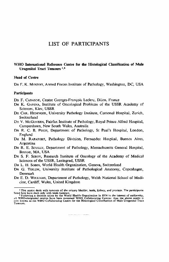
LIST OF PARTICIPANTS
WHO International Reference Centre for the Histological Classification of Male Urogenital Tract Tumours 1•2
Head of Centre
DR F. K. MosTOFI, Armed Forces Institute of Pathology, Washington, DC, USA
Participants
DR F. CABANNE, Centre Georges-Fran9ois Leclerc, Dijon, France DR K. GANINA, Institute of Oncological Problems of the USSR Academy of
Sciences, Kiev, USSR DR CHR. HEDINGER, University Pathology Institute, Cantonal Hospital, Zurich,
Switzerland DR V. McGOVERN, Fairfax Institute of Pathology, Royal Prince Alfred Hospital,
Camperdown, New South Wales, Australia DR R. C. B. PuoH, Department of Pathology, St Paul's Hospital, London,
England DR M. RAPAPORT, Pathology Division, Fernandez Hospital, Buenos Aires,
Argentina DR R. E. SCULLY, Department of Pathology, Massachusetts General Hospital,
Boston, MA, USA DR S. F. SEROV, Research Institute of Oncology of the Academy of Medical
Sciences of the USSR, Leningrad, USSR DR L. H. SOBIN, World Health Organization, Geneva, Switzerland DR G. TEILUM, University Institute of Pathological Anatomy, Copenhagen,
Denmark DR E. D. WILLIAMS, Department of Pathology, Welsh National School of Medi
cine, Cardiff, Wales, United Kingdom
1 This centre deals with tumours of the urinary bladder, testis, kidney, and prostate. The participants listed here have dealt only with testis tumours.
'Following a decision taken by the World Health Organization in 1974 in the interest of uniformity, all WHO-designated centres have been re-named WHO Collaborating Centres: thus the above centre is now known as the WHO Collaborating Centre for the Histological Classification of Male Urogenital Tract Tumours.
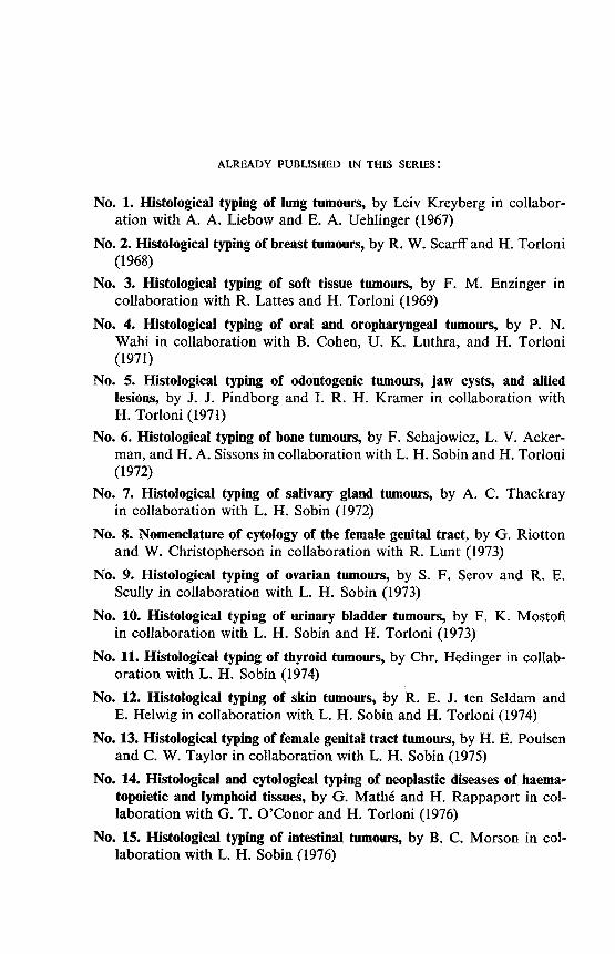
ALREADY PUBLISHED IN THIS SERIES:
No. 1. Histological typing of lung tumours, by Leiv Kreyberg in collaboration with A. A. Liebow and E. A. Uehlinger (1967)
No. 2. Histological typing of breast tumours, by R. W. Scarff and H. Torloni (1968)
No. 3. Histological typing of soft tissue tumours, by F. M. Enzinger in collaboration with R. Lattes and H. Torloni (1969)
No. 4. Histological typing of oral and oropharyngeal tumours, by P. N. Wahi in collaboration with B. Cohen, U. K. Luthra, and H. Torloni (1971)
No. 5. Histological typing of odontogenic tumours, jaw cysts, and allied lesions, by J. J. Pindborg and I. R. H. Kramer in collaboration with H. Torloni (1971)
No. 6. Histological typing of bone tumours, by F. Schajowicz, L. V. Ackerman, and H. A. Sissons in collaboration with L. H. Sobin and H. Torloni (1972)
No. 7. Histological typing of salivary gland tumours, by A. C. Thackray in collaboration with L. H. Sobin (1972)
No. 8. Nomenclature of cytology of the female genital tract, by G. Riotton and W. Christopherson in collaboration with R. Lunt (1973)
No. 9. Histological typing of ovarian tumours, by S. F. Serov and R. E. Scully in collaboration with L. H. Sobin (1973)
No. 10. Histological typing of urinary bladder tumours, by F. K. Mostofi in collaboration with L. H. Sobin and H. Torloni (1973)
No. 11. Histological typing of thyroid tumours, by Chr. Hedinger in collaboration with L. H. Sobin (1974)
No. 12. Histological typing of skin tumours, by R. E. J. ten Seldam and E. Helwig in collaboration with L. H. Sobin and H. Torloni (1974)
No. 13. Histological typing of female genital tract tumours, by H. E. Poulsen and C. W. Taylor in collaboration with L. H. Sobin (1975)
No. 14. Histological and cytological typing of neoplastic diseases of haematopoietic and lymphoid tissues, by G. Mathe and H. Rappaport in collaboration with G. T. O'Conor and H. Torloni (1976)
No. 15. Histological typing of intestinal tumours, by B. C. Morson in collaboration with L. H. Sobin (1976)
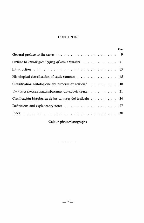
CONTENTS
General preface to the series . . . . . . . .
Preface to Histological typing of testis tumours
Introduction . . . . . . . . . . . . .
Histological classification of testis tumours
Classification histologique des tumeurs du testicule
f HCTOJIOfHtJeCKaSI KJiaccmpHKaQHSI orryxoneii SIHtJKa
Clasificaci6n histol6gica de los tumores del testiculo
Definitions and explanatory notes
Index ...... .
Colour photomicrographs
-7-
Page
9
11
13
15
18
21
24
27
38
ACKNOWLEDGEMENTS
The authors thank Dr I. Sesterhenn for her valuable contribution in the preparation of this publication. The photomicrographs reproduced in this volume were taken by Mr L. Duckett and Mr C. Edwards, Armed Forces Institute of Pathology, Washington, DC, USA.
GENERAL PREFACE TO THE SERIES
Among the prerequisites for comparative studies of cancer are international agreement on histological criteria for the classification of cancer types and a standardized nomenclature. At present, pathologists use different terms for the same pathological entity, and furthermore the same term is sometimes applied to lesions of different types. An internationally agreed classification of tumours, acceptable alike to physicians, surgeons, radiologists, pathologists and statisticians, would enable cancer workers in all parts of the world to compare their findings and would facilitate collaboration among them.
In a report published in 1952,1 a subcommittee of the WHO Expert Committee on Health Statistics discussed the general principles that should govern the statistical classification of tumours and agreed that, to ensure the necessary flexibility and ease in coding, three separate classifications were needed according to ( 1) anatomical site, ( 2) histological type, and ( 3) degree of malignancy. A classification according to anatomical site is available in the International Classification of Diseases.2
The question of establishing a universally accepted classification by histological type has received much attention during the last 20 years and a particularly valuable Atlas of Tumor Pathology-already numbering more than 40 volumes-is being published in the USA by the Armed Forces Institute of Pathology under the auspices of the National Research Council. An Illustrated Tumour Nomenclature in English, French, German, Latin, Russian, and Spanish has also been published by the International Union Against Cancer (UICC).
In 1956 the WHO Executive Board passed a resolution 3 requesting the Director-General to explore the possibility that WHO might organize centres in various parts of the world and arrange for the collection of human tissues and their histological classification. The main purpose of such centres would be to develop histological definitions of cancer types and to facilitate the wide adoption of a uniform nomenclature. This resolution was endorsed by the Tenth World Health Assembly in May 1957 4 and the following month a Study Group on Histological Classification of Cancer Types met in Oslo to
1 Wld Hlth Org. teclm. Rep. Ser., 1952, No. 53, p. 45. • World Health Organization (1967) Manual of the International Statistical Classification of Diseases,
Injuries, and Causes of Death, 1965 revision, Geneva. •Off.Ree. Wld Hlth Org., 1956, 68, 14 (Resolution EB17.R40). •Off.Ree. Wld Hlth Org., 1957, 79, 467 (Resolution WHAI0.18).
-9-
10 INTERNATIONAL HISTOWGICAL CLASSIFICATION OF TUMOURS
advise WHO on its implementation. The Group recommended criteria for selecting tumour sites for study and suggested a procedure for the drafting of histological classifications and testing their validity. Briefly, the procedure is as follows :
For each tumour site, a tentative histopathological typing and classification is drawn up by a group of experts, consisting of up to ten pathologists working in the field in question. An international reference centre and a number of collaborating laboratories are then designated by WHO to evaluate the proposed classification. These laboratories exchange histological preparations, accompanied by clinical information. The histological typing is then made in accordance with the proposed classification. Subsequently, one or more technical meetings are called by WHO to facilitate an exchange of opinions and the classification is amended to take account of criticisms.
In addition to preparing the publication and the photomicrographs for it, the reference centre produces up to JOO sets of microscope slides showing the major histological types for distribution to national societies of pathology.
Since 1958, WHO has established 23 centres covering tumours of the lung ; breast ; soft tissues ; oropharynx ; bone ; ovaries ; salivary glands ; thyroid; skin ; male urogenital tract ; jaws ; female genital tract ; stomach and oesophagus ; intestines ; central nervous system ; liver, biliary tract and pancreas ; upper respiratory tract ; eye ; and endocrine glands ; as well as oral precancerous conditions ; the leukaemias and lymphomas; comparative oncology ; and exfoliative cytology. This work has involved more than 300 pathologists from over 50 countries. A number of these centres have completed their work, and most of their classifications have already been published ( see page 6).
The World Health Organization is indebted to the many pathologists who have participated and are participating in this large undertaking. The pioneer work of many other international and national organizations in the field of histological classification of tumours has greatly facilitated the task undertaken by WHO. Particular gratitude is expressed to the National Cancer Institute, USA, which, through the National Research Council and the USA National Committee for the International Council of Societies of Pathology, is providing financial support to accelerate this work. Finally, WHO wishes to record its appreciation of the valuable help it has received from the International Council of Societies of Pathology (!CSP) in proposing participants and in undertaking to distribute copies of the classifications, with corresponding sets of microscope slides, to national societies of pathology all over the world.
PREFACE TO HISTOLOGICAL TYPING OF TESTIS TUMOURS
The WHO Centre for the Histological Classification of Male Urogenita/ Tract Tumours was established at the Armed Forces Institute of Pathology, Washington, DC, USA.
At a meeting in Zurich in 1971 attended by Dr Chr. Hedinger, Zurich, Dr V. McGovern, Sydney, Dr F. K. Mostofi, Washington, DC, and Dr L. H. Sobin, WHO, a tentative classification of testis tumours was drafted. This was then evaluated by the Centre and its collaborators, a list of which will be found on page 5.
The Centre prepared and distributed microscope specimens from selected cases of testis tumours to the participants for histological typing according to the tentative classification. In all, 250 cases were thus studied. The classification was reviewed at a meeting held in 1973 attended by the participants.
The photomicrographs reproduced as colour plates in this book are also available as a collection of transparencies intended especially for teaching purposes. To help pathologists who might wish to know the corresponding terms in French, Russian, and Spanish, translations of the classification into these languages are also given, immediately following the English version.
It will, of course, be appreciated that the classification reflects the present state of knowledge, and modifications are almost certain to be needed as experience accumulates. Although the present classification has been adopted by the members of the group, it necessarily represents a view from which some pathologists may wish to dissent. It is nevertheless hoped that, in the interests of international cooperation, all pathologists will try to use the classification as put forward. Criticism and suggestions for its improvement will be welcomed; these should be sent to the World Health Organization, Geneva.
The publications in the series International Histological Classification of Tumours are not intended to serve as textbooks but rather to promote the adoption of a uniform terminology of tumours that will facilitate and improve communication among cancer workers. For this reason the literature references have intentionally been kept to a minimum and readers should refer to standard works on the subject for extensive bibliographies.
-11-

INTRODUCTION
This classification is based primarily on the presence of morphologically identifiable cell types and patterns that can be correlated with the clinical behaviour of the tumour. Controversial histogenetic terms have been avoided whenever possible. Because of the many similarities between testicular tumours and those of the ovary, an attempt has been made to follow closely the WHO histological typing of ovarian tumours. However, important differences in the incidence, age distribution, and clinical behaviour of the various homologous tumours excludes the possibility of a classification identical with that of the ovarian tumours.
Tumours of the testis often have a very diverse histological structure and it is therefore essential that orchidectomy specimens be examined carefully. Many representative blocks of tumour tissue should be prepared after extensive slicing of the specimen, and the non-neoplastic part of the testis, the rete testis, the epididymis, the lower cord and the upper cord, at the level of surgical resection, should be included in the sections. The specimen should not be discarded until the clinician and pathologist are agreed that the response of the patient to treatment is characteristic of the tumour that was diagnosed. In the event of an unfavourable response further sectioning of the testis may be necessary, as hitherto undiscovered types of neoplastic tissue may have been responsible for the unexpected clinical course.
Occasionally the pathologist is confronted with a germ cell tumour in a specimen from an extratesticular site, and in such cases the testes may reveal no evidence of tumour on clinical examination. Although metastases from cancers of most organs reproduce the appearance of the primary tumour to varying degrees, those from testicular germ cell tumours may be different or of a more aggressive type than the primary tumour. Thus, in a case of a metastatic embryonal carcinoma, the testis after surgical removal or at autopsy may prove to contain an apparently pure seminoma or a teratoma. In other instances the only finding is a scar in association with a focus of germ cell tumour or malignant germ cells within tubules. Such scars are often referred to as regressed or burned-out tumours. Cases in which no diagnosable tumour is found in the testis or the testis is not available for examination have to be categorized accordingly, i.e., primary site not microscopically confirmed.
Another situation that may be encountered is the presence of a germ cell tumour, e.g., a seminoma, in the retroperitoneum, without clinical evidence of testicular involvement. In some such cases the patient may be
-13 -

14 INTERNATIONAL HISTOLOGICAL CLASSIFICATION OF TUMOURS
apparently cured without orchidectomy, but in other instances, particularly in association with retroperitoneal masses, the testis may contain an occult or regressed tumour, which may become clinically apparent several years later. For reporting purposes, cases of retroperitoneal and mediastinal tumour cannot be assumed to be examples of metastatic testicular cancer unless a tumour is demonstrated in the testis.
It must be emphasized that structurally abnormal testes such as cryptorchids, dysgenetic testes, and the testes of testicular feminization are characterized by an unusually high incidence of tumours and tumour-like conditions. In cryptorchids, foci of seminiferous tubules often form nodules that may be confused with Sertoli cell tumours. Also, cryptorchids are particularly prone to develop germ cell tumours, most commonly seminomas, even after an orchidopexy has been performed. Dysgenetic testes are often characterized by confusing microscopical patterns of germ cell arrangement, some of which are possibly precancerous; these testes are often involved by gonadoblastomas and germ cell tumours.
The age of the patient provides a clue to the most likely type of testicular tumour that may be present. The majority occur between the ages of 20 and 50 years. Before the age of puberty seminoma is extremely uncommon and yolk sac tumours and the better differentiated types of teratoma are the usual tumours. Malignant lymphoma is typically a tumour of the older patient, but may also occur in younger individuals.
Synonyms are listed only if they have been widely used or if they are considered to be important for an understanding of the disease. In such cases, the preferred term is given first, followed by the synonym in square brackets.
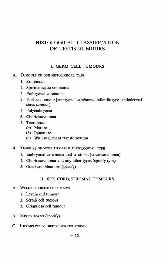
HISTOLOGICAL CLASSIFICATION OF TESTIS TUMOURS
I. GERM CELL TUMOURS
A. TUMOURS OF ONE HISTOLOGICAL TYPE
1. Seminoma
2. Spermatocytic seminoma
3. Embryonal carcinoma
4. Yolk sac tumour [embryonal carcinoma, infantile type; endodermal sinus tumour]
5. Polyembryoma
6. Choriocarcinoma
7. Teratomas (a) Mature (b) Immature (c) With malignant transformation
B. TuMOURS OF MORE THAN ONE HISTOLOGICAL TYPE
1. Embryonal carcinoma and teratoma [teratocarcinoma]
2. Choriocarcinoma and any other types (specify type)
3. Other combinations (specify)
II. SEX CORD/STROMAL TUMOURS
A. WELL-DIFFERENTIATED FORMS
1. Leydig cell tumour
2. Sertoli cell tumour
3. Granulosa cell tumour
B. MIXED FORMS (specify)
C. INCOMPLETELY DIFFERENTIATED FORMS
- 15
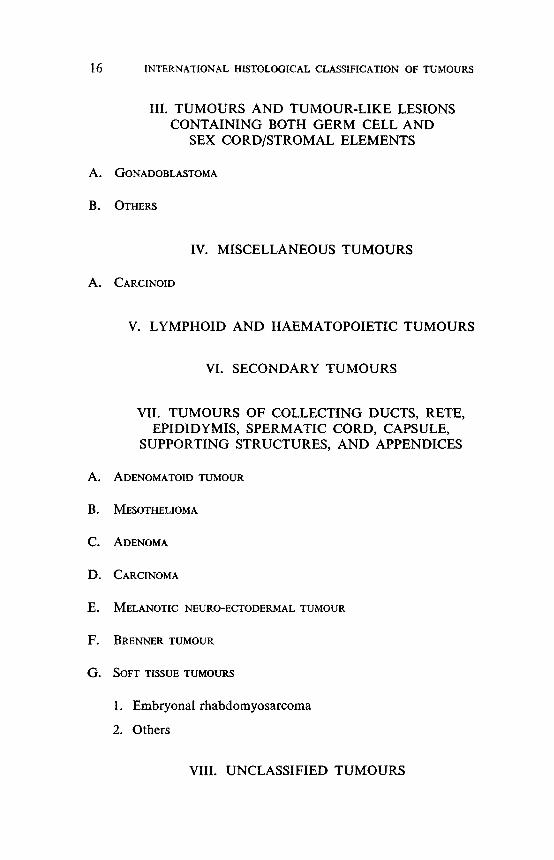
16 INTERNATIONAL HISTOLOGICAL CLASSIFICATION OF TUMOURS
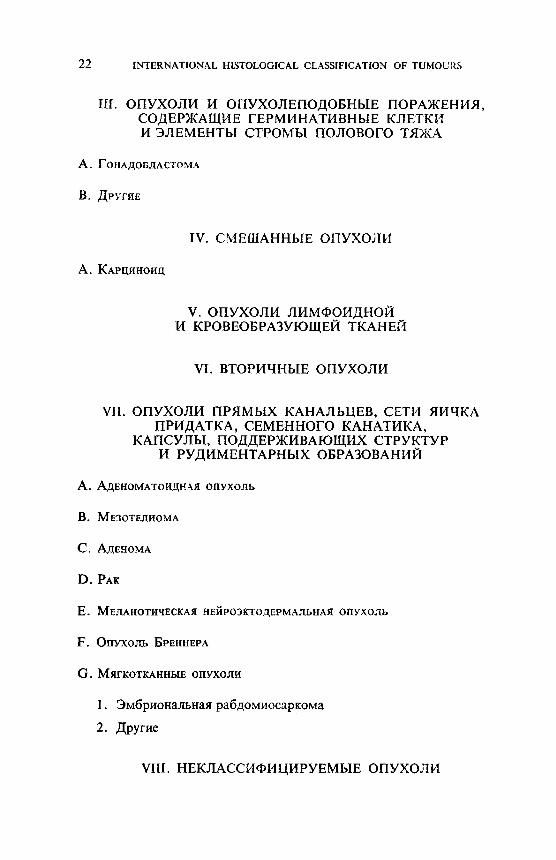
III. TUMOURS AND TUMOUR-LIKE LESIONS CONTAINING BOTH GERM CELL AND
SEX CORD/STROMAL ELEMENTS
A. GONADOBLASTOMA
B. OTHERS
IV. MISCELLANEOUS TUMOURS
A. CARCINOID
V. LYMPHOID AND HAEMATOPOIETIC TUMOURS
VI. SECONDARY TUMOURS
VIL TUMOURS OF COLLECTING DUCTS, RETE, EPIDIDYMIS, SPERMATIC CORD, CAPSULE,
SUPPORTING STRUCTURES, AND APPENDICES
A. ADENOMATOID TUMOUR
B. MESOTHELIOMA
C. ADENOMA
D. CARCINOMA
E. MELANOTIC NEURO-ECTODERMAL TUMOUR
F. BRENNER TUMOUR
G. SOFT TISSUE TUMOURS
I. Embryonal rhabdomyosarcoma
2. Others
VIII. UNCLASSIFIED TUMOURS

HISTOLOGICAL TYPING OF TESTIS TUMOURS 17
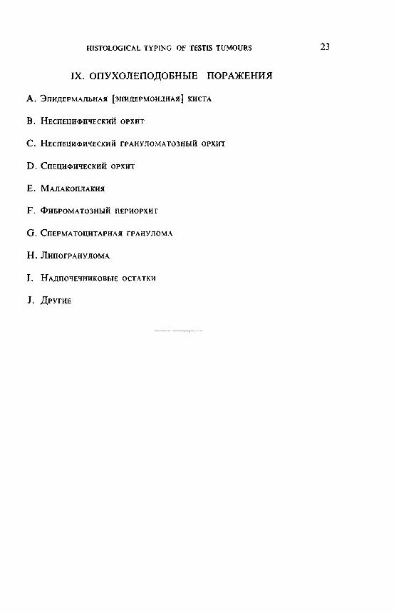
IX. TUMOUR-LIKE LESIONS
A. EPIDERMAL [EPIDERMOID] CYST
B. NONSPECIFIC ORCHITIS
C. NONSPECIFIC GRANULOMATOUS ORCHITIS
D. SPECIFIC ORCHITIS
E. MALAKOPLAKIA
F. FIBROMATOUS PERIORCHITIS
G. SPERM GRANULOMA
H. LIPOGRANULOMA
I. ADRENAL RESTS
J. OTHERS

CLASSIFICATION HISTOLOGIQUE DES TUMEURS DU TESTICULE
I. TUMEURS GERMINALES
A. TUMEURS A UNE SEULE COMPOSANTE HISTOLOGIQUE
I. Serninome
2. Seminome spermatocytaire
3. Carcinome embryonnaire
4. Turneur vitelline [carcinome embryonnaire, type infantile; turneur du sinus endodermique]
5. Polyembryome
6. Choriocarcinome
7. Teratome (dysembryome] (a) mature (b) immature ( c) cancerise
B. TUMEURS A PLUSIEURS COMPOSANTES HISTOLOGIQUES
I. Carcinome embryonnaire et teratome [teratocarcinome]
2. Choriocarcinome et tout autre type de tumeurs (le preciser)
3. Autres combinaisons (les preciser)
II. TUMEURS DES CORDONS SEXUELS ET DU STROMA GONADIQUE
A. FORMES BIEN DIFFERENCIEES
I. Tumeur a cellules de Leydig
2. Tumeur a cellules de Sertoli
3. Tumeur de la granulosa
B. FORMES INTRIQUEES (les preciser)
C. FORMES INCOMPLETEMENT DIFFERENCIEES
-18-
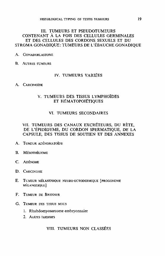
HISTOLOGICAL TYPING OF TESTIS TUMOURS
III. TUMEURS ET PSEUDOTUMEURS CONTENANT A LA FOIS DES CELLULES GERMINALES
19
ET DES CELLULES DES CORDONS SEXUELS ET DU STROMA GONADIQUE: TUMEURS DE L'EBAUCHE GONADIQUE
A. GONADOBLASTOME
B. AUTRES TUMEURS
IV. TUMEURS V ARIE.ES
A. CARCINOIDE
V. TUMEURS DES TISSUS LYMPHO'iDES ET HEMATOPOJETIQUES
VI. TUMEURS SECONDAIRES
VII. TUMEURS DES CANAUX EXCRETEURS, DU R.ETE, DE L'EPIDIDYME, DU CORDON SPERMATIQUE, DE LA CAPSULE, DES TISSUS DE SOUTIEN ET DES ANNEXES
A. TUMEUR ADENOMATOiDE
B. MESOTHELIOME
C. ADENOME
D. CARCINOME
E. TUMEUR MELANOTIQUE NEURO-ECTODERMIQUE [PROGONOME
MELANOTIQUE]
F. TUMEUR DE BRENNER
G. TUMEUR DES TISSUS MOUS
1. Rhabdomyosarcome embryonnaire
2. Autres tumeurs
VIII. TUMEURS NON CLASS.BES
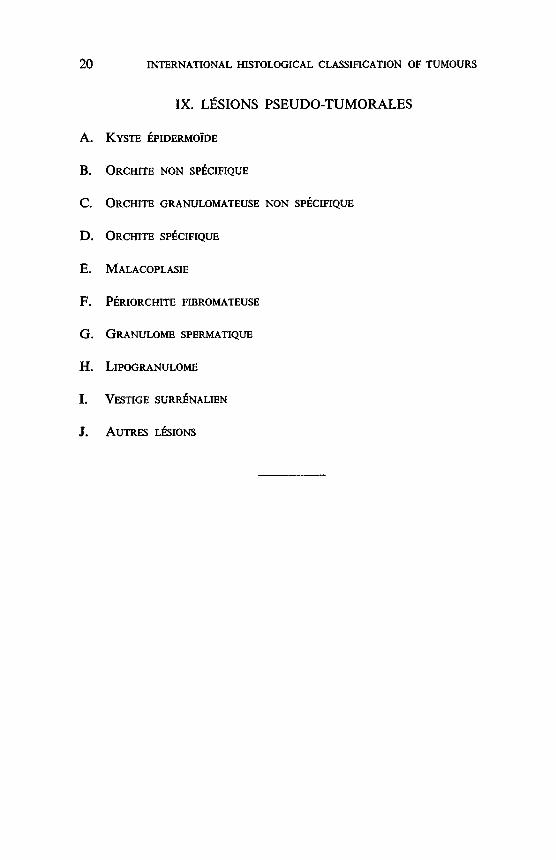
20 INTERNATIONAL HISTOLOGICAL CLASSIFICATION OF TUMOURS
IX. LESIONS PSEUDO-TUMORALES
A. K YSTE EPIDERMOIDE
B. ORCHITE NON SPECIFIQUE
C. 0RCHITE GRANULOMATEUSE NON SPECIFIQUE
0. 0RCHITE SPECIFIQUE
E. MALACOPLASIE
F. PERIORCHITE FIBROMATEUSE
G. GRANULOME SPERMATIQUE
H. LIPOGRANULOME
I. VESTIGE SURRENALIEN
J. AUTRES LESIONS
rMCTOnorMqECKiUI KnACCM~MKAQM~ OIIYXOnE:R ~MqKA
I. rEPM0HOrEHHhIE OilYXOJill
A. OIIYXOJIH OJ).HOro rHCTOJIOrH'IECKoro THIIA
1. CeMHHOMa
2. CnepMaTOUHTapHaH ceMHHOMa
3. 3M6pHOHa.JlhHl>IH paK
4. Onyxom. )l(eJITO'IHOro MellIKa [3M6pHOHa.JlhHblii paK HH(l>aHTHJib-
HOro THna; onyXOJib 3HJ).OJJ,epMaJibHOro CHHycaJ
5. IloJIH3M6pHoMa
6. XopHOH31IHTeJIHOMa
7. TepaTOMbI
(a) 3penaH
( b) He3penaH
(c) c 3JIOKa'lecrneHHOii TpaecqlOpMa~eii
B. OnYXOJIH EOJIEE 'IEM OJ).HOfO rHCTOJIOfH'IECKOfO THilA
1. 3M6pHOHaJibHblii paK H TepaTOMa [TepaToKap~HOMa)
2. XopHOH3IlHTeJIHOMa H JJ.pyrne BHJ).bl (yTO'IHBTb Kame)
3 . .IJ:pyrne KOM6HHanm1 (yTOqHBTb KaKHe)
II. OilYXOJill CTPOMbI IlOJIOBoro TJDKA
A. XoPomo J).H<l><l>EPEHUHPOBAHHbIE cl>OPMbI
1. OnyxonH H3 KJieTOK JleiiJJ,Hra
2. OnyxonH H3 KJieTOK CepTOJIH
3. rpaHyJie3oKJieTo'IHhre onyxonH
B. CMEllIAHHbIE <l>OPMbl (YTOqHHTb KAKHE)
C. HEIIOJIHOCTbIO J).B<l><l>EPEHUHPOBAHHblE cl>OPMbI
-21-
22 INTERNATIONAL HISTOLOGICAL CLASSIFICATION OF TUMOURS
III. OI1YXOJ1J1 J1 OCTYXOJIEITO.fl:OEHhIE IIOPA)KEHlHI, CO.fl:EP)KAIIJJIE rEPMI1HAT11BHhIE KJIETKH I1 3JIEMEHThl CTPOMhl flOJIOBOro TJDKA
A. roHA,Ll;OB.'lACTOMA
B . .fl:PYfllE
IV. CMEUIAHHbIE OilYXOJlH
A. KAPIJ;RHOHJJ;
V. OI1YXOJil1 Jll1M<l>OH.fl:HOJ1: H KPOBEOEPA3YIOIIJ:El1 TKAI·fEl1
VI. BTOPl1l.JHhIE OCTYXOJIH
VII. OilYXOJUI flPJCMbIX KAHAJibl(EB, CETH JCW-IKA ITPM.fl:ATKA, CEMEHHoro KAHATHKA,
KAITCYJibl, f10.fl:.fl:EP)KI1BAIOIIJ:l1.X CTPYKTYP If PY.fl:HMEHTAPHhIX OEPA30BAHHH
A. AJJ;EHOMA TOR~HAll OIIYXOJlb
B. ME30TEJIROMA
c. A,.a;EHOMA
D. PAK
E. MEJlAHOTit:qECKAH HEHP03KT0,1J;EPMAJlhHAH OIIYXOJlb
F. 0IIYXOJih bPEHHEPA
G. MsmcoTKAHHhIE 011Yxom1
I. 3M6pHOHam,HaH pa6p;OMMOCapKOMa
2 . .fl:pyrne
vm. HEKJIACCl1<l>I1l...(I1PYEMbIE OIIYXOm1
HISTOLOGICAL TYPING OF TESTIS TUMOURS 23
IX. OIIYXOJIEilO.[(OEHbIE ITOPA)KEHIUI
A. 3IIH)].EPMAJlbHASI [311H)].EPMOlf)lHASI] KHCTA
B. HECIIEI.l;If<l>ll'IECKHH OPXHT
C. HECIIEU:H<l>H'IECKHH. rPAHYJIOMAT03KblH OPXHT
D. CIIEU:H<l>H'IECKHH OPXHT
E. MAJIAKOIIJIAKHSI
F. <l>HEPOMA T03Kl>IR IIEPHOPXHT
G. CnEPMATOU:HTAPHAS[ rPAHYJ10!v!A
H. JIHIIOrPAHYJIOMA
l. HA,!J.IIO'IE'IHHKOB&IE OCTATKH
J . .[(PYrHE

CLASIFICACION HISTOLOGICA DE LOS TUMORES DEL TESTICULO
I. TUMORES DE CELULAS GERMINATIVAS
A. TUMORES DE UN SOLO TIPO HISTOLOOICO
1. Seminoma
2. Seminoma espermatocitico
3. Carcinoma embrionario
4. Tumor de saco vitelino [carcinoma embrionario, tipo infantil; tum or del seno endodermico]
5. Poliembrioma 6. Coriocarcinoma
7. Teratomas (a) Maduro (b) lnmaduro (c) Con transformaci6n maligna
B. TUMORES DE MAS DE UN TIPO HISTOLOGICO
1. Carcinoma embrionario y teratoma [teratocarcinoma]
2. Coriocarcinoma y cualquier otro tipo (especificar tipo)
3. Otras combinaciones (especificar)
II. TUMORES DE LOS CORDONES SEXUALES Y DEL ESTROMA
A. FORMAS BIEN DIFERENCIADAS
1. Tumor de celulas de Leydig
2. Tumor de celulas de Sertoli
3. Tumor de celulas de la granulosa
B. FORMAS MIXTAS (especificar)
C. FORMAS INCOMPLETAMENTE DIFERENCIADAS
-24-
HISTOLOGICAL TYPING OF TESTIS TUMOURS
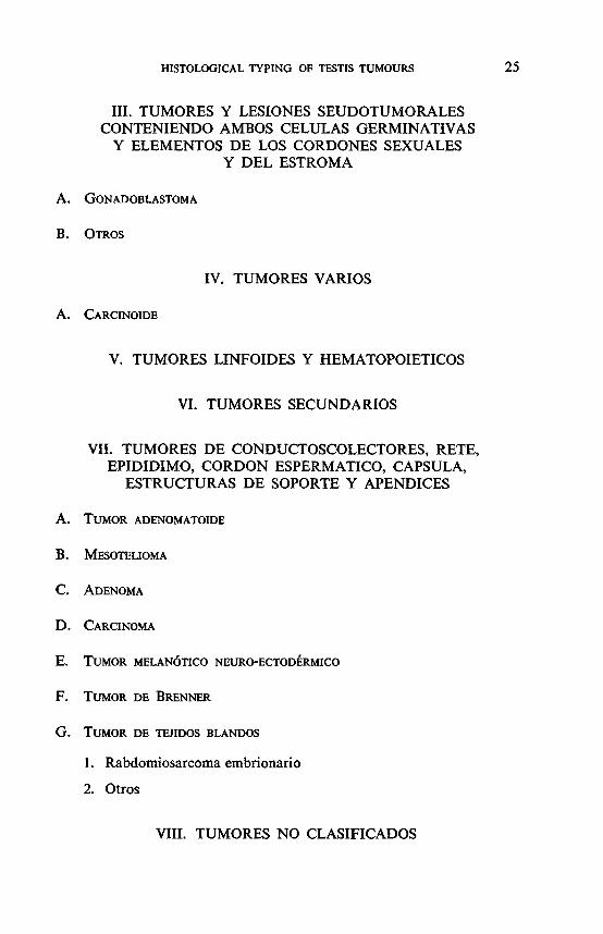
III. TUMORES Y LESIONES SEUDOTUMORALES CONTENIENDO AMBOS CELULAS GERMINATIVAS
Y ELEMENTOS DE LOS CORDONES SEXUALES Y DEL ESTROMA
A. GONADOBLASTOMA
B. 0TROS
IV. TUMORES VARIOS
A. CARCINOIDE
V. TUMORES LINFOIDES Y HEMATOPOIETICOS
VI. TUMORES SECUNDARIOS
VII. TUMORES DE CONDUCTOSCOLECTORES, RETE, EPIDIDIMO, CORDON ESPERMATICO, CAPSULA,
ESTRUCTURAS DE SOPORTE Y APENDICES
A. TUMOR ADENOMATOIDE
B. MESOTELIOMA
C. AoENOMA
D. CARCINOMA
E. TUMOR MELAN6TICO NEURO-ECTODERMICO
F. TUMOR DE BRENNER
G. TUMOR DE TEJIDOS BLANDOS
l. Rabdomiosarcoma embrionario
2. Otros
VIII. TUMORES NO CLASIFICADOS
25
26 INTERNATIONAL HISTOLOGICAL CLASSIFICATION OF TUMOURS
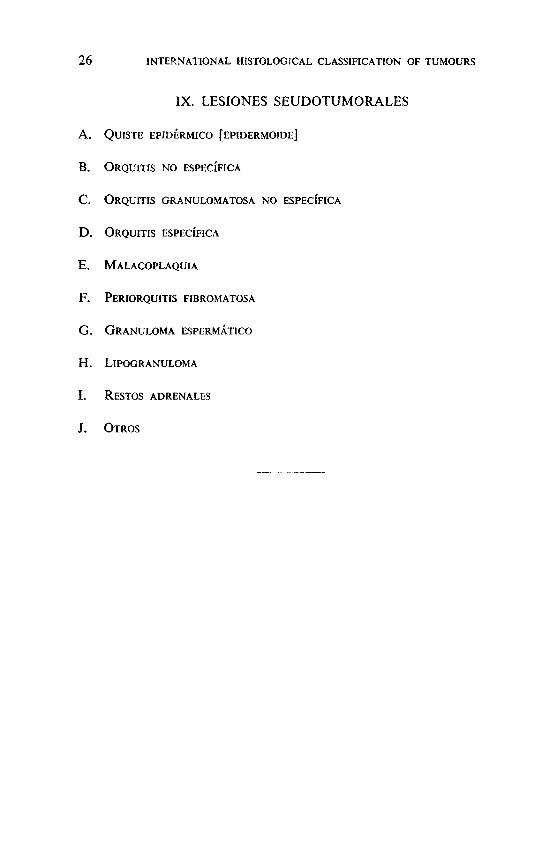
IX. LESIONES SEUDOTUMORALES
A. QUISTE EPlDERMICO [EPIDERMOIDE]
B. 0RQUITfS NO ESPECIFICA
C. 0RQUITIS GRANULOMA TOSA NO ESPECIFICA
0. 0RQUITIS ESPECIFJCA
E. MALACOPLAQUIA
F. PERIORQUITrS FIBROMATOSA
G. GRANULOMA ESPERMAT!CO
H. LIPOGRANULOMA
I. RESTOS ADRENALES
J. 0TROS

DEFINITIONS AND EXPLANATORY NOTES
I. GERM CELL TUMOURS
The large majority of primary testicular tumours originate from germ cells and contain one or more of the following histological types: seminoma, spermatocytic seminoma, embryonal carcinoma, yolk sac tumour, teratoma, and choriocarcinoma. The classification of germ cell tumours must take into consideration the potential of neoplastic germ cells to form a wide range of cells, tissues, or organs recapitulating embryonic and extra-embryonic development.
Nearly half the germ cell tumours of the testis contain more than one tumour type and the metastases may be of different types from the primary growth.
One of the component types of tumour may be quite small and yet determine the clinical course. It is therefore necessary to take many blocks of tissue for adequate histological sampling.
Germ cell tumours are also found in the ovary, the retroperitoneum, the mediastinum, and the pineal region.
A. TUMOURS OF ONE HISTOLOGICAL TYPE
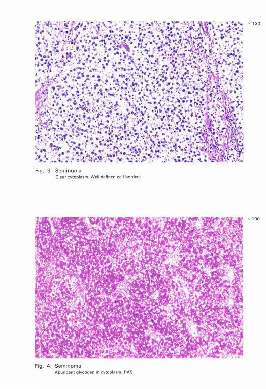
I. Seminoma (Fig. 1-14): A tumour of fairly uniform cells, typically with clear cytoplasm and well-defined cell borders, resembling primitive germ cells.
The cytoplasm typically contains glycogen, a feature that may be helpful in distinguishing this tumour from spermatocytic seminoma and lymphoma. A characteristic feature of seminoma is the lymphocytic infiltration of the stroma. A variable amount of fibrous tissue may be present, resulting in an alveolar pattern. Occasionally the stromal reaction consists of tuberculoid granulomas with Langhans' giant cells.
-27-
28 INTERNATIONAL HISTOLOGICAL CLASSIFICATION OF TUMOURS
The tumour may be in the form of circumscribed masses or it may be diffusely infiltrative. Neoplastic cells may be seen within adjacent testicular tubules. Such an intratubular seminoma is occasionally observed in association with other germ cell tumours.
Tumours with many mitoses (i.e., 3 per high power field) and/or considerable nuclear variation have been designated anaplastic and are claimed to have a higher incidence of metastases. Attention should be drawn to such tumours by recording their degree of mitotic activity.
Tumour giant cells or syncytial masses resembling syncytiotrophoblast may be present. Although the latter may sometimes be shown to contain chorionic gonadotrophin, their presence should not be interpreted as indicating choriocarcinoma unless they are associated with cytotrophoblast.
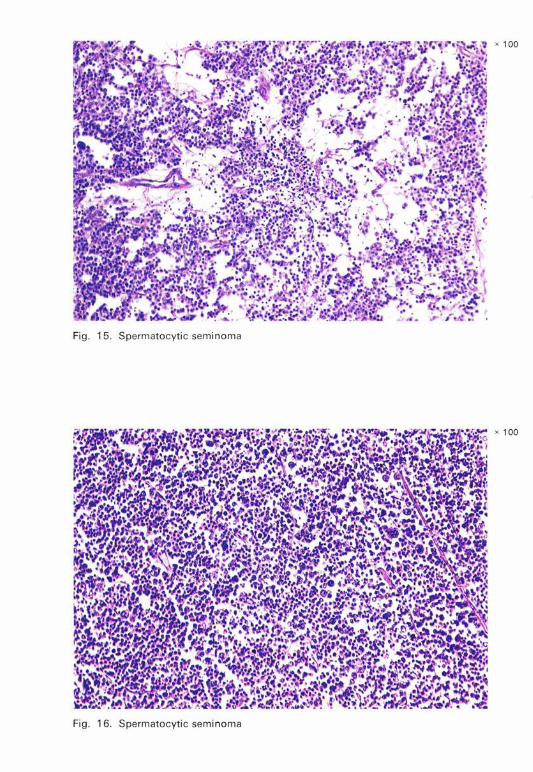
2. Spermatocytic seminoma (Fig. 15-19): A distinctive tumour composed of cells that vary in size from lymphocyte-like cells to giant cells of about 100 µ.m in diameter. The bulk of the tumour, however, is composed of cells of intermediate size.
The cells of this tumour have eosinophilic cytoplasm and round nuclei. The nuclei of larger cells may have a filamentous or spireme pattern not unlike that seen in spermatogenesis. Mitoses are often very numerous, but metastases are seldom reported.
Other features in which this tumour differs from classical seminoma are the absence of intracellular glycogen and the absence of a lymphocytic or granulomatous stroma.
3. Embryonal carcinoma (Fig. 20-34): A tumour composed of cells of primitive epithelial appearance, often with clear cytoplasm, growing in a variety of pattems-acinar, tubular, papillary, and solid.
The stroma may contain primitive mesenchyme but its presence does not indicate a diagnosis of teratoma. Lymphocytic and granulomatous reactions are infrequent.
Solid embryonal carcinomas may simulate seminoma. The absence of a lymphocytic stromal infiltrate is helpful in distinguishing between the two. The cell borders in embryonal carcinoma are usually much less distinct than those in seminoma. Some areas of embryonal carcinoma may resemble cytotrophoblast.
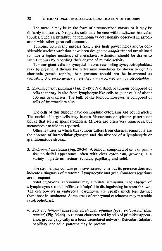
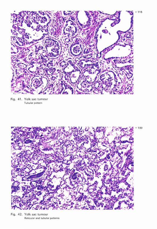
4. Yolk sac tumour [embryonal carcinoma, infantile type; endodermal sinus tumour] (Fig. 35-44): A tumour characterized by cells of primitive appearance, growing typically in a loose vacuolated network. Reticular, tubular, papillary, and solid patterns may be present.

HISTOLOGICAL TYPING OF TESTIS TUMOURS 29
The cells vary from columnar or cuboidal to flattened endothelial-like cells and may have a clear vacuolated cytoplasm. The tumour contains varying amounts of mucin, glycogen, and fat.
Distinctive perivascular structures resembling the endodermal sinuses of the rat placenta (Fig. 43), cysts resembling yolk sac vesicles, and both intracellular and extracellular PAS-positive hyaline globules may be present.
Elongated cells with some of the features of smooth muscle may be present in the stroma, but this should not be interpreted as representing a teratomatous element.
Yolk sac tumour is the characteristic and most common testicular tumour of infants and young children and has also been known by the following terms: embryonal carcinoma, infantile type; endodermal sinus tumour; embryonal adenocarcinoma; clear cell adenocarcinoma; orchioblastoma. Comparable tumours may occur in adults, in which case they are usually associated with other germ cell tumours.
5. Polyembryoma (Fig. 45-47): A tumour composed predominantly of embryoid bodies.
Embryoid bodies are structures contammg a disk and cavities surrounded by loose mesenchyme simulating an embryo of about two weeks' gestation. Tubular structures (resembling endoderm) and syncytiotrophoblastic elements may be present.
Polyembryoma as defined is extremely rare. Embryoid bodies are found more often within embryonal carcinoma and teratoma.
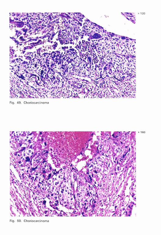
6. Choriocarcinoma (Fig. 48-50): A highly malignant tumour composed of elements identical with syncytiotrophoblast and cytotrophoblast.
This category is reserved for the very rare pure forms of this tumour. When choriocarcinoma occurs in combination with other germ cell tumours it should be assigned to category I.B.2. (see page 31).
The only criterion by which cytotrophoblastic cells can be recognized is their intimate relationship with syncytiotrophoblastic cells. Syncytial cells alone are not sufficient for the diagnosis. There is no definite villus formation of placental type, i.e., with stromal cores, although villus-like structures with syncytiotrophoblast surrounding masses of cytotrophoblast are sometimes seen, particularly at the advancing edge of the tumour. Villus formation is not essential for the diagnosis.
The tumour masses are frequently related to thin-walled vascular spaces and the tumour is usually very haemorrhagic.
Serum and urinary levels of chorionic gonadotrophin are almost invariably elevated in the presence of choriocarcinoma but this can also occur with other germ cell tumours in the absence of demonstrable choriocarcinoma.
30 I!'-.TERNATIONAL HISTOLOGICAL CLASSIFICATION OF TUMOURS
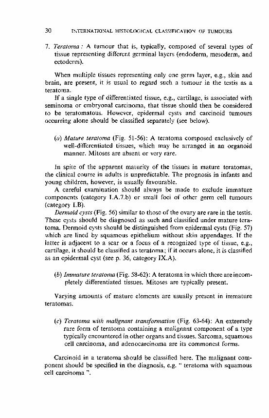
7. Teratoma: A tumour that is, typically, composed of several types of tissue representing different germinal layers (endoderm, mesoderm, and ectoderm).
When multiple tissues representing only one germ layer, e.g., skin and brain, are present, it is usual to regard such a tumour in the testis as a teratoma.
If a single type of differentiated tissue, e.g., cartilage, is associated with seminoma or embryonal carcinoma, that tissue should then be considered to be teratomatous. However, epidermal cysts and carcinoid tumours occurring alone should be classified separately (see below).
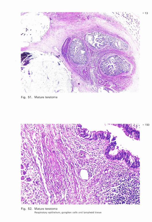
(a) Mature teratoma (Fig. 51-56): A teratoma composed exclusively of well-differentiated tissues, which may be arranged in an organoid manner. Mitoses are absent or very rare.
In spite of the apparent maturity of the tissues in mature teratomas, the clinical course in adults is unpredictable. The prognosis in infants and young children, however, is usually favourable.
A careful examination should always be made to exclude immature components (category I.A.7.b) or small foci of other germ cell tumours (category LB).
Dermoid cysts (Fig. 56) similar to those of the ovary are rare in the testis. These cysts should be diagnosed as such and classified under mature teratoma. Dermoid cysts should be distinguished from epidermal cysts (Fig. 57) which are lined by squamous epithelium without skin appendages. If the latter is adjacent to a scar or a focus of a recognized type of tissue, e.g., cartilage, it should be classified as teratoma; if it occurs alone, it is classified as an epidermal cyst (seep. 36, category IX.A).
(b) Immature teratoma (Fig. 58-62): A teratoma in which there are incompletely differentiated tissues. Mitoses are typically present.
Varying amounts of mature elements are usually present in immature teratomas.
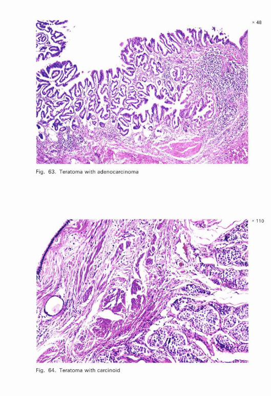
(c) Teratoma with malignant transformation (Fig. 63-64): An extremely rare form of teratoma containing a malignant component of a type typically encountered in other organs and tissues. Sarcoma, squamous cell carcinoma, and adenocarcinoma are its commonest forms.
Carcinoid in a teratoma should be classified here. The malignant component should be specified in the diagnosis, e.g. " teratoma with squamous cell carcinoma ".
HISTOLOGICAL TYPING OF TESTIS TUMOURS 31
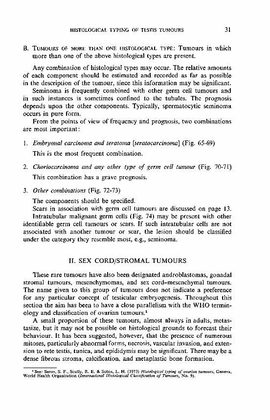
B. TUMOURS OF MORE THAN ONE HISTOLOGICAL TYPE: Tumours in which more than one of the above histological types are present.
Any combination of histological types may occur. The relative amounts of each component should be estimated and recorded as far as possible in the description of the tumour, since this information may be significant.
Seminoma is frequently combined with other germ cell tumours and in such instances is sometimes confined to the tubules. The prognosis depends upon the other components. Typically, spermatocytic seminoma occurs in pure form.
From the points of view of frequency and prognosis, two combinations are most important:
I. Embryonal carcinoma and teratoma [teratocarcinoma] (Fig. 65-69)
This is the most frequent combination.
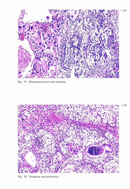
2. Choriocarcinoma and any other type of germ cell tumour (Fig. 70-71)
This combination has a grave prognosis.
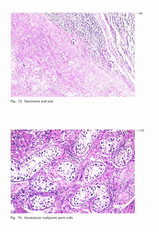
3. Other combinations (Fig. 72-73)
The components should be specified. Scars in association with germ cell tumours are discussed on page 13. Intratubular malignant germ cells (Fig. 74) may be present with other
identifiable germ cell tumours or scars. If such intratubular cells are not associated with another tumour or scar, the lesion should be classified under the category they resemble most, e.g., seminoma.
II. SEX CORD/STROMAL TUMOURS
These rare tumours have also been designated androblastomas, gonadal stromal tumours, mesenchymomas, and sex cord-mesenchymal tumours. The name given to this group of tumours does not indicate a preference for any particular concept of testicular embryogenesis. Throughout this section the aim has been to have a close parallelism with the WHO terminology and classification of ovarian tumours.1
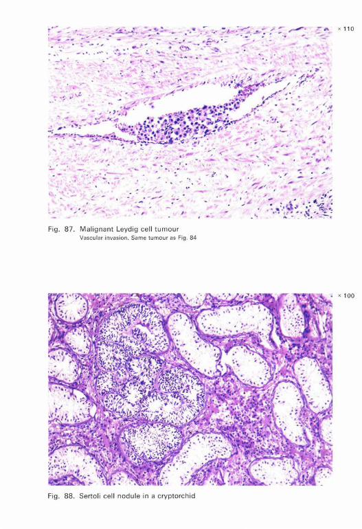
A small proportion of these tumours, almost always in adults, metastasize, but it may not be possible on histological grounds to forecast their behaviour. It has been suggested, however, that the presence of numerous mitoses, particularly abnormal forms, necrosis, vascular invasion, and extension to rete testis, tunica, and epididymis may be significant. There may be a dense fibrous stroma, calcification, and metaplastic bone formation.
1 See: Serov, S. F., Scully, R. E. & Sobin, L. H. (1973) Histological typing of ovarian tumours, Geneva, World Health Organization (International Histological Classification of Tumours, No. 9).
32 INTERNATIONAL HISTOLOGICAL CLASSIFICATION OF TUMOURS
A. WELL-DIFFERENTIATED FORMS
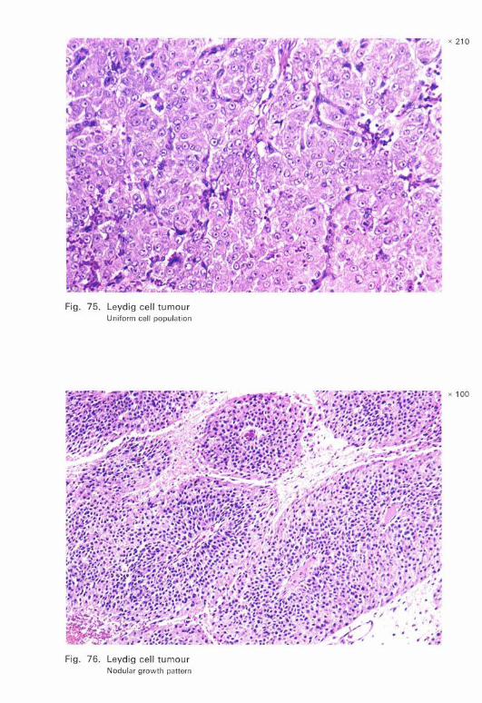
1. Leydig cell tumour [interstitial cell tumour] (Fig. 75-87): A tumour composed of Leydig cells, which corresponds to the hilus cell tumour of the ovary.
When present, Reinke's crystals are helpful in the identification of Leydig cells. Lipofuscin is often present in Leydig cells.
Leydig cell tumours have to be distinguished from the nodular aggregations of Leydig cells found in the testes of persons with Klinefelter's syndrome.
In children these tumours are associated with macrogenitosomia. In adults gynaecomastia and other signs of feminization may be present.
Tumours and tumour-like proliferations of cells resembling both Leydig and adrenocortical cells, typically bilateral, may occur in congenital adrenogenital syndrome. Clinical and biochemical data are necessary to determine whether the lesion is a manifestation of the adrenogenital syndrome.
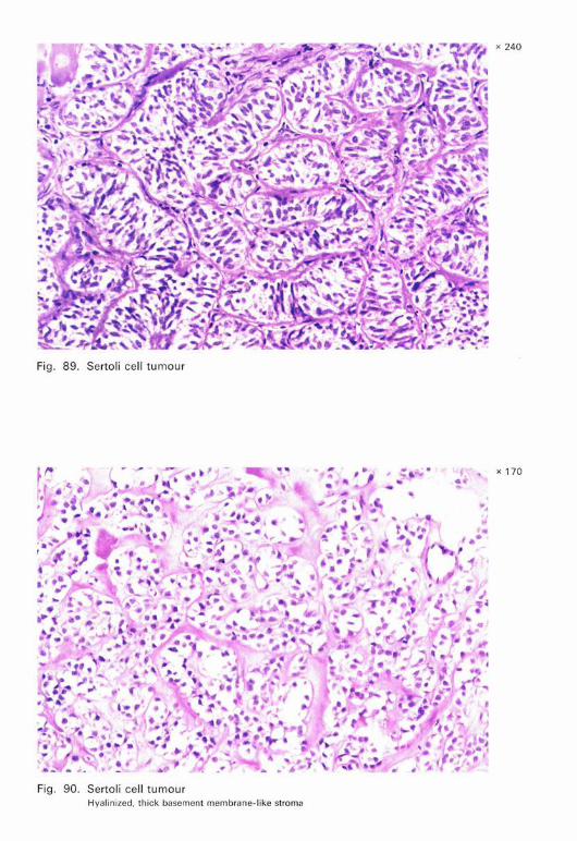
2. Sertoli cell tumour (Fig. 89-94): A tumour composed of Sertoli cells arranged in well-defined tubules.
These tumours correspond to the tubular androblastoma or Sertoli cell tumour of the ovary, although the morphological similarity is not exact in many instances.
The tubules forming this tumour may have a distinct lumen or they may appear solid as in the prepubertal testis, and their cells may contain varying amounts of lipid. When the intratubular Sertoli cells are distended with lipid the tumour corresponds to the " tubular androblastoma with lipid storage " of the ovary. Sometimes the intratubular cells have eosinophilic granular cytoplasm.
Coiled tubules lined by Sertoli cells found in undescended testes (Fig. 88) are not neoplastic and should not be mistaken for a Sertoli cell tumour.
A number of adults with Sertoli cell tumours have gynaecomastia and other signs of feminization.
In the rare syndrome of testicular feminization (androgen-insensitivity syndrome) the patients have an XY genotype and are well-developed phenotypic females whose testes are usually found in the groin or abdomen. The testes may contain tumour-like masses of Sertoli cells and Leydig cells that could perhaps be more properly regarded as hamartomas. Sertoli cell lesions (" tubular adenomas ") of very large size (up to 25 cm in diameter) can also occur. About 25 % of patients over the age of 30 years develop seminoma.
3. Granu/osa cell tumour (Fig. 95)
This is morphologically similar to the ovarian counterpart. It is exceedingly rare in the testis.
HISTOLOGICAL TYPING OF TESTIS TUMOURS 33
B. MIXED FORMS (Fig. 96-98)
These include combinations of differentiated cell types, e.g., Sertoli and Leydig cell tumours. Their components should be specified.
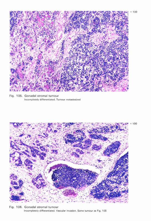
C. INCOMPLETELY DIFFERENTIATED FORMS (GONADAL STROMAL TUMOURS, ANDROBLASTOMA) (Fig. 99-106)
These include intermediate and poorly differentiated tumours similar to those found in the ovary.1
The tumours of this group can be composed of undifferentiated tissue, in which attempts at tubule formation may be discerned. Elements resembling theca cells may constitute part or all of the tumour.
About 10% of these tumours are clinically malignant.
III. TUMOURS AND TUMOUR-LIKE LESIONS CONTAINING BOTH GERM CELL AND SEX
CORD/STROMAL ELEMENTS
There is a predisposition in dysgenetic gonads and undescended testes to the development of tumours or tumour-like proliferations of both germ cell and sex cord/stromal elements.
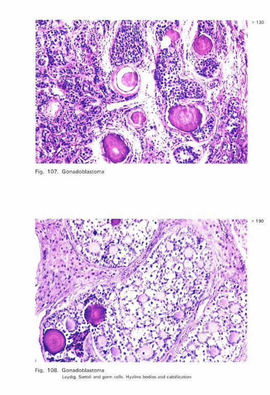
A. GONADOBLASTOMA (Fig. 107-109): A tumour composed of two principal cell types, large germ cells similar to those of seminoma and small cells resembling immature Sertoli and granulosa cells; elements resembling Leydig cells may also be present.
Hyaline bodies that simulate Call-Exner bodies are typically present, and foci of calcification are common. In some tumours the nests composed of the two major cell types are circumscribed, but in others the germ cells transgress the margins of the nests and grow as a seminoma. Certain tumours appear in the form of a seminoma or a more highly malignant type of germ cell tumour with only small foci of gonadoblastoma within them or at their margins. The type of germ cell tumour should be specified because of therapeutic implications. The occurrence of two distinct tumours, e.g., Leydig cell tumour and seminoma, should not be considered as a gonadoblastoma.
Gonadoblastomas arise almost exclusively in patients with dysgenetic gonads, most of whom are phenotypic females and almost all of whom are chromatin-negative and have a Y-chromosome.
1 See: Serov, S. F., Scully, R. E. & Sobin, L. H. (1973) Histological typing of ovarian tumours, Geneva, World Health Organization (International Histological Classification of Tumours, No. 9).

34 INTERNATIONAL HISTOLOGICAL CLASSIFICATION OF TUMOURS
Cases without known chromosomal abnormalities have been seen. These have a more diffuse growth pattern than the typical gonadoblastoma. Hyaline bodies are inconspicuous or absent. These tumours have been referred to as " atypical gonadoblastomas " (Fig. 109).
B. OTHERS
In some instances structures may occur resembling primitive gonads without distinct tumour formation. These have been regarded as hamartomas.
IV. MISCELLANEOUS TUMOURS
A. CARCINOID (Fig. 110)
This may be a component of a teratoma and should then be classified in category I.A. 7. and appropriately designated; however, it may occur in pure form either in a primary growth (classified in category IV.A.) or as a metastasis (classified in category VI). The distinction between the last two may be difficult.
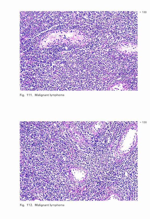
V. LYMPHOID AND HAEMATOPOIETIC TUMOURS (Fig. lll-ll4)
Testicular enlargement may be the initial manifestation of lymphomas or plasmacytoma. This category is reserved for such tumours.
Characteristic features of testicular lymphomas are extensive intertubular infiltration, bilaterality, and paratesticular involvement. Reticulosarcoma is sometimes mistaken for seminoma or embryonal carcinoma. The nuclear/ cytoplasmic ratio is greater in reticulosarcoma than in seminoma or embryonal carcinoma.
VI. SECONDARY TUMOURS (Fig. 115-116)
Rarely, testicular enlargement due to metastatic tumour is the first indication of extragonadal cancer. Any tumour that does not have the typical appearance of a testicular tumour should be suspected of being a secondary tumour.
VII. TUMOURS OF COLLECTING DUCTS, RETE, EPIDIDYMIS, SPERMATIC CORD, CAPSULE,
SUPPORTING STRUCTURES, AND APPENDICES
A variety of uncommon tumours is found in these sites. The topography of the tumour is important in determining its nature, and multiple sections may be necessary. This is particularly the case in lesions of the appendices.
HISTOLOGICAL TYPING OF TESTIS TUMOURS 35
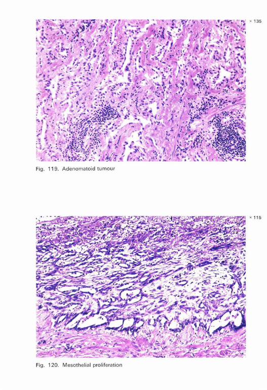
A. ADENOMATOID TUMOUR (Fig. 117-119): A benign tumour in which there are numerous spaces and clefts lined or traversed by cells that may resemble endothelium, epithelium, or mesothelium. The fibrous stroma is often hyalinized and there may be lymphocytic infiltrates. Smooth muscle is often present but should not be taken as evidence of invasion. The tumour is seen in the epididymis more frequently than in the capsule and spermatic cord.
B. MESOTHELIOMA (Fig. 121-123)
Apart from adenomatoid tumour, which is regarded as mesothelial, neoplastic proliferations of mesothelium occur that may be benign or malignant, but sometimes the distinction between these two may be difficult.
Hyperplasias, often papillary, and sequestrations of mesothelium in inflammatory lesions must not be mistaken for neoplasms (Fig. 120).
C. ADENOMA (Fig. 124-126): A benign epithelial tumour forming glandular or papillary structures.
Adenomas occur in the collecting ducts, rete and epididymis. Those in the epididymis are usually composed of glycogen-rich clear cells.
Amongst the tumours of this type are those that occur in the von HippelLindau complex.
Adenomas should not be mistaken for metastatic carcinoma.
D. CARCINOMA (Fig. 127-130): A malignant epithelial tumour forming glandular or papillary structures.
Carcinomas occur in the collecting ducts, rete and epididymis. Because of their extreme rarity, these tumours can be diagnosed only
when the possibility that the tumour is not a secondary has been excluded.
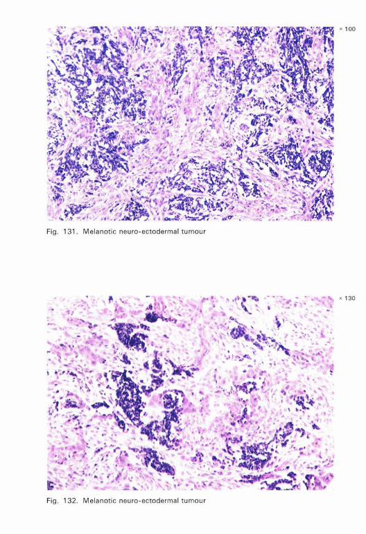
E. MELANOTIC NEURO-ECTOOERMAL TUMOUR (Fig. 131-132): A tumour arising in the epididymis and occasionally in the testis. The tumour consists of varying proportions of two cell types-epithelium-like cells, often arranged in strands, and small, darkly staining lymphocyte-like cellsin a cellular fibrous stroma. Melanin is found within the epithelium-like cells, and to a lesser extent within the lymphocyte-like cells.
This tumour is also known as retinal anlage tumour, melanotic hamartoma, and melanotic progonoma.
F. BRENNER TUMOUR
This tumour is identical with its ovarian counterpart but is exceedingly rare in the testis.
36 INTERNATIONAL HISTOLOGICAL CLASSIFICATION OF TUMOURS
G. SOFT TISSUE TUMOURS
Soft tissue tumours are classified according to the scheme devised by the International Reference Centre for the Histological Classification of Soft Tissue Tumours.1
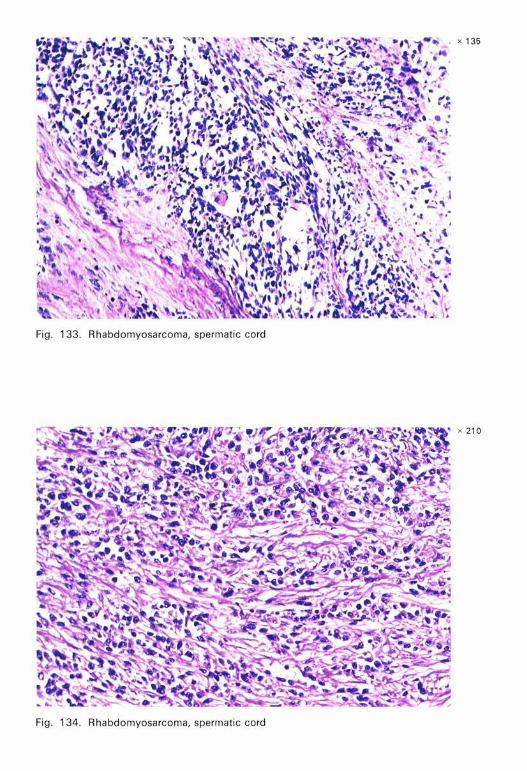
1. Embryonal rhabdomyosarcoma (Fig. 133-134)
This is the most important of the soft tissue tumours at this site. It occurs particularly in infants, children, and young adults.
In some cases cross-striations of the neoplastic cells are readily recognized on staining either with haematoxylin and eosin or with phosphotungstic acid haematoxylin. In others, less differentiated, there may be no demonstrable striations but characteristic rounded cells with eosinophilic cytoplasm may be found among the elongated spindle cells that form the bulk of the tumour.
2. Others (Fig. 135-138)
Of the other soft tissue tumours, lipoma, malignant fibrous histiocytoma, liposarcoma, fibrosarcoma, and leiomyosarcoma are the most common.
VIII. UNCLASSIFIED TUMOURS
These are primary benign or malignant tumours that cannot be placed in any other category.
IX. TUMOUR-LIKE LESIONS
A. EPIDERMAL [EPIDERMOID] CYST (Fig. 57): A cyst lined by keratinized stratified squamous epithelium without skin appendages. The cyst contains keratohyaline material.
If the cyst is adjacent to a scar or a focus of a recognized type of tissue, it should be classified as teratoma. If it occurs alone, it should be classified as an epidermal cyst.
B. NONSPECIFIC ORCHITIS
Tumour-like enlargement of the testis may result from acute or chronic nonspecific orchitis.
1 Enzinger, F. M., Lattes, R. & Torloni, H. (1969) Histological typing of soft tissue tumours, Geneva, World Health Organization (International Histological Classification of Tumours, No. 3).
HISTOLOGICAL TYPING OF TESTIS TUMOURS 37
C. NONSPECIFIC GRANULOMATOUS ORCHITIS (Fig. 139)
Granulomas are present within degenerating tubules and also between the tubules.
0. SPECIFIC ORCHITIS (Fig. 140-141)
Tuberculous orchitis is usually an epididymo-orchitis. Syphilitic orchitis, however, may be confined to the body of the testis. Other infections such as brucellosis and rickettsial and viral infections, e.g. mumps, may be mistaken clinically for tumours.
E. MALAKOPLAKIA (Fig. 142-143)
In this condition large histiocytes containing Michaelis-Guttman bodies are the predominant constituents. In some cases special stains, e.g., PAS, von Kossa, or iron, may be required to demonstrate the inclusions.
F. FIBROMA TOUS PERIORCHITIS (Fig. 144): A diffuse or focal fibroblastic proliferation, often hyalinized and generally following inflammation or trauma, which may simulate neoplasm.
G. SPERM GRANULOMA (Fig. 145-146): A granulomatous lesion, usually in the epididymis, resulting from the extravasation of spermatozoa that sometimes occurs in the cord following vasectomy. There are numerous phagocytes containing spermatozoa.
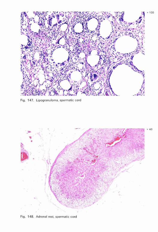
H. LIPOGRANULOMA (Fig. 147): A reactive sclerosing lesion due to the injection of oily material. It may simulate liposarcoma.
I. ADRENAL RESTS (Fig. 148): Encapsulated nodules of adrenal cortical cells situated on the surface of the spermatic cord, in the region of the rete testis, or in the testicular parenchyma. These cells reproduce the architecture of the adrenal cortex.
J. OTHERS
Other lesions that may simulate tumours are gonadal splenic fusion (Fig. 149), supernumerary testis, intratesticular haemorrhage, nodules of Leydig cells in the spermatic cord, mesonephric rests, Miillerian rests, and cysts of various types (e.g., mesothelial cysts (Fig. 150)).
Adenoma ..... Adenomatoid tumour Adrenal rest
INDEX
Androblastoma, see sex cord/stromal tumours Brenner tumour Carcinoid .. . Carcinoma .. . Choriocarcinoma Clear cell adenocarcinoma, see yolk sac tumour Dermoid cyst . . . . . . . . . . . . . . . Embryonal adenocarcinima, see yolk sac tumour Embryonal carcinoma . . . . . . . . . . . . Embryonal carcinoma, infantile type, see yolk sac tumour Endodermal sinus tumour, see yolk sac tumour Epidermal [epidermoid] cyst Germ cell tumours . . . Gonadal splenic fusion Gonadal stromal tumours Gonadoblastoma . . . . Granulosa cell tumour Intratubular malignant germ cells Leydig cell tumour . . . . . . Lipogranuloma . . . . . . . . Lymphoid and haematopoietic tumours Lymphomas ........... . Malakoplakia . . . . . . . . . . . Melanotic hamartoma, see melanotic neuro-ectodcrmal tumour Melanotic neuro-ectodermal tumour . . . . . . . . . . . Melanotic progonoma, see melanotic neuro-ectodermal tumour Mesenchymomas, see sex-cord/stromal tumours Mesothelioma ......... . Orchioblastoma, see yolk sac tumour Orchitis, nonspecific
nonspecific granulomatous specific ..... .
Periorchitis, fibromatous . Plasmacytoma . Polyembryoma . Reticulosarcoma
-38-
Page Figures
35 124-126 35 117-119 37 148
35 34 110 35 127-130 29 48-50
30 56
28 20-34
36 57 27 37 149 33 99-106 33 107-109 33 95 31 74 32 75-87 37 147 34 111-114 34 111-113 37 142-143
35 131-132
35 121-123
36 36 139 37 140-141 37 144 34 114 29 45-47 34
HISTOLOGICAL TYPING OF TESTIS TUMOURS
Rhabdomyosarcoma Secondary tumours Scars .... . Seminoma ... .
spermatocytic . Sertoli and Leydig cell tumour Sertoli cell tumour . . . . . Sex cord-mesenchymal tumours, see sex cord/stromal tumours Sex cord/stromal tumours Soft tissue tumours Sperm granuloma Teratocarcinoma . Teratoma .... Teratoma, immature
mature ..... with malignant transformation
Tumour-like lesions . Unclassified tumours Yolk sac tumour . .
39
Page Figures
36 133-134 34 115-116 13 73 27 1-14 28 15-19 33 96-98 32 89-94
31 36 133-138 37 145-146 31 65-69 30 30 58-62 30 51-56 30 63-64 36 36 28 35-44
Unless otherwise stated, the preparations shown in the photomicrographs reproduced on the following pages were stained with haematoxylin-eosin.
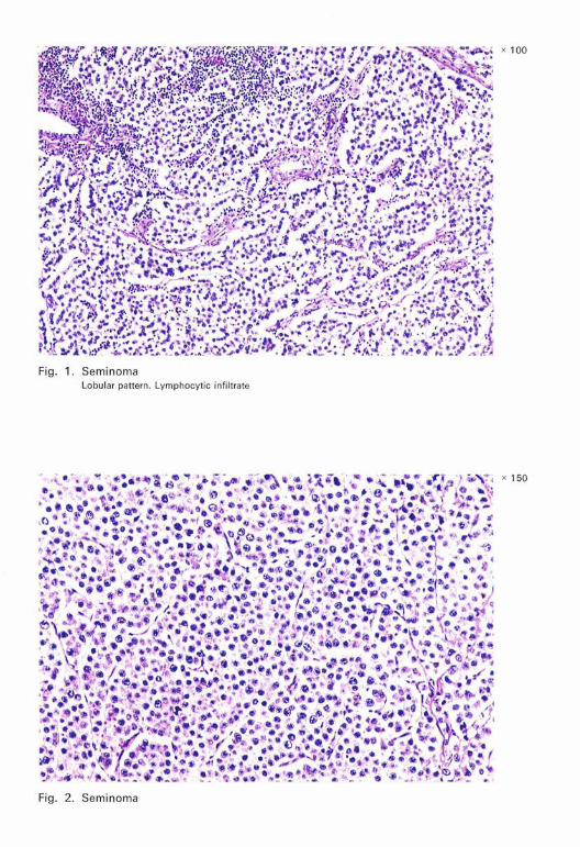
x 100
Fig. 1. Seminoma Lobular pattern. Lymphocytic infiltrate
x 150
Fig . 2. Seminoma
x 130
Fig. 3. Seminoma Clear cytoplasm. Well defined cell borders
x 100
Fig. 4. Seminoma Abundant glycogen in cytoplasm. PAS
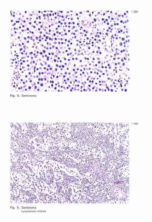
Fig . 5. Seminoma
Fig. 6. Seminoma Lymphocyt ic infi ltrate
x 250
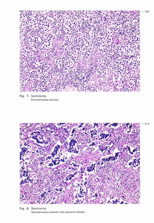
Fig. 7. Seminoma Granulomatous reaction
Fig. 8. Seminoma Granulomatous reaction and extensive fibrosis
x 100
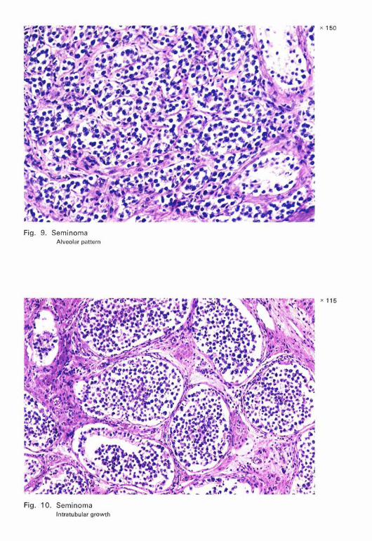
Seminoma Alveolar pattern
Fig. 10. Seminoma lntratubular growth
x 150
x 115

Fig. 11. Seminoma Interstitial infiltration
Fig. 12. Seminoma Numerous mitoses
x 100
x 200
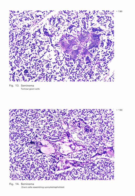
Fig. 13. Seminoma Tumour g iant cells
Fig. 14. Seminoma Giant cells resembling syncytiotrophoblast
x 130
x 130
x 100
Fig. 15. Spermatocytic seminoma
Fig . 16. Spermatocytic seminoma
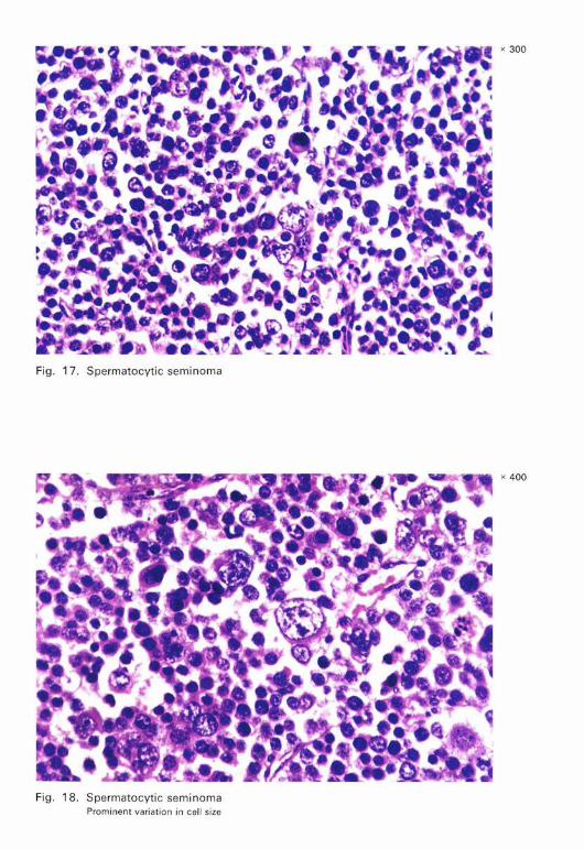
Fig. 17. Spermatocytic seminoma
Fig. 18. Spermatocytic seminoma Prominent variation in cell size
x 300
x 400
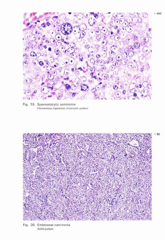
Fig. 19. Spermatocytic seminoma Fil amentous (spireme) chromatin pattern
Fig. 20. Em bryo nal ca rci no ma Solid pattern
x 400
x 80

Fig. 21. Embryonal carcinoma Solid pattern . Indistinct cell borders
Fig. 22. Embryonal carcinoma Solid and acinar patterns
x 300
x 105
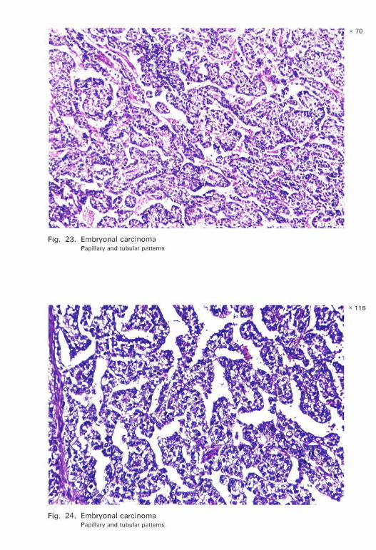
Fig. 23. Embryonal carcinoma Papillary and tubular patterns
Fig. 24. Embryonal carcinoma Papillary and tubular patterns
x 70
x 115
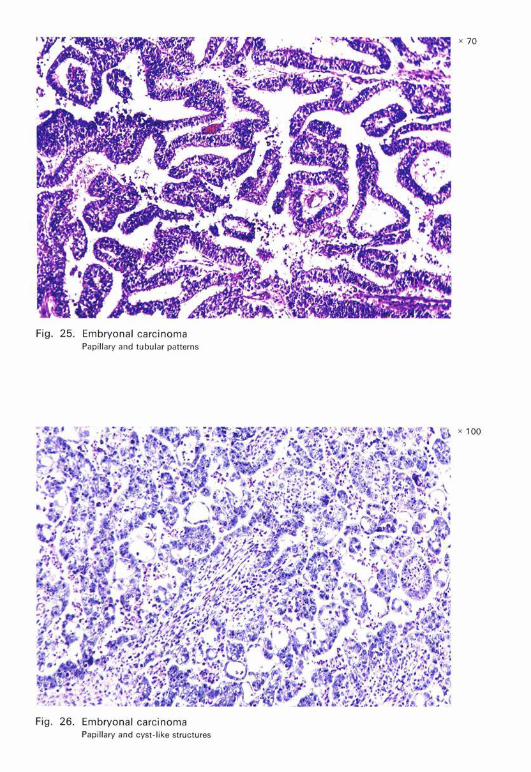
Fig . 25. E mbryonal carcinoma Papill ary and tub I u ar patterns
Embryonal carcinoma Papillary and cyst-l'k i e stru ctures
x 70
x 100
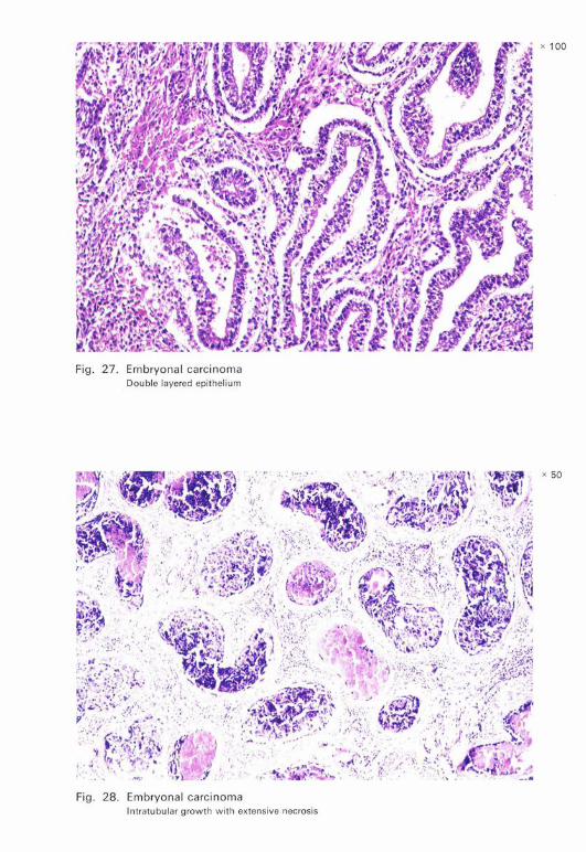
Fig. 27. Embryonal carcinoma Double layered epithelium
Fig. 28. Embryonal carci noma lntratubu lar growth with extensive necrosis
x 100
x 50
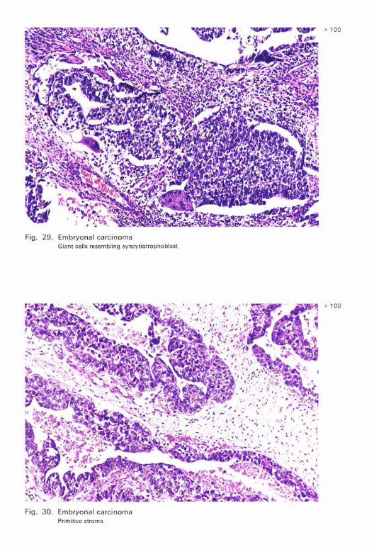
Fig. 29. Embryonal carcinoma Giant cells resembling syncytiotrophoblast
Fig . 30. Embryonal carci noma Primitive stroma
x 100
x 100
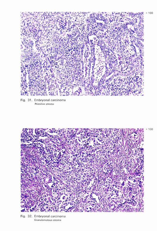
Fig . 31 . Embryonal ca rcinoma Primitive stroma
Fig. 32. Embryonal ca rcinoma Granulomatous stroma
x 100
x 100
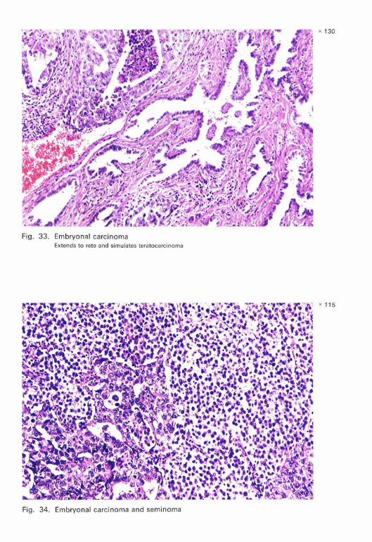
Fig. 33. Embryonal carcinoma Extends to rete and simulates teratocarcinoma
x 115
Fig. 34. Embryonal carcinoma and seminoma

x 100
Fig. 35. Yolk sac tumour Tubular and papillary structures
x 100
Fig. 36. Yolk sac tumour

x 110
Fig. 37. Yolk sac tumour Solid, tubular and reticular patterns. Adult patient
x 100
Fig. 38. Yolk sac tumour

Fig. 39. Yolk sac tumour Tubul ar stru ctures and loose mesenchymal tissue
Fi g. 40 . Yolk sac tumour Reticular pattern
x 115
x 100
tumour Fig. 42. Yolk sac tubular patterns
Reticular and

Fig. 43. Yolk sac tumour Schiller- Duva l body
Fig. 44. Yolk sac tumour Oedematous stroma
x 130
x 130
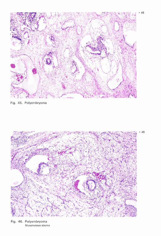
Fig. 45. Polyembryoma
Fi g. 46 . Polyembryoma Myxomatous stroma
x 45
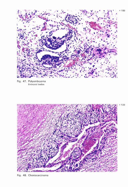
Fig. 47. Polyembryoma Embryoid bodies
Fig. 48. Choriocarcinoma
x 150
x 120
x 120
Fig. 49 . Choriocarcinoma
x 160
Fig. 50. Choriocarcinoma
"f i
.. '
\ '·
Fig. 51 . Mature teratoma
x 150
Fig. 52. M ature teratoma Respiratory epithelium, ganglion cells and lymphoid tissue
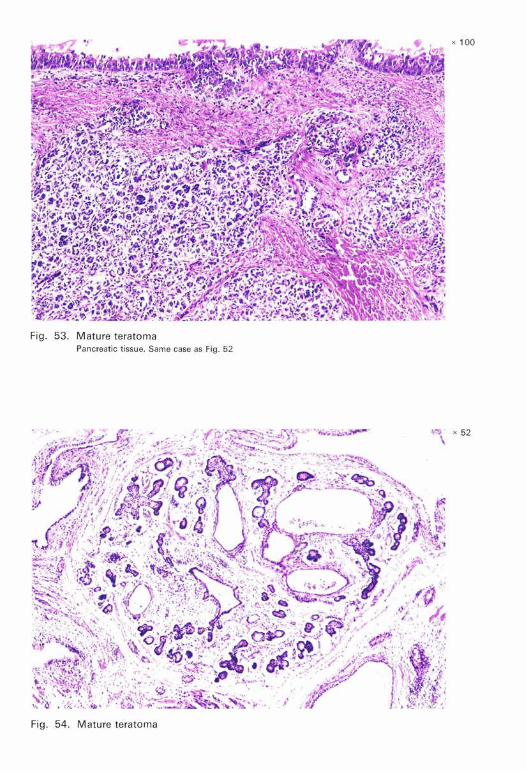
Fig. 53. Mature teratoma Pancreatic tissue. Same case as Fig. 52
x 52
Fig . 54. Mature teratoma

Fig, 55, Mature teratoma Within spermatic vein
J, ,' /,
'~ .{· .s:. :; . ,, , ., . ,,
'I
/' '..:. · . . \''\ /
.... ~ . ./ /, :,
.',t _:· . , ,:• ·
.,, r- :· : .
Fig, 56, Mature teratoma, dermoid cyst
x 40
x 100
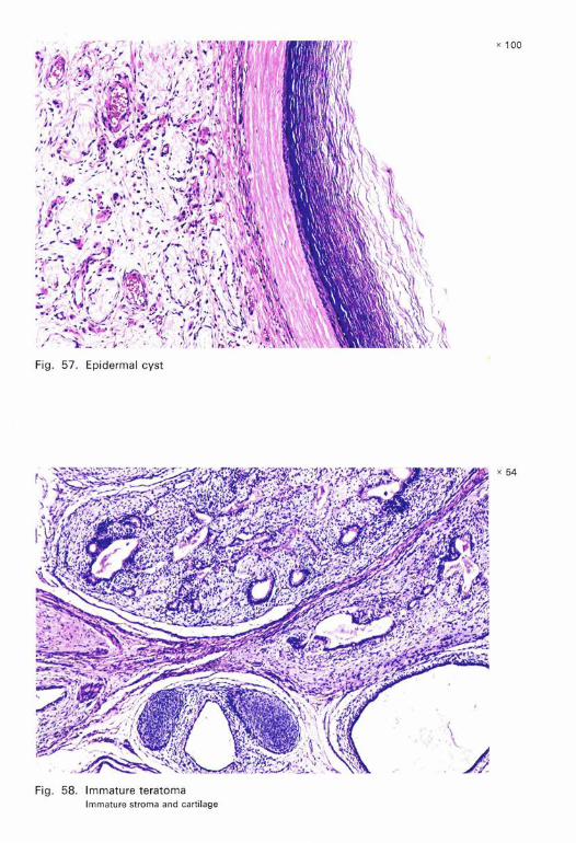
Fig. 57. Epidermal cyst
Fig . 58. Immature teratoma Immature stroma and cartilage
x 100
x 54

x 48
Fig. 59. Immature teratoma
x 100
Fig. 60. Immature teratoma
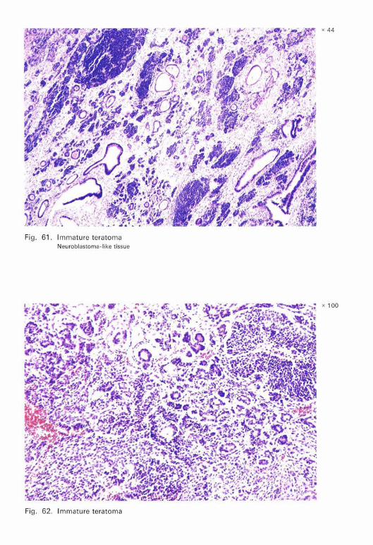
Fig. 61 . Immature teratoma Neuroblastoma- like t issue
x 44
x 100
x 48
Fig. 63. Teratoma with adenocarc inoma
x 110
Fig. 64. Teratoma with carcinoid

Fig. 65. Embryonal carcinoma and teratoma [teratocarcinoma] Embryonal carcinoma beneath epithelial structure (teratoma)
Fi g. 66. Embryona l ca rcinoma and teratoma [teratocarcinoma] Embryonal carcinoma at right, epithelium (teratoma) at left
x 100
x 130

Fig. 67. Embryonal carcinoma and teratoma [teratocarcinoma]
Fi g . 68. Embryonal carcinoma and teratoma [ teratocarcinoma ] Giant cells resembling syncytiotrophoblast
x 100
x 100
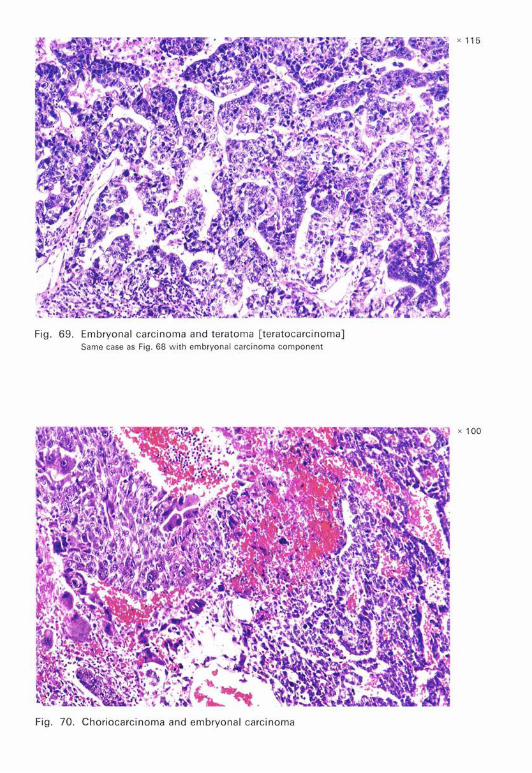
Fig. 69. Embryona l ca rcinoma and teratoma [ teratoca rc inoma] Same case as Fig. 68 with embryona l ca rci noma component
Fig. 70. Chorioca rcinoma and embryonal carcinoma
x 100
x 125
Fig. 71. Choriocarcinoma and teratoma
x 100
Fig. 72. Teratoma and seminoma
x 60
Fig. 73. Seminoma and scar
x 130
Fig. 74. lntratubular malignant germ cells
x 21 0
Leydig cell tumou Uniform cell r population
x 100
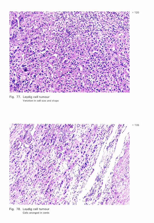
Fig. 77. Leydig cell tumour Variation in cel l size and shape
Fig. 78. Leydig cell tumour Cells arranged in cords
x 120
x 105
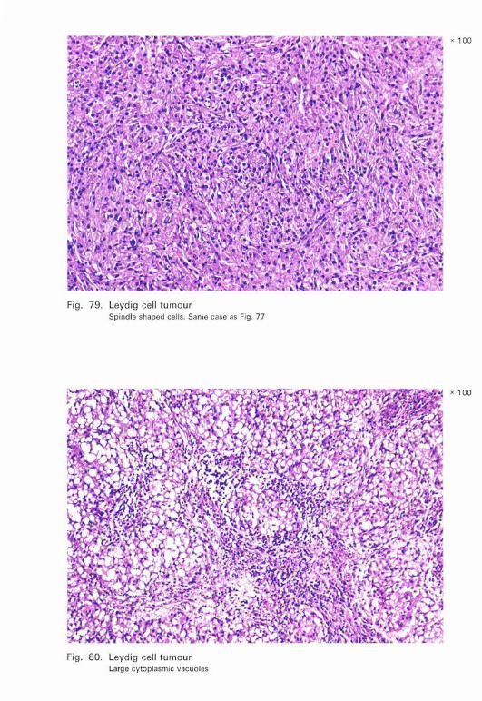
Fig . 79. Leydig cell tumour Spindle shaped cells. Same case as Fig. 77
Fig. 80. Leydig cell tumour Large cytoplasmic vacuoles
x 100
x 100
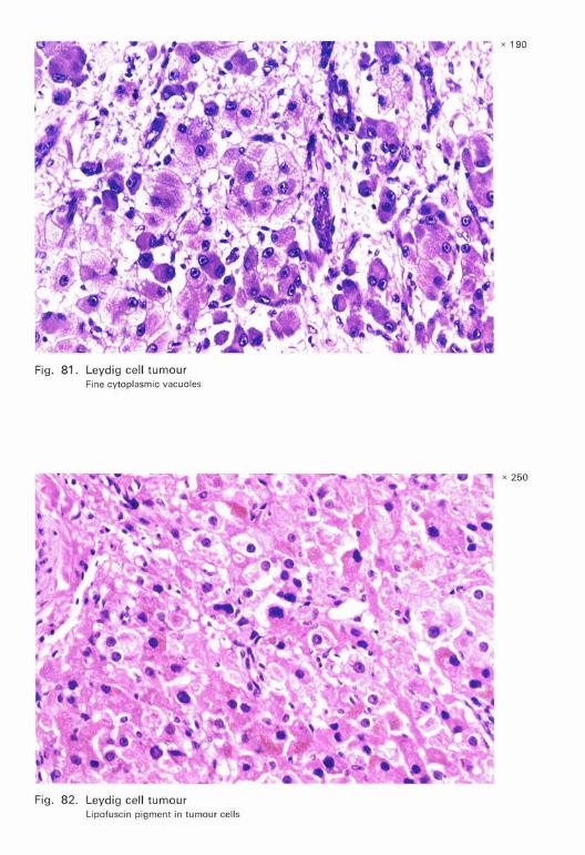
Fig. 81. Leydig cell tumour Fin e cytoplasmic vacuoles
Fig. 82. Leydig cell tumour Lipofuscin pigment in tumour cells
x 250

Fig . 83. Leydig cell tumour Reinke's crystals
Fig. 84. M alignant Leydig cell tumour Necrosis. Tumour metastasized
x 250
x 100

Fig . 85 . Malignant Leydig cell tumour Pl eomorphism. Sa me tu mour as Fig . 84
Fig. 86 . Malignant Leydig cell tumour Mitoses . Same tumour as Fig . 84
x 100
x 250
r ·' ~ .,..'.'., ., : . .. ~, .:, .. -.:-· -··
. . .. . :. ... . , - . · .. ... • · s .
-·
.;; .... .: ...,, . ~·
. .. . • 1 / .
[.
:.,.· ·-.. '
,:
...
- .. ~ ,,~ --
. .
~-, ,
:·
•• ' 1
--, /'
Fig. 87. M alignant Leydig cell tumour Vascu lar invasion. Same tumour as Fig. 84
Fig. 88. Sertoli ce ll nodule in a cryptorchid
- t · . ...
. , ..
.. ,;: ,- .,,....,... .. -., .. .,,,.
·-...
.-"•
. . ., ~..,,, ,·
,. r
. '
x 110
x 100
x 240
Fig. 89. Sertoli cell tumour
, ! '"• . ,. . x 170 .. .. .,. ..
"'• ii
Fig. 90. Sertoli cell tumour Hya linized, thick basement membrane- like stroma
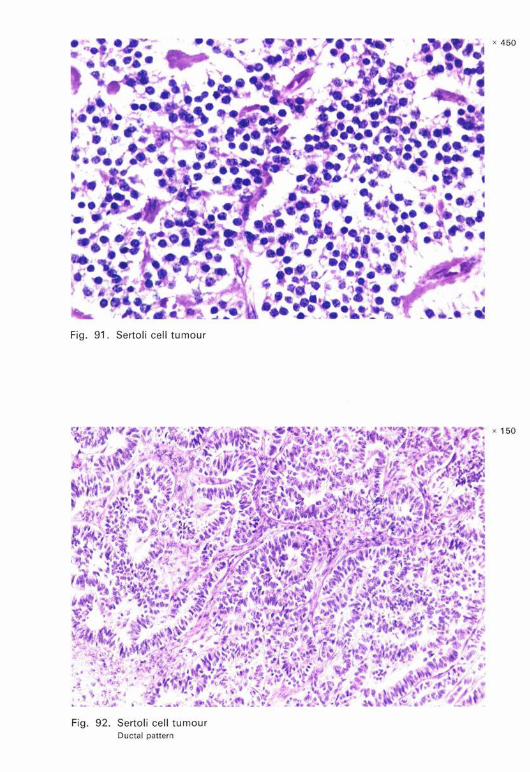
Fig. 91. Sertoli cell tumour
Fig. 92. Sertoli cell tumour Ductal pattern
x 450
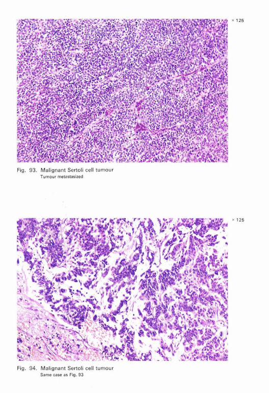
Fig. 93. Malignant Sertoli cell tumour Tumour metastasized
Fig. 94. M alignant Sertoli ce ll tumour Same case as Fig. 93
x 125
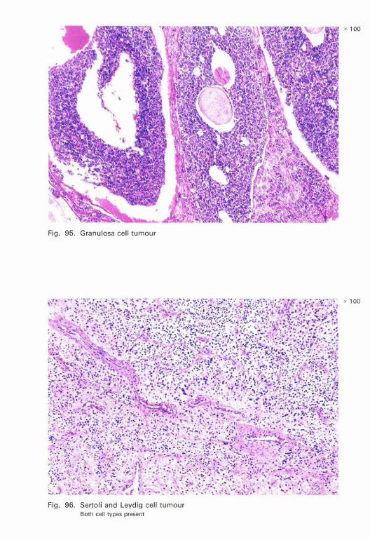
Fig. 95. Granulosa cell tumour
Fig. 96. Sertoli and Leydig cell tumour Both cell types present
x 100
x 100

Fig. 97. Sertoli and Leydig cell tumour lntratubular cells with eosinophilic cytoplasm resembling Leydig cells. Same case as Fig. 96.
Fig . 98. Sertoli and Leyd ig cel l tumour Mixture of Sertoli and Leyd ig cells
x 100
x 130
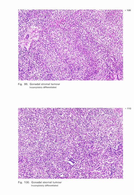
Fig . 99. Gonadal stromal tumour Incompl etely differentiated
Fi g. 100. Gonadal stromal tumour Incompletely differentiated
x 100

x 100
Fig . 101. Gonada l stromal tumour Parti al differentiation to granulosa cell elements. Same case as Fig. 100
x 100
Fig . 102. Gonadal stromal tumour Partial differentiation to theca and Sertoli cel l elements
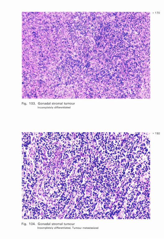
Fig . 103. Gonadal stromal tumour Incompletely differentiated
Fig. 104. Gonadal stromal tumour Incompletely d ifferentiated. Tumour metastasized
x 150
x 100
Fig. 105. Gonadal stromal tumour Incomplete ly differentiated. Tu mour metastasized
x 100
Fig . 106. Gonadal stromal tumour Incompletely differentiated. Vascular invasion. Same tum our as Fi g. 105
x 130
Fig. 107. Gonadoblastoma
x 190
Fig. 108. Gonadob lastoma Leyd ig , Serto l i and germ cells. Hyaline bodies and calcification
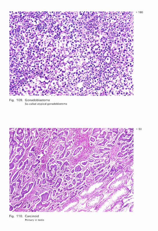
Fig. 109. Gonadoblastoma So-called atypical gonadoblastoma
Fig . 110. Carcinoid Primary in testis
x 180
x 50
x 100
Fig. 111. Malignant lymphoma
x 120
Fig. 112. Malignant lymphoma

x 150
Fig. 11 3. M alignant lymphoma
x 150
Fig. 114 . Plasmacytoma
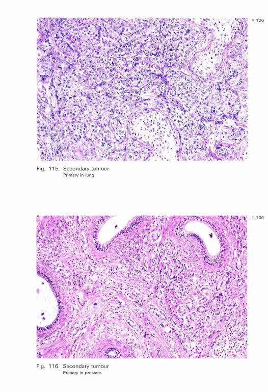
Fig. 115. Secondary tumour Primary in Jung
Fig. 116. Secondary tumour Primary in prostate
x 100
x 100

x 100
Fig. 117. Adenomatoid tumour
x 100
Fig. 118. Adenomatoid tumour
x 135
't~''--'~"" ,1.
Fig. 119. Adenomatoid tumour
Fig. 120. M esothelial proliferation
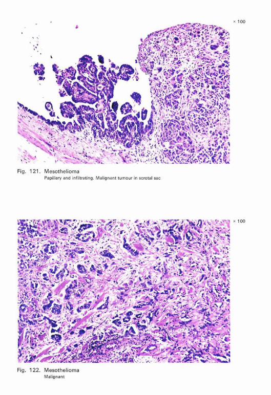
Fig. 121. Mesothelioma Papillary and infiltrating. Malignant tumour in scrotal sac
Fig. 122. Mesothelioma M alignant
x 100
x 100
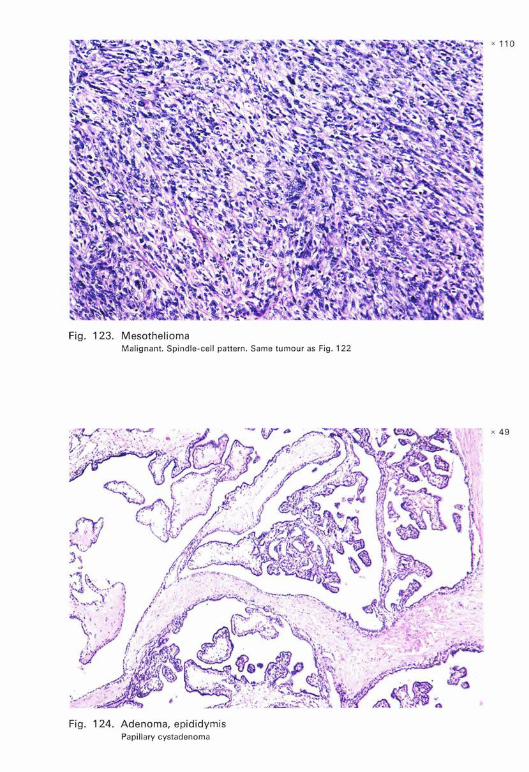
Fig. 123. Mesothelioma M alignant. Spindle-cell pattern. Same tumour as Fig. 122
Fig. 124. Adenoma, epididymis Papillary cystadenoma
x 110
x 49
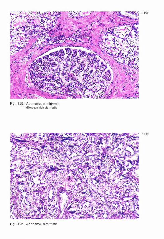
Fig. 125. Adenoma, epididymis Glycogen- ri ch clear cells
Fig . 126. Adenoma, rete testis
x 100
x 115

Fig. 127. Carcinoma, collecting ducts and rete testis Resembles Sertoli cell tumour
Fig . 128. Carcinoma, col lecting ducts and rete testis Same case as Fig. 127
x 130
x 145
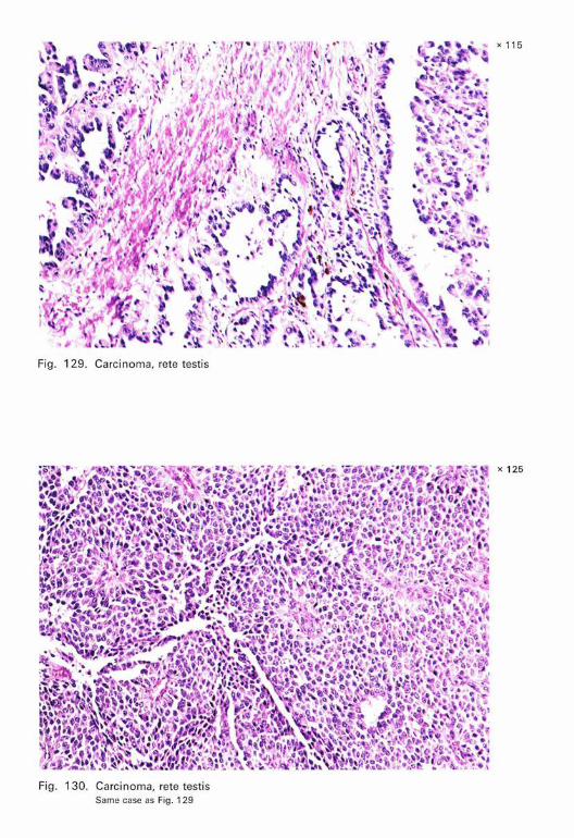
Fig. 129. Carcinoma, rete testis
Fig. 130. Carcinoma, rete testis Same case as Fig . 129
x 115
x 125
x 100
Fig. 131 . Melanotic neuro-ectodermal tumour
x 130
Fig . 132. M elanotic neuro-ectodermal tumour
x 135
Fig. 133. Rhabdomyosarcoma, spermatic cord
x 210
Fig. 134. Rhabdomyosarcoma, spermatic cord
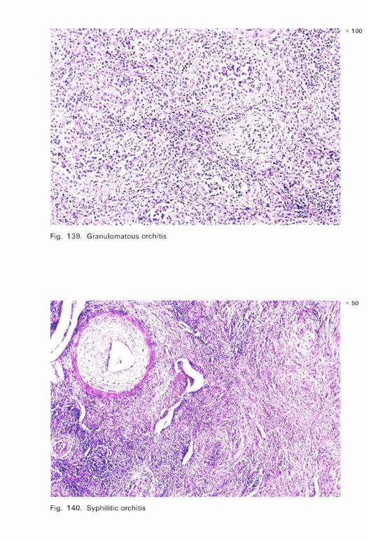
Fig. 139. Granulomatous orch itis
x 50
Fig. 140. Syphilitic orchitis
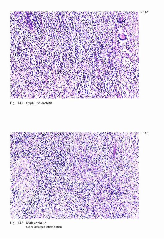
Fig. 142. Malakoplakia Granulomatous inflammation
x 110
x 11 5
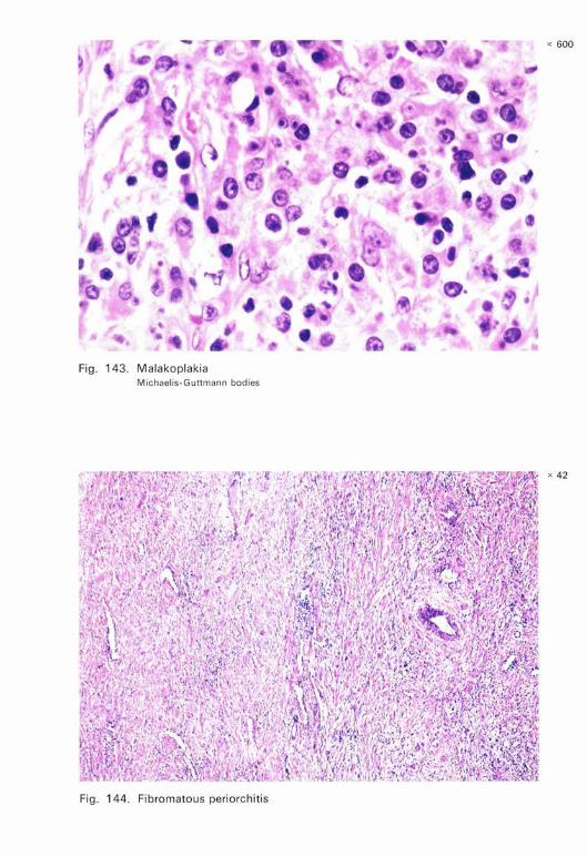
Fig. 143. Malakoplakia Michaelis-Guttmann bodies
Fig. 144. Fibromatous periorchitis
x 600
x 42

x 100
Fig. 145. Sperm granuloma
x 180
Fig . 146. Sperm granuloma High magnificat ion of Fig. 145
x 100
Fig. 147. Lipogranuloma, spermatic cord
x 40
\ • '\
t
flt'~ ' .I
.. I
Fig. 148. Adrenal rest. spermatic cord

Fig . 149. Gonadal splenic fusion
,:·
Fig. 150. Mesothelial cyst, tunica
, , ,/ ,.,,. .,
x 56
x 130
..


