HAL Id: hal-03329422 https://hal.archives-ouvertes.fr/hal-03329422 Submitted on 30 Aug 2021 HAL is a multi-disciplinary open access archive for the deposit and dissemination of sci- entific research documents, whether they are pub- lished or not. The documents may come from teaching and research institutions in France or abroad, or from public or private research centers. L’archive ouverte pluridisciplinaire HAL, est destinée au dépôt et à la diffusion de documents scientifiques de niveau recherche, publiés ou non, émanant des établissements d’enseignement et de recherche français ou étrangers, des laboratoires publics ou privés. Arbuscular Mycorrhizal Symbiosis Triggers Major Changes in Primary Metabolism Together With Modification of Defense Responses and Signaling in Both Roots and Leaves of Vitis vinifera Mary-Lorène Goddard, Lorène Belval, Isabelle Martin, Lucie Roth, Hélène Laloue, Laurence Deglene-Benbrahim, Laure Valat, Christophe Bertsch, Julie Chong To cite this version: Mary-Lorène Goddard, Lorène Belval, Isabelle Martin, Lucie Roth, Hélène Laloue, et al.. Arbuscular Mycorrhizal Symbiosis Triggers Major Changes in Primary Metabolism Together With Modification of Defense Responses and Signaling in Both Roots and Leaves of Vitis vinifera. Frontiers in Plant Science, Frontiers, 2021, 12, 10.3389/fpls.2021.721614. hal-03329422
Transcript
HAL Id: hal-03329422https://hal.archives-ouvertes.fr/hal-03329422
Submitted on 30 Aug 2021
HAL is a multi-disciplinary open accessarchive for the deposit and dissemination of sci-entific research documents, whether they are pub-lished or not. The documents may come fromteaching and research institutions in France orabroad, or from public or private research centers.
L’archive ouverte pluridisciplinaire HAL, estdestinée au dépôt et à la diffusion de documentsscientifiques de niveau recherche, publiés ou non,émanant des établissements d’enseignement et derecherche français ou étrangers, des laboratoirespublics ou privés.
Arbuscular Mycorrhizal Symbiosis Triggers MajorChanges in Primary Metabolism Together With
Modification of Defense Responses and Signaling inBoth Roots and Leaves of Vitis vinifera
To cite this version:Mary-Lorène Goddard, Lorène Belval, Isabelle Martin, Lucie Roth, Hélène Laloue, et al.. ArbuscularMycorrhizal Symbiosis Triggers Major Changes in Primary Metabolism Together With Modificationof Defense Responses and Signaling in Both Roots and Leaves of Vitis vinifera. Frontiers in PlantScience, Frontiers, 2021, 12, �10.3389/fpls.2021.721614�. �hal-03329422�
Arbuscular Mycorrhizal SymbiosisTriggers Major Changes in PrimaryMetabolism Together WithModification of Defense Responsesand Signaling in Both Roots andLeaves of Vitis viniferaMary-Lorène Goddard 1,2, Lorène Belval 1‡, Isabelle R. Martin 1†, Lucie Roth 1,2†,
Vine and wine are very ancient and represent both a part ofhuman history and a significant socioeconomic sector (Reynolds,2017). Today, grapevine is a very important crop worldwide, witha global production area of 6.8 Mha in 2018, generating a globalvalue of 31.4 billion euros for world wine trade (OIV, 2018).In the context of global warming, grapevine culture, like othercultivated plants, is confronted with multiple stresses, especiallyheat stress and drought (Songy et al., 2019). In addition toreduced growth and yield, it is well known that these stresseswill lead to the weakening of the plant that could explainthe recent explosion of grapevine dieback syndromes such astrunk diseases.
In order to gain better resistance to multiple stresses,the grapevine culture could take advantage of symbiosisthrough association with arbuscular mycorrhizal fungi (AMF).AMF belong to the Glomeromycota phylum and are soilmicroorganisms that are able to establish symbiosis with mostterrestrial plants (Trouvelot et al., 2015). In this mutualisticassociation, a plant supplies fungi with carbohydrates and lipids,and fungal colonization increases the root absorption surface,thus improving plant access to water and minerals. Theseexchanges take place in root cortical cells where specializedstructures called “arbuscules” are developed.
It is well established that association with AMF reducesthe need of fertilizers in plants by enhancing the uptake ofnutrients such as nitrogen and phosphate (Trouvelot et al.,2015). In grapevines, it has also been recently demonstrated thatmycorrhization with Funneliformis mosseae (F. mosseae)increases shoot growth and root system branching ofacclimatized plantlets, correlated with high expression oftwo grapevine phosphate transporters (Valat et al., 2018). It isalso known that AMF association can improve plant toleranceto abiotic stresses. In grapevines, colonization with AMF couldresult in more efficient water uptake and better resistance towater stress (Trouvelot et al., 2015). Indeed, higher mycorrhizalcolonization was evidenced in drier soil areas (Donkó et al.,2014). In addition, colonization of 1-year-old grapevines byAMF increased the water flux and photosynthesis comparedto non-colonized plants (Nogales et al., 2020). In the samestudy, Nogales et al. (2020) further investigated responses ofmycorrhized cv. Touriga Nacional grapevines to heat stress.They reported that colonization with Rhizophagus irregularis (R.irregularis) could help the plant to sustain its growth after heatshocks. In addition to water stress tolerance, AMF symbiosishas the potential to improve grapevine tolerance to salinity andheavy metals (Trouvelot et al., 2015).
Interestingly, it has been reported that several plant–AMF associations also result in better resistance to bioticstresses. Resistance mechanisms involve both competition forcolonization sites and induction of plant defense responses.Indeed, root inoculation with AMF triggers the so-called“mycorrhiza-induced-resistance” (MIR), which is systemicprotection against a wide range of pathogens (Cameron et al.,2013). Although MIR has been shown to be efficient against awide range of diseases in diverse plants, this kind of resistancehas mainly been involved in the case of root diseases ingrapevines (Petit and Gubler, 2006; Nogales et al., 2009; Haoet al., 2012). Concerning resistance to foliar diseases, Bruissonet al. (2016) showed that mycorrhization of several grapevinevarieties (Chasselas, Pinot Noir, and the interspecific hybridDivico) with R. irregularis triggered a higher expression of genesinvolved in stilbene biosynthesis; a higher bioactive stilbenecontent after leaf inoculation with Botrytis cinerea (B. cinerea) orPlasmopara viticola (P. viticola) was also recorded. Priming ofdefense reactions in leaves induced by AMF root colonization,therefore, supports the hypothesis of a role of mycorrhizationin improving resistance to foliar diseases in grapevines. In arecent study, Cruz-Silva et al. (2021) have further revealed thatmycorrhization of Vitis vinifera cv. Cabernet Sauvignon with R.irregularis altered the expression of several P. viticola effectorsafter infection, suggesting that AMF could enhance resistance todowny mildew.
Regarding all the beneficial effects provided by AMFassociations, understanding the effect of mycorrhization ongrapevine metabolism appears essential. Indeed, it is known thatmetabolic status is crucial to plant resistance to both abioticand biotic stresses. In the Tempranillo variety, which is widelycultivated in Mediterranean areas, mycorrhizal symbiosis with acommercial inoculum of five AMFs triggered primary metaboliteprofile alterations in berries, with an increase in glucose andamino acids (Torres et al., 2019). Torres et al. (2018a) also foundthat AMF colonization of Tempranillo increased anthocyanincontents in the berries of fruit cuttings and modulatedABA metabolism, especially under high temperatures. Still, inTempranillo grapevines, mycorrhization with a mixture of fiveAMFs induced the accumulation of phenolics, hydroxycinnamicacids, and carotenoids in leaves, although with different levelsdepending on the clone analyzed (Torres et al., 2018b). Finally,in recent studies, a significant increase in volatile organiccompounds was reported in V. vinifera Cabernet Sauvignonroots (Velásquez et al., 2020a) and V. vinifera Sangiovese leaves(Velásquez et al., 2020b) after colonization with F. mosseae. TheseVOCs included terpenes and monoterpene alcohols related toplant defense (Velásquez et al., 2020a).
Frontiers in Plant Science | www.frontiersin.org 2 August 2021 | Volume 12 | Article 721614
Few studies have focused on both transcriptomic andmetabolomic changes induced by mycorrhization in roots andleaves of grapevines. Balestrini et al. (2017) compared the effectof a single AMF (F. mosseae) and of a mixed inoculum containingbacteria and fungi on the transcriptome reprogramming of110R rootstock roots. Although the mixed inoculum eliciteda more important transcriptional regulation, the expressionof genes involved in nutrient transport, transcription factors,and cell wall-related genes was significantly altered in the twoconditions. In this study, in order to better understand theeffect of mycorrhization on grapevine metabolism and defensereactions, we combined a non-targeted metabolomic approachand a targeted transcriptomic study to analyze changes inducedin both the roots and leaves of V. vinifera cv. Gewurztraminerfollowing colonization with R. irregularis DAOM 197198. ThisAMF strain has been sequenced and is commercially availablefor winegrowers. The non-targeted metabolomic study used GC-MS and LC-MS to highlight the effect of Ri colonization on rootand leaf primary and specialized metabolites. The consequenceof mycorrhization was further analyzed by targeted analysisof the contents in defense hormones jasmonates and salicylicacid and by the study of the expression of genes involved indefense responses, sugar transport andmetabolism, and jasmonicacid biosynthesis.
MATERIALS AND METHODS
Plant Culture and MycorrhizationTwo-node segments of V. vinifera (L.) cv. Gewurztraminerclone 643 were collected in the winter of 2018–2019 in theexperimental vineyard from INRAE (Colmar, France), treatedwith Beltanol-L, and briefly conserved at 4◦C. Wood cuttingswere previously dipped with indole-3-butyric acid (IBA, 1mg/ml,Sigma Aldrich, MO, USA) and then planted in individual 1-Lpots (four cuttings/pot) for rooting in a 1:1 sterile mix of sandand perlite. The pots were then placed in a growth chamber at27◦C with a 16 h photoperiod (150 µEm−2·s−1 light irradiance,25/20◦C day/night). Plants were watered to saturation two timesa week with tap water for 3 weeks; then, with a low Pi nutritivesolution [2.5 mM Ca(NO3)2, 2.5mM KNO3, 0.1mM KH2PO4,1mM MgSO4, 50µM EDTA-Fe(III)-Na, 10µM H3BO3, 2µMMnCl2, 1µM ZnSO4, 0.5µMCuSO4, and 0.05µM Na2MoO4].After 5–6 weeks of culture, well-rooted plants with homogenousgrowth were transplanted individually (1 plant per pot) in 1 Lpots containing a 1:1 sterile mix of sand and perlite. Plants werewatered to saturation two times a week, alternately with tapwater and low Pi solution. After 10 days, one batch of 12 plantswas inoculated with R. irregularis DAOM197198 (Ri) producedunder axenic conditions (Agronutrition, Carbonne, France): theRi suspension was diluted with tap water to contain 50 spores/mland 1,000 spores (20ml) were gently poured at the base of theplant stem. Plants of the non-inoculated control plant batch (14plants) were reated with 20ml of diluted spore conservationsolution without spores. Two months after mycorrhization, theplants were carefully removed from the culture pots. Rootsystems were separated from the aerial parts and rinsed in tapwater. Fully expanded leaves, full length roots, and root tips were
harvested and snap frozen in liquid N2 for transcriptomic andmetabolomic analysis.
Evaluation of AMF ColonizationNon-lignified roots (∼1 g) were randomly harvested fromeach plant and stored in a lactoglycerol solution (lacticacid/glycerol/demineralized H2O 1:1:1) at 4◦C. For coloration,roots were put in 15-ml polypropylene tubes and cleared for20min at 90◦C in 10% (w/v) KOH, and then five droplets ofH2O2 solution (30% v/v) were added under agitation. Roots weresubsequently rinsed gently two times with demineralized water,and then immersed in an ink solution (5% Black Schaeffer Skritink/8% acetic acid) for 5min at 90◦C. Roots were then rinsedthree times with demineralized water and discolored with aceticacid (8%) for 15min at room temperature. After washing withdemineralized water, the samples were stored in lactoglycerol at4◦C. Thirty fragments, ca. 10 mm long, were randomly collectedfrom each sample, mounted 10 per slide in glycerol, and observedat × 200 magnification. Mycorrhization efficiency parameterswere evaluated according to Trouvelot et al. (1986): frequency(F): percentage of colonized fragments, intensity (M): estimationof the proportion of hyphae in the root cortex, (a): arbusculeabundance in the mycorrhized root part, and (A): arbusculeabundance in the whole root system.
Green Supermix, 0.2mM of forward and reverse primers, and10 ng of reverse transcribed RNA in a final volume of 25 µl.Thermal cycling conditions were 30 s at 95◦C, followed by 40cycles of 15 s at 94◦C, 30 s at 60◦C, and 30s at 72◦C. Thespecificity of the individual PCR amplification was checkedusing a heat dissociation curve from 55 to 95◦C followingthe final cycle of the PCR and by sequencing the final PCRproducts. The results obtained for each gene of interest werenormalized to the expression of two reference genes (VvEF1αand VvActin). Relative expression (2−11CT) compared to thesample with the lowest expression (highest CT value) wasalso calculated as described in Schmittgen and Livak (2008).Mean values and standard deviations were obtained from 3technical and 12 biological replicates. Primers used for real-time quantitative PCR are listed in Supplementary Table 1.Preliminary analysis of PR gene expression was performedwith the NeoViGen96 chip on three independent biologicalreplicates for each condition as described in Dufour et al.(2016).
Metabolomic AnalysisExtractionWe analyzed 14 independent biological replicates for theNM condition and 11 independent biological replicates for
Frontiers in Plant Science | www.frontiersin.org 3 August 2021 | Volume 12 | Article 721614
the Ri condition. Leaves and whole roots were ground inliquid nitrogen and freeze-dried. About 10 and 5mg of theserespective tissues were precisely weighted and transferred to1.5ml polypropylene microtubes for two successive extractionsof primary and specialized metabolites from the same startingmaterial. For primary metabolite extraction, the samples wereextracted with 600 µl for leaves and 300 µl for roots ofextract solution composed of 50mM potassium phosphate bufferpH 6 and 10 mg/L of phenoxyacetic acid as an internalstandard in an ultrasound bath during 30min (power 9, 20◦Cas starting temperature). After centrifugation (20,000 g, 20min,20◦C), supernatants were transferred to a filtration plate (96-well plates Acroprep 1ml, 45µm GHP membrane, Pall LifeScience, Portsmouth, United Kingdom) and filtered under avacuum with a vacuummanifold (Pall Corporation, Portsmouth,United Kingdom) to be collected on a collector plate (a 96-well PP plate, 1.2ml, VWR 732-2890/391-0077, Fontenay-sous-Bois, France). This operation was performed two times for eachsample, and filtrates were gathered in the same proportions priorto GC-MS analyses.
After primary metabolite extraction, the remaining pellet waswashed with ultrapure water, freeze-dried, and re-lyophilizedfor specialized metabolite extraction. The second extraction wascarried out in the same conditions but with 300 µl of LC-MSgrade methanol (Fisher Scientific, Illkirch, France), containing 5-methylsalicylic acid as an internal standard at 5 mg/L for bothleaves and roots. After two extractions, filtrates were equallypooled and analyzed by LC-MS. The methods GC-MS and LC-MS have been described in detail in Labois et al. (2020).
GC-MS AnalysisGC-MS analysis was carried out on 30 µl of each aqueous extractafter freeze-drying and derivatization reaction, which consistsof subsequent addition of methoxyamine hydrochloride solution(20 µl, 30 mg/ml in anhydrous pyridine) followed by MSTFA(80 µl). The samples were respectively incubated during 90 and30min at 37◦C and 600 rpm.
GC-MS analysis was performed on GC-2010 gaschromatography, coupled with the GC-QP2010 mass detector(Shimadzu Corporation, Tokyo, Japan) in the acquisitionconditions already described by Labois et al. (2020).
After data file conversion, thanks to the ABF file converter(Reifycs), raw data were processed using MS-DIAL 4.0 (version4.48) software (http://prime.psc.riken.jp/) for peak detection andintegration and sample alignment. Identification of compoundswas performed, thanks to an owner commercial compoundlibrary and the mass spectral library NIST 17 by comparison ofretention time and a spectrum profile.
HPLC-MS AnalysisMethanolic extracts were analyzed on the High-PerformanceLiquid Chromatography Agilent 1,100 series coupled to theAgilent 6,510 accurate-mass Quadrupole-Time of Flight (Q-TOF) mass spectrometer with an electrospray ionization(ESI) interface in a negative ionization mode (AgilentTechnologies, Santa Clara, CA, USA). Separation and detectionof compounds were performed as described in Labois et al.
(2020). Deconvolution, integration, and alignment wereperformed, thanks to the Profinder software (version B.08.00,Agilent Technologies, Santa Clara, CA, USA). Compoundannotation was carried out with commercially availablestandards. Additional compounds were putatively identifiedusing the molecular formula calculated from the exact mass andan isotope profile, relative retention times, and mass spectrafrom Metlin (https://metlin.scripps.edu).
Extraction and Quantitative HPLC-MSAnalysis of Jasmonates and Salicylic AcidFresh leaves and whole roots were manually ground in liquidnitrogen. Extraction and quantification of jasmonates wereperformed as described in Widemann et al. (2013) and Smirnovaet al. (2017). Approximately 90mg of powder, exactly weighted,was extracted with 900 µl of extraction solution (methanol:water: acetic acid (70:29:0.5), containing two internal standards:9,10-dihydrojasmonoylisoleucine (dihydro-JA-Ile, 2µM) andprostaglandine A1 (PGA1, 50 nM). The samples were agitatedon a wheel for 30min at 4◦C and then centrifuged at maximumspeed for 15min at 4◦C. About 450 µl of supernatant wastransferred in a 1.5-ml microtube and concentrated to 250 µlin a speed vac at 30◦C. The samples were stored overnight at−20◦C and then centrifuged for 10min at maximal speed topellet insoluble material. Approximately 150 µl of the particle-free supernatant was transferred to an amber glass vial to beinjected in LC-MS/MS.
Jasmonates [jasmonic acid (JA), jasmonoyl-isoleucine (JA-Ile), 12-oxophytodienoic acid (12-OPDA)] and salicylic acid(SA) were identified and quantified with a UHPLC system Eluteconnected to a mass spectrometer Impact II Q-TOF-MS/MSequipped with an electrospray ESI source Apollo II operating ina negative mode (Bruker Daltonics GmbH, Bremen, Germany).The raw samples were separated, thanks to the reversed-phaseC18 Bruker Intensity Solo HPLC column (2-µmparticle size, 100Å pore size, and of 2 × 100-mm dimensions) (BRHSC18022100,Bruker Daltonics GmbH, Bremen, Germany). The mobile phaseswere ultrapure water with 0.1% formic acid (eluent A) andMeOH with 0.1% formic acid (eluent B). They were used ina gradient expressed as B percentage: 2min at 1.%, a lineargradient during 15min until 99%, 3min at 99%, followed by5min in the initial conditions (1% B) for equilibration beforea new injection. The total flow rate was 0.25 ml/min, injectionvolume was 10 µl, and the oven column and the autosamplertemperature were 35 and 8◦C, respectively. The MS detectorwas internally calibrated before starting the analysis batch andadditionally at the beginning of each injection by infusing a 10-mM sodium formate solution in iPrOH: H2O (1:1, v/v). The ESIparameters were as follows: capillary voltage,−3,500V; end-plateoffset, −500V; nebulizer gas, 29 psi; dry gas, 8 L/min; and drytemperature, 200◦C. The spectra rate was 2Hz over a 30–1,000m/z mass range of both auto MS/MS and bbCID scan modes.To determine fragments of each parent metabolite, MS/MSspectra were recorded with a collision energy ramp from 20 to50 eV. In the broadband collision-induced dissociation (bbCID)mode, each parent ion was fragmented by alternating low- and
Frontiers in Plant Science | www.frontiersin.org 4 August 2021 | Volume 12 | Article 721614
high-collision energy, namely, 6 and 50 eV, respectively. Dataacquisition was achieved with the Bruker Compass HyStar 5.1and otofControl 5.2 software, and data treatment was performedwith the DataAnalysis 5.3 and TASQ 2021 (Bruker DaltonicsGmbH, Bremen, Germany).
Qualifier ions were determined from data obtained inan auto MS/MS acquisition mode in DataAnalysis andlisted in Supplementary Table 2 together with chromatographicretention times, assigned internal standards, and quantifier ionsto achieve quantification in TASQ. Absolute quantification wascarried out, thanks to external calibration from commerciallyavailable standards (serial dilutions were performed from a 200nM stock solution (SA and PGA1) and a 100 nM stock solution(12-OPDA, JA, JA-Ile, dh-JA-Ile) in methanol. Calibrants wereanalyzed in triplicates in the same acquisitionmethod as samples.Final results were given in nanogram of each compound relatedto gram of fresh weight raw material after normalization by theinternal standard and fresh weight of raw material.
Statistical AnalysisFor all GC-MS and LC-MS data, the area of each compoundwas normalized with respect to the raw material weight and theinternal standard area. Only metabolites present in 80% of thesamples of each condition were considered. Statistical analyseswere performed with the MetaboAnalyst 5.0 online platform(https://www.metaboanalyst.ca (Chong et al., 2019). Normalizedareas were transformed using the base 10 logarithm and mean-centering. Metabolites significantly different between NM and Riconditions were identified with a fold change of at least +/– 1.5and a p value <0.05 (Wilcoxon test analysis).
Transcriptomic data were analyzed using a non-parametrictest (Kruskall and Wallis) with the R 4.0.0 software.
RESULTS
Plant Growth Parameters and Analysis ofMycorrhizationAnalysis of plant growth parameters 2 months aftermycorrhization showed that shoot length, internode length,and leaf mass were not significantly different between non-mycorrhized (NM) and mycorrhized (Ri) plants. However, Riplants were characterized by a higher mass of the root system(although not statistically significant) as well as a higher rootmass/leaf mass ratio (Figure 1).
Microscopic observations of cleared and stained rootsrevealed the presence of mycorrhizal structures in roots from Riplants. In contrast, no fungal structure was found in the roots ofNM plants (Figure 2A). In the colonized plants, mycorrhizationfrequency (F) ranged from 93 to 100%; intensity (I) rangedfrom 69 to 94%, and arbuscule content in colonized roots (a)ranged from 66 to 98%, showing a high level of root colonization(Figure 2B). To check the functionality of the mycorrhiza, westudied the expression of the grapevine phosphate transporterVvPht1.2 and the R. irregularis hexose transporter RiMST2.VvPht1.2 expression was previously reported to be highlyinduced, following grapevine colonization with F. mosseae (Valatet al., 2018). RiMST2 is a major component for hexose uptake by
(Ri) or not (NM) with R. irregularis. Shoot length, internode length, mass of the
seven first leaves, and whole root system mass (fresh weights) were measured
2 months after mycorrhization. Results are means ± SE of 12 biological
replicates. Asterisks indicate significant difference between NM and Ri
condition (Kruskall and Wallis, p < 0.05).
the AM fungus and is essential for functional symbiosis (Helberet al., 2011). As shown in Figure 2C, an intense expression ofthese twomarker genes was detected in mycorrhized roots. Theseresults show that grapevine roots are efficiently colonized by R.irregularis and the well functioning of mycorrhiza in our system.
Changes in Primary and SpecializedMetabolites Induced by Colonization WithR. irregularisTo determine the metabolic responses in grapevine roots andleaves upon mycorrhization with Ri, we used a non-targetedapproach. Two extraction-analysis methods were performed inorder to cover the maximum metabolite diversity. Raw material(leaves and roots of the same plants) was firstly extracted with aphosphate buffer and analyzed by GC-MS; the remaining pelletwas subsequently extracted in MeOH and analyzed with LC-MS. Impact of mycorrhization onmetabolomic response was firstanalyzed by PCA on GC-MS and LC-MS data. To get insightinto the metabolite differences between NM and Ri samples, wefurther analyzed the mean relative levels of each metabolite in thedifferent conditions. Metabolites significantly different betweenNM and Ri conditions were identified with a fold change ofat least +/– 1.5 and a p value <0.05 or 0.1 (Wilcoxon test).Metabolic changes triggered by Ri colonization in roots andleaves are detailed in the following subsections.
Metabolomic Responses in RootsFor metabolites analyzed by GC-MS and mostly primarymetabolites, a total of 83 compounds (comprising 34identified and 23 putative compounds) were detected thatbelonged to sugars, sugar alcohols, sugar acids, polyols, smallorganic acids, amino acids, aliphatic acids, and phenolics(Supplementary Table 3). Colonization of grapevine roots
Frontiers in Plant Science | www.frontiersin.org 5 August 2021 | Volume 12 | Article 721614
FIGURE 2 | Plant mycorrhization parameters (A,B) and expression of
colonization marker genes in grapevine roots (B). (A) Representative pictures
of Ri (right) and NM (left) roots. Pictures were taken with a Nikon
ALPHAPHOT-2 microscope (X100 magnification). Bar = 5µm. (B) F
(frequency): percentage of colonized fragments, M (intensity): proportion of
hyphae in the root cortex, a: arbuscule content in colonized roots, A:
arbuscule abundance in the whole root system (from Trouvelot et al., 1986).
Data are the mean ± SD of 12 biological replicates. (C) Expression of the
grapevine Pht1.2 phosphate transporter and the R. irregularis RiMST2 hexose
transporter in roots of NM and Ri plants. Expression was studied by RT-qPCR
in root tips 2 months after mycorrhization. Transcript levels were normalized to
V. vinifera ACTIN and EF1α transcript levels. Fold change (2−11CT method)
indicates normalized expression levels in Ri roots compared to the mean of the
normalized expression levels measured in NM roots. Data are the mean ± SD
of 12 biological replicates. Asterisks indicate significant difference in gene
expression between NM and Ri conditions (Kruskall and Wallis, p <0.001).
with Ri resulted in significant changes in the content ofprimary metabolites since PCA analysis revealed a separationbetween NM and Ri conditions (Supplementary Figure 1).Twenty-nine metabolites were significantly impacted bythe mycorrhization (Figure 3). Among identified primarymetabolites, Ri colonization triggered enhanced contents offructose, trehalose, succinic, and tartaric acids. In contrast,mycorrhizal roots had decreased levels in most of the sugaracids and sugars identified, i.e., galactose, glucose, mannose,sucrose, erythronic, and threonic acids, and also in myoinositol.A decrease in shikimic acid and epicatechin contents was alsodetected in mycorrhizal roots (Figure 3).
Concerning metabolites analyzed by LC-MS, a total of194 compounds (comprising 13 confirmed and 55 putativemetabolites) were detected belonging to fatty acids, stilbenes,tannins, terpenes, flavonoids, anthraquinones, and aromatics(Supplementary Table 4). 3D-PCA of LC-MS data showed aseparation between the NM and Ri (Supplementary Figure 2).Twenty-one metabolites were significantly altered by themycorrhization (Figure 4). Interestingly, among metabolites
FIGURE 3 | GC-MS metabolites significantly altered by the mycorrhization in
roots. Contents are expressed as normalized areas for each metabolite. Data
are the mean ± SE of 14 biological replicates for the NM condition and 11
biological replicates for the Ri condition. Metabolites significantly different
between NM and Ri conditions were identified with a fold change of at least
+/– 1.5 and a p value < 0.05 (Wilcoxon test).
significantly altered by mycorrhization, several fatty acids(arachidonic acid, eicosapentaenoic acid, and putativehydroxytetracosenoic acid) were solely detected in Ri root
Frontiers in Plant Science | www.frontiersin.org 6 August 2021 | Volume 12 | Article 721614
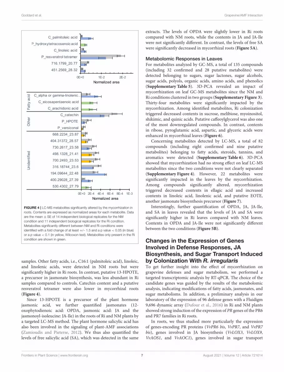
FIGURE 4 | LC-MS metabolites significantly altered by the mycorrhization in
roots. Contents are expressed as normalized areas for each metabolite. Data
are the mean ± SE of 14 independent biological replicates for the NM
condition and 11 independent biological replicates for the Ri condition.
Metabolites significantly different between NM and Ri conditions were
identified with a fold change of at least +/– 1.5 and a p value < 0.05 (in blue)
or a p value < 0.1 (in yellow, Wilcoxon test). Metabolites only present in the Ri
condition are shown in green.
samples. Other fatty acids, i.e., C16:1 (palmitoleic acid), linoleic,and linolenic acids, were detected in NM roots but weresignificantly higher in Ri roots. In contrast, putative 13-HPOTE,a precursor in jasmonate biosynthesis, was less abundant in Risamples compared to controls. Catechin content and a putativeresveratrol tetramer were also lower in mycorrhizal roots(Figure 4).
Since 13-HPOTE is a precursor of the plant hormonejasmonic acid, we further quantified jasmonates (12-oxophytodienoic acid: OPDA, jasmonic acid: JA and thejasmonoyl-isoleucine: JA-Ile) in the roots of Ri and NM plants bya targeted LC-MS method. The plant hormone salicylic acid hasalso been involved in the signaling of plant-AMF associations(Zamioudis and Pieterse, 2012). We thus also quantified thelevels of free salicylic acid (SA), which was detected in the same
extracts. The levels of OPDA were slightly lower in Ri rootscompared with NM roots, while the contents in JA and JA-Ilewere not significantly different. In contrast, the levels of free SAwere significantly decreased in mycorrhizal roots (Figure 5A).
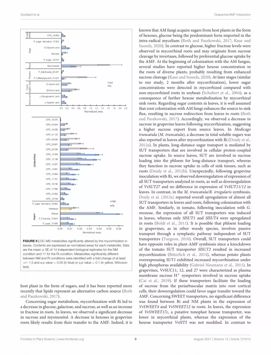
Metabolomic Responses in LeavesFor metabolites analyzed by GC-MS, a total of 135 compounds(including 32 confirmed and 28 putative metabolites) weredetected belonging to sugars, sugar lactones, sugar alcohols,sugar acids, polyols, organic acids, amino acids, and phenolics(Supplementary Table 5). 3D-PCA revealed an impact ofmycorrhization on leaf GC-MS metabolites since the NM andRi conditions clustered in two groups (Supplementary Figure 3).Thirty-four metabolites were significantly impacted by themycorrhization. Among identified metabolites, Ri colonizationtriggered decreased contents in sucrose, melibiose, myoinositol,shikimic, and quinic acids. Putative caffeoylglycerol was also oneof the most downregulated compounds. In contrast, contentsin ribose, pyroglutamic acid, aspartic, and glyceric acids wereenhanced in mycorrhizal leaves (Figure 6).
Concerning metabolites detected by LC-MS, a total of 82compounds (including eight confirmed and nine putativemetabolites) belonging to fatty acids, steroids, tannins, andaromatics were detected (Supplementary Table 6). 3D-PCAshowed that mycorrhization had no strong effect on leaf LC-MSmetabolites since the two conditions were not clearly separated(Supplementary Figure 4). However, 22 metabolites weresignificantly impacted in the leaves by the mycorrhization.Among compounds significantly altered, mycorrhizationtriggered decreased contents in ellagic acid and increasedcontents in linoleic acid, linolenic acid, and putative EOTE,another jasmonate biosynthesis precursor (Figure 7).
Interestingly, further quantification of OPDA, JA, JA-Ile,and SA in leaves revealed that the levels of JA and SA weresignificantly higher in Ri leaves compared with NM leaves.Contents in OPDA and JA-Ile were not significantly differentbetween the two conditions (Figure 5B).
Changes in the Expression of GenesInvolved in Defense Responses, JABiosynthesis, and Sugar Transport Inducedby Colonization With R. irregularisTo get further insight into the effect of mycorrhization ongrapevine defenses and sugar metabolism, we performed atargeted transcriptomic analysis by RT-qPCR. The choice of thecandidate genes was guided by the results of the metabolomicanalysis, indicating modifications of fatty acids, jasmonates, andsugar metabolisms. In addition, a preliminary analysis in ourlaboratory of the expression of 96 defense genes with a Fluidigm9,696 dynamic array (Dufour et al., 2016) in Ri and NM plantsshowed strong induction of the expression of PR genes of the PR6and PR7 families in Ri roots.
In roots, we thus studied more particularly the expressionof genes-encoding PR proteins (VvPR6 bis, VvPR7, and VvPR7bis), genes involved in JA biosynthesis (VvLOX3, VvLOX9,VvAOS1, and VvAOC1), genes involved in sugar transport
Frontiers in Plant Science | www.frontiersin.org 7 August 2021 | Volume 12 | Article 721614
FIGURE 5 | Levels of jasmonates (OPDA, JA, and JA-Ile) and salicylic acid (SA) in roots (A) and leaves (B) of Ri and NM grapevines, 2 months after mycorrhization.
Data are mean ± SE of 8 independent biological replicates. Asterisks indicate significant differences between NM and Ri conditions (Kruskall and Wallis, ***p < 0.001,
Candidate genes studied in leaves were VvWRKY2 (defensesignalization), genes involved in sugar transport and metabolism(VvSWEET17c, VvHT1, VvHT5, VvSUC11, VvSUC12, VvSUC27,VvCIN2, and VvWINV), and genes involved in JA biosynthesis(VvLOX3, VvLOX9, VvAOS5, and VvAOC1).
It should be noticed that preliminary analysis of the expressionof PR genes that could be regulated by the SA (PR1 and PR2) orJA (proteinase inhibitor PIN and PR14) defense hormones wasnot significantly affected by the mycorrhization in both roots andleaves (Supplementary Figure 5).
Transcriptomic Responses in RootsColonization of grapevines with Ri triggered a strong inductionof VvPR6bis, VvPR7, and VvPR7bis expression in roots. Onthe other hand, mycorrhization resulted in significant lowerexpression of several genes involved in JA biosynthesis (VvLOX3,VvLOX9, VvAOS1, and VvAOC1, Figure 8). Similarly, severalgenes involved in sucrose transport were downregulated inRi roots (VvSUC11, VvSUC12, and VvSUC27) (Figure 8). Theexpression of the hexose transporter VvSWEET4 also seemsdownregulated in Ri roots, although the difference betweenNM and Ri roots is not significant. In contrast, expression
of a SWEET transporter putatively involved in disaccharidetransport (VvSWEET12) and of cytoplasmic and cell wallinvertases (VvCIN2 and VvWINV) was not significantly alteredby mycorrhization (Supplementary Figure 6).
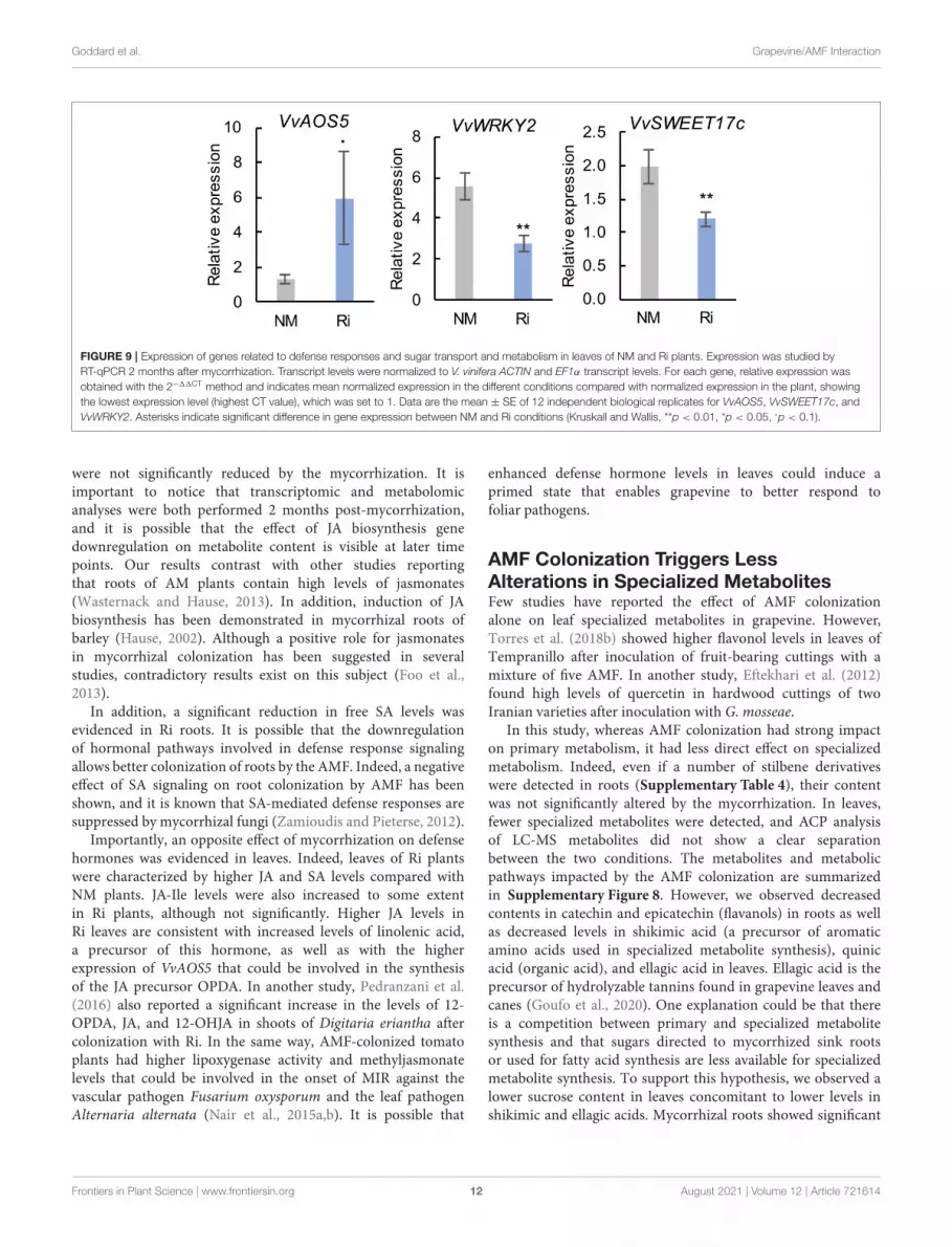
Transcriptomic Responses in LeavesIn contrast to roots, mycorrhization had less impact onthe expression of genes involved in sugar transport and JAbiosynthesis since there was no significant difference in theexpression of most analyzed genes (Supplementary Figure 7).However, we observed some induction of the expression ofVvAOS5. In addition, root colonization by Ri resulted insignificantly lower expression of VvWRKY2 and of the putativehexose vacuolar transporter VvSWEET17c (Figure 9).
DISCUSSION
AMF Colonization Triggers StrongReprogramming of Sugar Metabolism inGrapevineIn this study, we show that colonization of grapevine roots withRi leads to major changes in primary metabolites, especiallysugars and lipids. These results are in accordance with theconcept of nutrient exchanges between plants and AM fungi.Indeed, it is assumed that AM fungi acquire carbon from their
Frontiers in Plant Science | www.frontiersin.org 8 August 2021 | Volume 12 | Article 721614
FIGURE 6 | GC-MS metabolites significantly altered by the mycorrhization in
leaves. Contents are expressed as normalized areas for each metabolite. Data
are the mean ± SE of 14 independent biological replicates for the NM
condition and 11 for the Ri condition. Metabolites significantly different
between NM and Ri conditions were identified with a fold change of at least
+/– 1.5 and a p value < 0.05 (in blue) or a p value < 0.1 (in yellow, Wilcoxon
test).
host plant in the form of sugars, and it has been reported morerecently that lipids represent an alternative carbon source (Rothand Paszkowski, 2017).
Concerning sugar metabolism, mycorrhization with Ri led toa decrease in glucose, galactose, and sucrose, as well as an increasein fructose in roots. In leaves, we observed a significant decreasein sucrose and myoinositol. A decrease in hexoses in grapevineroots likely results from their transfer to the AMF. Indeed, it is
known that AM fungi acquire sugars from host plants in the formof hexoses, glucose being the predominant form imported in theintra-radical mycelium (Roth and Paszkowski, 2017; Kaur andSuseela, 2020). In contrast to glucose, higher fructose levels wereobserved in mycorrhizal roots and may originate from sucrosecleavage by invertases, followed by preferential glucose uptake bythe AMF. At the beginning of colonization with the AM fungus,several studies have reported higher hexose concentration inthe roots of diverse plants, probably resulting from enhancedsucrose cleavage (Kaur and Suseela, 2020). At later stages (similarto our study, 2 months after mycorrhization), lower sugarconcentrations were detected in mycorrhized compared withnon-mycorrhized roots in soybean (Schubert et al., 2004), as aconsequence of further hexose metabolization by mycorrhizalsink roots. Regarding sugar contents in leaves, it is well assumedthat root colonization with AM fungi enhances the source to sinkflux, resulting in sucrose redirection from leaves to roots (Rothand Paszkowski, 2017). Accordingly, we observed a decrease insucrose in grapevine leaves following mycorrhization, suggestinga higher sucrose export from source leaves. In Medicagotruncatula (M. truncatula), a decrease in total soluble sugars wasalso reported in leaves after mycorrhization with Ri (Doidy et al.,2012a). In plants, long-distance sugar transport is mediated bySUT transporters that are involved in cellular proton-coupledsucrose uptake. In source leaves, SUT are involved in sucroseloading into the phloem for long-distance transport, whereasthey function in sucrose uptake in cells of sink tissues, such asroots (Doidy et al., 2012b). Unexpectedly, following grapevineinoculation with Ri, we observed downregulation of expression ofall SUT transporters analyzed in roots, as well as downregulationof VvSUT27 and no difference in expression of VvSUT11/12 inleaves. In contrast, in the M. truncatula/R. irregularis symbiosis,Doidy et al. (2012a) reported overall upregulation of almost allSUT transporters in leaves and roots, following colonization withthe AMF. Similarly, in tomato, following inoculation with G.mosseae, the expression of all SUT transporters was inducedin leaves, whereas only SlSUT1 and SlSUT4 were upregulatedin roots (Boldt et al., 2011). It is possible that phloem loadingin grapevines, as in other woody species, involves passivetransport through a symplastic pathway independent of SUTtransporters (Turgeon, 2010). Overall, SUT transporters couldhave opposite roles in plant-AMF symbiosis since a knockdownof the tomato SUT transporter SlSUT2 resulted in increasedmycorrhization (Bitterlich et al., 2014), whereas potato plantsoverexpressing SUT1 exhibited increased mycorrhization underhigh phosphorus availability (Gabriel-Neumann et al., 2011). Ingrapevines, VvSUC11, 12, and 27 were characterized as plasmamembrane sucrose H+ symporters involved in sucrose uptake(Cai et al., 2019). If these transporters facilitate the loadingof sucrose from the periarbuscular matrix into root corticalcells, their downregulation could favor sugar transfer toward theAMF. Concerning SWEET transporters, no significant differencewas found between Ri and NM plants in the expression ofVvSWEET4 and VvSWEET12 in roots. In leaves, the expressionof VvSWEET17c, a putative tonoplast hexose transporter, waslower in mycorrhizal plants, whereas the expression of thehexose transporter VvHT5 was not modified. In contrast to
Frontiers in Plant Science | www.frontiersin.org 9 August 2021 | Volume 12 | Article 721614
FIGURE 7 | LC-MS metabolites significantly altered by the mycorrhization in leaves. Contents are expressed as normalized areas for each metabolite. Data are the
mean ± SE of 14 independent biological replicates for the NM condition and 11 for the Ri condition. Metabolites significantly different between NM and Ri conditions
were identified with a fold change of at least +/– 1.5 and a p value < 0.05 (in blue) or a p value < 0.1 (in yellow, Wilcoxon test).
our results, Manck-Götzenberger and Requena (2016) reportedmajor transcriptional changes of SWEETs in potato roots inducedby the AM fungus R. irregularis. In addition to sugar transporters,extracellular invertases would play a role in supplying the AMfungus with hexoses (Schaarschmidt et al., 2006). However,no difference in the expression of both cytoplasmic invertaseor cell wall invertase was found between mycorrhizal andnon-mycorrhizal roots. It is possible that basal plant invertaseactivity is sufficient for apoplastic sucrose hydrolysis in Ri-colonized roots.
Our results thus show a different regulation pattern forSUTs and SWEETs transporters in grapevines compared withother plants and suggest a different regulation of carbonpartitioning in grapevines compared with herbaceous species.Although we observed overall downregulation of sugar contentsin both roots and leaves of Ri grapevines, it has to benoticed that it did not lead to a negative impact ongrapevine growth.
AMF Colonization Triggers StrongReprogramming of Fatty Acid Metabolismin GrapevineOther primary metabolites strongly impacted in mycorrhizalroots belong to the lipid family, in agreement with the fact thatlipids constitute a main carbon store in AMF (Keymer et al.,
2017). It has been recently demonstrated that, in addition tosugars, lipids are also major nutrients synthesized by plants andtransferred to AMFs, which are fatty acid auxotrophs (Jiang et al.,2017; Luginbuehl et al., 2017). In addition, lipids are essential forthe establishment of symbiosis, either as signals or as constituentsof the periarbuscular membrane (Vijayakumar et al., 2016). Uponarbuscule formation, a specific lipid biosynthesis pathway isinduced in plant cells to synthesize high levels of palmitic acid(C16:0), a precursor of 16:0 β-monoacylglycerol, which can beexported to the symbiont across the periarbuscular membrane(Wipf et al., 2019). Subsequently, AMF are able to elongateand desaturate fatty acids provided by the plant (Trépanieret al., 2005). In this study, higher contents in palmitoleic acid(C16:1) were found in Ri roots, and several C20–24 fattyacids (arachidonic acid, eicosapentaenoic acid, and putativehydroxytetracosenoic acid) were only detected in mycorrhizedroots. C16:1 fatty acids, which are monounsaturated fatty acidsproduced by desaturation of palmitic acid have been alreadydescribed as good indicators of fungal AM development (VanAarle and Olsson, 2003; Trépanier et al., 2005). C20 fatty acidsare likely produced by the AMF from fatty acids provided bythe plant. Indeed, in the Lotus japonicus/R. irregularis symbiosis,arachidonic acid (C20:4) and eicosapentaenoic acid (C20:5) wereshown to be synthesized by the symbiont (Vijayakumar et al.,2016).
Frontiers in Plant Science | www.frontiersin.org 10 August 2021 | Volume 12 | Article 721614
FIGURE 8 | Expression of genes related to defense responses and sugar transport and metabolism in roots of NM and Ri plants. Expression was studied by
RT-qPCR in root tips 2 months after mycorrhization. Transcript levels were normalized to V. vinifera ACTIN and EF1α transcript levels. For each gene, relative
expression was obtained with the 2−11CT method and indicates mean normalized expression in the different conditions compared with normalized expression in the
plant showing the lowest expression level (highest CT value), which was set to 1. Data are the mean ± SE of 12 independent biological replicates for VvLOX3,
VvLOX9, VvAOS1, VvSUC11, VvSUC12, and VvSUC27. Data are the mean ± SE of six independent biological replicates for VvSWEET4. Asterisks indicate significant
difference in gene expression between NM and Ri conditions (Kruskall and Wallis, ***p < 0.001, **p < 0.01, *p < 0.05, ·p < 0.1).
Furthermore, we show that AMF colonization results insignificantly higher contents in linoleic (C18:2) and linolenic(C18:3) acids in roots and in higher levels in putative EOTE,linoleic, and linolenic acids in leaves. Besides their role as acarbon source for the AMF, fatty acids, and especially unsaturatedfatty acids, play a key role in plant defense (Lim et al., 2017).Interestingly, Xing and Chin (2000) have shown that eggplantswith enhanced levels of palmitoleic acid (C16:1) and C16:3due to overexpression of a yeast 19 desaturase have betterresistance to Verticillium dahliae. In addition, increased levelsof C18:2 and C18:3 lead to better resistance to Colletotrichum
gloeosporioides in avocado (Wang et al., 2004) and Pseudomonas
syringae in tomato (Yaeno et al., 2004). In another study inbean, Ongena et al. (2004) reported that the accumulationof C18:2 and C18:3 fatty acids is associated with enhancedresistance to B. cinerea triggered by rhizobacteria (Ongena et al.,2004).
AMF Colonization Results in ContrastingEffect on JA and SA Metabolism in Rootsand Leaves of GrapevineWhereas the levels of several fatty acids involved in JAbiosynthesis (linoleic and mostly linolenic acid) are enhancedin Ri roots, mycorrhization results in some extent indownregulation of the JA pathway. Indeed, the expressionof several enzymes involved in JA biosynthesis (VvLOX3,VvLOX9, VvAOS1, and VvAOC1) was lower in Ri roots, anda lower content in OPDA was detected. It is known thatlinoleic and linolenic acids can be synthesized by the plantor the AMF in roots and could be precursors for longerchain fatty acids (C20) produced by the fungus (Vijayakumaret al., 2016). It is possible that the α-linolenic acid pool isdirected toward the synthesis of AMF specific C20 fatty acids(eicosapentaenoic acid) and less available for the JA biosynthesisin roots. However, contents in JA and in the active form JA-Ile
Frontiers in Plant Science | www.frontiersin.org 11 August 2021 | Volume 12 | Article 721614
FIGURE 9 | Expression of genes related to defense responses and sugar transport and metabolism in leaves of NM and Ri plants. Expression was studied by
RT-qPCR 2 months after mycorrhization. Transcript levels were normalized to V. vinifera ACTIN and EF1α transcript levels. For each gene, relative expression was
obtained with the 2−11CT method and indicates mean normalized expression in the different conditions compared with normalized expression in the plant, showing
the lowest expression level (highest CT value), which was set to 1. Data are the mean ± SE of 12 independent biological replicates for VvAOS5, VvSWEET17c, and
VvWRKY2. Asterisks indicate significant difference in gene expression between NM and Ri conditions (Kruskall and Wallis, **p < 0.01, *p < 0.05, ·p < 0.1).
were not significantly reduced by the mycorrhization. It isimportant to notice that transcriptomic and metabolomicanalyses were both performed 2 months post-mycorrhization,and it is possible that the effect of JA biosynthesis genedownregulation on metabolite content is visible at later timepoints. Our results contrast with other studies reportingthat roots of AM plants contain high levels of jasmonates(Wasternack and Hause, 2013). In addition, induction of JAbiosynthesis has been demonstrated in mycorrhizal roots ofbarley (Hause, 2002). Although a positive role for jasmonatesin mycorrhizal colonization has been suggested in severalstudies, contradictory results exist on this subject (Foo et al.,2013).
In addition, a significant reduction in free SA levels wasevidenced in Ri roots. It is possible that the downregulationof hormonal pathways involved in defense response signalingallows better colonization of roots by the AMF. Indeed, a negativeeffect of SA signaling on root colonization by AMF has beenshown, and it is known that SA-mediated defense responses aresuppressed by mycorrhizal fungi (Zamioudis and Pieterse, 2012).
Importantly, an opposite effect of mycorrhization on defensehormones was evidenced in leaves. Indeed, leaves of Ri plantswere characterized by higher JA and SA levels compared withNM plants. JA-Ile levels were also increased to some extentin Ri plants, although not significantly. Higher JA levels inRi leaves are consistent with increased levels of linolenic acid,a precursor of this hormone, as well as with the higherexpression of VvAOS5 that could be involved in the synthesisof the JA precursor OPDA. In another study, Pedranzani et al.(2016) also reported a significant increase in the levels of 12-OPDA, JA, and 12-OHJA in shoots of Digitaria eriantha aftercolonization with Ri. In the same way, AMF-colonized tomatoplants had higher lipoxygenase activity and methyljasmonatelevels that could be involved in the onset of MIR against thevascular pathogen Fusarium oxysporum and the leaf pathogenAlternaria alternata (Nair et al., 2015a,b). It is possible that
enhanced defense hormone levels in leaves could induce aprimed state that enables grapevine to better respond tofoliar pathogens.
AMF Colonization Triggers LessAlterations in Specialized MetabolitesFew studies have reported the effect of AMF colonizationalone on leaf specialized metabolites in grapevine. However,Torres et al. (2018b) showed higher flavonol levels in leaves ofTempranillo after inoculation of fruit-bearing cuttings with amixture of five AMF. In another study, Eftekhari et al. (2012)found high levels of quercetin in hardwood cuttings of twoIranian varieties after inoculation with G. mosseae.
In this study, whereas AMF colonization had strong impacton primary metabolism, it had less direct effect on specializedmetabolism. Indeed, even if a number of stilbene derivativeswere detected in roots (Supplementary Table 4), their contentwas not significantly altered by the mycorrhization. In leaves,fewer specialized metabolites were detected, and ACP analysisof LC-MS metabolites did not show a clear separationbetween the two conditions. The metabolites and metabolicpathways impacted by the AMF colonization are summarizedin Supplementary Figure 8. However, we observed decreasedcontents in catechin and epicatechin (flavanols) in roots as wellas decreased levels in shikimic acid (a precursor of aromaticamino acids used in specialized metabolite synthesis), quinicacid (organic acid), and ellagic acid in leaves. Ellagic acid is theprecursor of hydrolyzable tannins found in grapevine leaves andcanes (Goufo et al., 2020). One explanation could be that thereis a competition between primary and specialized metabolitesynthesis and that sugars directed to mycorrhized sink rootsor used for fatty acid synthesis are less available for specializedmetabolite synthesis. To support this hypothesis, we observed alower sucrose content in leaves concomitant to lower levels inshikimic and ellagic acids. Mycorrhizal roots showed significant
Frontiers in Plant Science | www.frontiersin.org 12 August 2021 | Volume 12 | Article 721614
lower accumulation of a resveratrol tetramer, and it is possiblethat lower contents in antifungal stilbene in Ri roots favorbetter colonization by the AMF. No stilbene accumulation wasfound in leaves of mycorrhized grapevines. In another study,Bruisson et al. (2016) reported higher stimulation of stilbenebiosynthesis genes and accumulation of bioactive stilbenes inleaves of mycorrhized grapevines, but only after inoculation withdifferent types of pathogens.
Expression of Several Defense Genes IsStrongly Modulated by AMF Colonization inGrapevine, Especially in RootsInterestingly, colonization of grapevine roots with Ri triggered ahigh induction of the expression ofVvPR6 bis,VvPR7, andVvPR7bis in roots. PR proteins are inducible defense proteins expressedafter plant infection with pathogens, such as oomycetes, fungi,bacteria, and viruses (Sels et al., 2008). PR6 bis is a proteinaseinhibitor and belongs to a subclass of serine proteinase inhibitorsthat could play a role in plant defense by interacting withproteinases from bioagressors (Sels et al., 2008).
PR7 and 7 bis are plant proteases belonging to the subtilisin-like serine protease (subtilase) family of proteins involved in bothmutualistic symbiosis and plant/pathogen interactions (Takedaet al., 2007; Figueiredo et al., 2016). Induction of subtilase geneswas evidenced by transcriptome analysis during AM symbiosisas well as during root nodule symbiosis in different plant species(Takeda et al., 2007). Interestingly, it has been shown in Lotusjaponicus that apoplastic subtilases support the development ofarbuscularmycorrhiza. Indeed, inhibition of the subtilases SbtM1or SbtM3 by RNAi resulted in lower intra-radical hyphae andarbuscule development (Takeda et al., 2009). In grapevine, it waspreviously reported that subtilisin-like proteases are induced inrootstocks colonized by R. irregularis or F. mosseae (Cangahuala-Inocente et al., 2011; Balestrini et al., 2017). In addition, subtilasesmay also be linked to immune priming in plants (Figueiredoet al., 2014). Indeed, several subtilases were early induced ingrapevine leaves following inoculation with P. viticola, especiallyin resistant genotypes (Figueiredo et al., 2016), as well as inVitis pseudoreticulata inoculated with Erysiphe necator (Wenget al., 2014). In this study, it is likely that grapevine subtilisin-likePR7 and PR7 bis are involved in the development of arbuscularmycorrhiza, but high levels of PR expression could also conferbetter resistance to root pathogens. It should be noticed thatsince SA levels are downregulated in Ri roots, VvPR6 and VvPR7expression is likely regulated via SA-independent pathways. InLotus japonicus, it has been reported that inhibition of GAbiosynthesis or signaling repressed the AM-induced subtilisin-like SbtM1 (Takeda et al., 2015).
In leaves, mycorrhization alone had no strong effect on theexpression of defense genes. However, we found downregulationof WRKY2 expression in mycorrhized grapevine leaves. Thegrapevine transcription factor WRKY2 influences the ligninpathway and xylem development when expressed in tobacco, andtransgenic tobacco overexpressing VvWRKY2 exhibited reducedsusceptibility to several necrotrophic pathogens (Mzid et al.,2007; Guillaumie et al., 2010). However, several WRKY also
function as negative regulators of plant immunity (Jiang et al.,2019). More experiments are needed to precise the role of thistranscription factor in the grapevine response to AMF.
CONCLUSION
In conclusion, our study provides a picture of transcriptomic andmetabolomic changes induced byAMF colonization in both rootsand leaves of grapevine. As we are aware that this study has beenrealized in controlled conditions on non-grafted grapevine, it willbe interesting to complete these results by further studies closerto field conditions. However, our results reveal that enhancedlevels of several unsaturated fatty acids in grapevine roots andleaves, together with higher levels of SA and JA in leaves and PRprotein accumulation in roots, have the potential to confer betterresistance to various pathogens in mycorrhized plants, either bydirect or priming effects. Future work will also focus on theeffect of mycorrhization on grapevine tolerance to bioagressors,especially leaf pathogens.
DATA AVAILABILITY STATEMENT
The original contributions presented in the study are includedin the article/Supplementary Material, further inquiries can bedirected to the corresponding author/s.
AUTHOR CONTRIBUTIONS
JC, LB, and M-LG: conceived and designed experiments. IM,HL, JC, LB, LD-B, LR, LV, and M-LG: performed experiments.IM, JC, LR, LB, and M-LG: analyzed experiments. M-LG andCB: contributed materials/analysis tools. JC andM-LG: wrote thepaper. CB and JC: funding acquisition. All authors contributed tothe article and approved the submitted version.
FUNDING
This work was supported by Vitifutur Interreg project VOberrhein/Rhin supérieur, by VitEst Project financed by “FondsRégional pour la Coopération Scientifique” from the GrandEst Région (France) and by the Université de Haute Alsace(Vitiprotec project).
ACKNOWLEDGMENTS
We are grateful to the Agronutrition society (Carbonne,FRANCE) for providing the R. irregularis inoculum, to Pr.Daniel Wipf (Université de Bourgogne) for the root inkcoloration protocol and to INRAE Colmar for the V. viniferaGewurztraminer cuttings. We thank Yann Leva for his technicalsupport and Dr. Thierry Heitz (IBMP-CNRS, Strasbourg) forhelp and advices for jasmonate quantification.
Frontiers in Plant Science | www.frontiersin.org 13 August 2021 | Volume 12 | Article 721614
The Supplementary Material for this article can be foundonline at: https://www.frontiersin.org/articles/10.3389/fpls.2021.721614/full#supplementary-material
Supplementary Figures 1–4 | Three-dimensional principal component analysis
(3D PCA) of GC-MS and LC-MS metabolite levels in roots and leaves.
Supplementary Figure 5 | Expression of PR genes putatively regulated by SA
and JA in leaves and roots of NM and Ri plants.
Supplementary Figure 6 | Expression of genes related to sugar transport and
metabolism in roots of NM and Ri plants.
Supplementary Figure 7 | Expression of genes related to defense responses
and sugar transport and metabolism in leaves of NM and Ri plants.
Supplementary Figure 8 | Summary of the root and leaf metabolites in the
metabolic pathways significantly affected by the mycorrhization.
Supplementary Table 1 | Primers used in this study.
chromatographic retention times (RT), assigned internal standards, quantifier, and
qualifier ions.
Supplementary Tables 3–6 | A list of all GC-MS and LC-MS metabolites
identified in roots and leaves.
Supplementary Tables 7 and 8 | Melt curves of qPCR amplicons in roots
and leaves.
REFERENCES
Balestrini, R., Salvioli, A., Dal Molin, A., Novero, M., Gabelli, G., Paparelli, E., et al.(2017). Impact of an arbuscular mycorrhizal fungus versus a mixed microbialinoculum on the transcriptome reprogramming of grapevine roots.Mycorrhiza
27, 417–430. doi: 10.1007/s00572-016-0754-8Bitterlich, M., Krügel, U., Boldt-Burisch, K., Franken, P., and Kühn, C. (2014).
The sucrose transporter SlSUT2 from tomato interacts with brassinosteroidfunctioning and affects arbuscularmycorrhiza formation. Plant J. Cell Mol. Biol.
78, 877–889. doi: 10.1111/tpj.12515Boldt, K., Pörs, Y., Haupt, B., Bitterlich, M., Kühn, C., Grimm, B., et al. (2011).
Photochemical processes, carbon assimilation and RNA accumulation ofsucrose transporter genes in tomato arbuscular mycorrhiza. J. Plant Physiol.168, 1256–1263. doi: 10.1016/j.jplph.2011.01.026
Bruisson, S., Maillot, P., Schellenbaum, P., Walter, B., Gindro, K., and Deglène-Benbrahim, L. (2016). Arbuscular mycorrhizal symbiosis stimulates key genesof the phenylpropanoid biosynthesis and stilbenoid production in grapevineleaves in response to downy mildew and grey mould infection. Phytochemistry
of resistant grapevine are involved in stress resistance. Plant Mol. Biol. 100,111–132. doi: 10.1007/s11103-019-00847-5
Cameron, D. D., Neal, A. L., van Wees, S. C. M., and Ton, J. (2013). Mycorrhiza-induced resistance: more than the sum of its parts? Trends Plant Sci. 18,539–545. doi: 10.1016/j.tplants.2013.06.004
Cangahuala-Inocente, G. C., Da Silva, M. F., Johnson, J.-M., Manga, A., vanTuinen, D., Henry, C., et al. (2011). Arbuscular mycorrhizal symbiosis elicitsproteome responses opposite of P-starvation in SO4 grapevine rootstockupon root colonisation with two Glomus species. Mycorrhiza 21, 473–493.doi: 10.1007/s00572-010-0352-0
Chong, J., Wishart, D. S., and Xia, J. (2019). Using MetaboAnalyst 4.0 forcomprehensive and integrative metabolomics data analysis. Curr. Protoc.
Bioinforma. 68:e86. doi: 10.1002/cpbi.86Cruz-Silva, A., Figueiredo, A., and Sebastiana, M. (2021). First insights into
the effect of mycorrhizae on the expression of pathogen effectors duringthe infection of grapevine with Plasmoparaviticola. Sustainability 13:1226.doi: 10.3390/su13031226
Doidy, J., Grace, E., Kühn, C., Simon-Plas, F., Casieri, L., and Wipf, D. (2012b).Sugar transporters in plants and in their interactions with fungi. Trends PlantSci.17, 413–422. doi: 10.1016/j.tplants.2012.03.009
Doidy, J., van Tuinen, D., Lamotte, O., Corneillat, M., Alcaraz, G., and Wipf, D.(2012a). The Medicago truncatula sucrose transporter family: characterizationand implication of key members in carbon partitioning towards arbuscularmycorrhizal fungi.Mol. Plant 5, 1346–1358. doi: 10.1093/mp/sss079
Donkó, Á., Zanathy, G., Èros-Honti, Z., Villang,ó, S., and Bisztray, G. D.(2014). Changes of mycorrhizal colonization along moist gradient in avineyard of Eger (Hungary). Acta Univ. Sapientiae Agric. Environ. 6, 13–23.doi: 10.2478/ausae-2014-0008
Dufour, M.-C., Magnin, N., Dumas, B., Vergnes, S., and Corio-Costet, M.-F.(2016). High-throughput gene-expression quantification of grapevine defense
responses in the field using microfluidic dynamic arrays. BMC Genom. 17:957.doi: 10.1186/s12864-016-3304-z
Eftekhari, M., Alizadeh, M., and Ebrahimi, P. (2012). Evaluation of the totalphenolics and quercetin content of foliage in mycorrhizal grape (Vitis viniferaL.) varieties and effect of postharvest drying on quercetin yield. Ind. Crops Prod.38, 160–165. doi: 10.1016/j.indcrop.2012.01.022
Figueiredo, A., Monteiro, F., and Sebastiana, M. (2014). Subtilisin-like proteases inplant–pathogen recognition and immune priming: a perspective. Front. PlantSci. 5:739. doi: 10.3389/fpls.2014.00739
Figueiredo, J., Costa, G. J., Maia, M., Paulo, O. S., Malh,ó, R., Sousa Silva, M.,et al. (2016). Revisiting Vitis viniferasubtilase gene family: a possible rolein grapevine resistance against Plasmoparaviticola. Front. Plant Sci. 7:783.doi: 10.3389/fpls.2016.01783
Foo, E., Ross, J. J., Jones, W. T., and Reid, J. B. (2013). Plant hormones inarbuscular mycorrhizal symbioses: an emerging role for gibberellins. Ann. Bot.111, 769–779. doi: 10.1093/aob/mct041
Gabriel-Neumann, E., Neumann, G., Leggewie, G., and George, E. (2011).Constitutive overexpression of the sucrose transporter SoSUT1 in potato plantsincreases arbuscular mycorrhiza fungal root colonization under high, butnot under low, soil phosphorus availability. J. Plant Physiol. 168, 911–919.doi: 10.1016/j.jplph.2010.11.026
Goufo, P., Singh, R. K., and Cortez, I. (2020). A reference list of phenoliccompounds (including stilbenes) in grapevine (Vitis vinifera L.) roots, woods,canes, stems, and leaves. Antioxidants 9:398. doi: 10.3390/antiox9050398
Guillaumie, S., Mzid, R., Mechin, V., Leon, C., Hichri, I., Destrac-Irvine, A.,et al. (2010). The grapevine transcription factor WRKY2 influences the ligninpathway and xylem development in tobacco. Plant Mol. Biol. 72, 215–234.doi: 10.1007/s11103-009-9563-1
Hao, Z., Fayolle, L., van Tuinen, D., Chatagnier, O., Li, X., Gianinazzi, S.,et al. (2012). Local and systemic mycorrhiza-induced protection againstthe ectoparasitic nematode Xiphinema index involves priming of defencegene responses in grapevine. J. Exp. Bot. 63, 3657–3672. doi: 10.1093/jxb/ers046
Hause, B. (2002). Induction of jasmonate biosynthesis in arbuscularmycorrhizal barley roots. Plant Physiol. 130, 1213–1220. doi: 10.1104/pp.006007
Helber, N., Wippel, K., Sauer, N., Schaarschmidt, S., Hause, B., and Requena, N.(2011). A versatile monosaccharide transporter that operates in the arbuscularmycorrhizal fungus Glomussp is crucial for the symbiotic relationship withplants. Plant Cell 23, 3812–3823. doi: 10.1105/tpc.111.089813
Jiang, J., Xi, H., Dai, Z., Lecourieux, F., Yuan, L., Liu, X., et al. (2019). VvWRKY8represses stilbene synthase genes through direct interaction with VvMYB14to control resveratrol biosynthesis in grapevine. J. Exp. Bot. 70, 715–729.doi: 10.1093/jxb/ery401
Jiang, Y., Wang, W., Xie, Q., Liu, N., Liu, L., Wang, D., et al. (2017). Plants transferlipids to sustain colonization by mutualistic mycorrhizal and parasitic fungi.Science 356, 1172–1175. doi: 10.1126/science.aam9970
Kaur, S., and Suseela, V. (2020). Unraveling arbuscular mycorrhiza-inducedchanges in plant primary and secondary metabolome. Metabolites 10:335.doi: 10.3390/metabo10080335
Frontiers in Plant Science | www.frontiersin.org 14 August 2021 | Volume 12 | Article 721614
Keymer, A., Pimprikar, P., Wewer, V., Huber, C., Brands, M., Bucerius, S. L.,et al. (2017). Lipid transfer from plants to arbuscular mycorrhiza fungi. elife6:e29107. doi: 10.7554/eLife.29107.051
Labois, C., Wilhelm, K., Laloue, H., Tarnus, C., Bertsch, C., Goddard, M.-L.,et al. (2020). Wood metabolomic responses of wild and cultivated grapevineto infection with Neofusicoccum parvum, a trunk disease pathogen.Metabolites
10:232. doi: 10.3390/metabo10060232Lim, G.-H., Singhal, R., Kachroo, A., and Kachroo, P. (2017). Fatty acid–and
Luginbuehl, L. H., Menard, G. N., Kurup, S., Erp, H. V., Radhakrishnan,G. V., Breakspear, A., et al. (2017). Fatty acids in arbuscular mycorrhizalfungi are synthesized by the host plant. Science 356, 1175–1178.doi: 10.1126/science.aan0081
Manck-Götzenberger, J., and Requena, N. (2016). Arbuscular mycorrhizasymbiosis induces a major transcriptional reprogramming of the potato sweetsugar transporter family. Front. Plant Sci. 7:487. doi: 10.3389/fpls.2016.00487
Mzid, R., Marchive, C., Blancard, D., Deluc, L., Barrieu, F., Corio-Costet, M.-F., et al. (2007). Overexpression of VvWRKY2 in tobacco enhances broadresistance to necrotrophic fungal pathogens. Physiol. Plant.131, 434–447.doi: 10.1111/j.1399-3054.2007.00975.x
Nair, A., Kolet, S. P., Thulasiram, H. V., and Bhargava, S. (2015a). Role ofmethyl jasmonate in the expression of mycorrhizal induced resistance againstFusarium oxysporum in tomato plants. Physiol. Mol. Plant Pathol. 92, 139–145.doi: 10.1016/j.pmpp.2015.10.002
Nair, A., Kolet, S. P., Thulasiram, H. V., and Bhargava, S. (2015b). Systemicjasmonic acid modulation in mycorrhizal tomato plants and its role ininduced resistance against Alternaria alternata. Plant Biol. 17, 625–631.doi: 10.1111/plb.12277
Nogales, A., Aguirreolea, J., Santa María, E., Camprub,í, A., and Calvet,C. (2009). Response of mycorrhizal grapevine to Armillaria mellea
inoculation: disease development and polyamines. Plant Soil 317, 177–187.doi: 10.1007/s11104-008-9799-6
Nogales, A., Ribeiro, H., Nogales-Bueno, J., Hansen, L. D., Gonçalves, E. F.,Coito, J. L., et al. (2020). Response of mycorrhizal’ TourigaNacional ‘varietygrapevines to high temperatures measured by calorespirometry and near-infrared spectroscopy. Plants 9:1499. doi: 10.3390/plants9111499
OIV (2018). Statistical Report onWorld Vitiviniculture.Available online at: https://www.oiv.int/en/oiv-life/oiv-2018-report-on-the-world-vitivinicultural-situation.
Ongena, M., Duby, F., Rossignol, F., Fauconnier, M.-L., Dommes, J., and Thonart,P. (2004). Stimulation of the lipoxygenase pathway is associated with systemicresistance induced in bean by a nonpathogenic Pseudomonas strain.Mol. Plant-
Microbe Interact. MPMI 17, 1009–1018. doi: 10.1094/MPMI.2004.17.9.1009Pedranzani, H., Rodríguez-Rivera, M., Gutiérrez, M., Porcel, R., Hause, B.,
and Ruiz-Lozano, J. M. (2016). Arbuscular mycorrhizal symbiosis regulatesphysiology and performance of Digitariaeriantha plants subjected to abioticstresses by modulating antioxidant and jasmonate levels. Mycorrhiza 26,141–152. doi: 10.1007/s00572-015-0653-4
Petit, E., and Gubler, W. D. (2006). Influence of Glomus intraradices on blackfoot disease caused by Cylindrocarponmacrodidymum on Vitisrupestris undercontrolled conditions. Plant Dis. 90, 1481–1484. doi: 10.1094/PD-90-1481
Reynolds, A. G. (2017). “The Grapevine, Viticulture, and Winemaking: ABrief Introduction,” in Grapevine Viruses: Molecular Biology, Diagnostics and
Management (New York, NY: Springer International Publishing), 3–29.Roth, R., and Paszkowski, U. (2017). Plant carbon nourishment of
Schaarschmidt, S., Roitsch, T., and Hause, B. (2006). Arbuscularmycorrhiza induces gene expression of the apoplastic invertase LIN6in tomato (Lycopersiconesculentum) roots. J. Exp. Bot. 57, 4015–4023.doi: 10.1093/jxb/erl172
Schmittgen, T. D., and Livak, K. J. (2008). Analyzing real-time PCRdata by thecomparativeCT method. Nat. Protocols 3, 1101–1108.doi: 10.1038/nprot.2008.73
Schubert, A., Allara, P., and Morte, A. (2004). Cleavage of sucrose in roots ofsoybean (Glycine max) colonized by an arbuscular mycorrhizal fungus. NewPhytol. 161, 495–501. doi: 10.1046/j.1469-8137.2003.00965.x
Sels, J., Mathys, J., De Coninck, B. M. A., Cammue, B. P. A., and De Bolle, M. F. C.(2008). Plant pathogenesis-related (PR) proteins: a focus on PR peptides. PlantPhysiol. Biochem. 46, 941–950. doi: 10.1016/j.plaphy.2008.06.011
Smirnova, E., Marquis, V., Poirier, L., Aubert, Y., Zumsteg, J., Ménard, R., et al.(2017). Jasmonic acid oxidase 2 hydroxylates jasmonic acid and represses basaldefense and resistance responses against Botrytis cinerea infection. Mol. Plant
10, 1159–1173. doi: 10.1016/j.molp.2017.07.010Songy, A., Fernandez, O., Clément, C., Larignon, P., and Fontaine, F. (2019).
Grapevine trunk diseases under thermal and water stresses. Planta 249,1655–1679. doi: 10.1007/s00425-019-03111-8
Takeda, N., Handa, Y., Tsuzuki, S., Kojima, M., Sakakibara, H., and Kawaguchi,M. (2015). Gibberellins interfere with symbiosis signaling and gene expressionand alter colonization by arbuscular mycorrhizal fungi in Lotus japonicus. PlantPhysiol 167, 545–557. doi: 10.1104/pp.114.247700
Takeda, N., Sato, S., Asamizu, E., Tabata, S., and Parniske, M. (2009). Apoplasticplant subtilases support arbuscularmycorrhiza development in Lotus japonicus.Plant J. 58, 766–777. doi: 10.1111/j.1365-313X.2009.03824.x
Torres, N., Antolín, M. C., Garmendia, I., and Goicoechea, N. (2018b). Nutritionalproperties of Tempranillo grapevine leaves are affected by clonal diversity,mycorrhizal symbiosis and air temperature regime. Plant Physiol. Biochem.130,542–554. doi: 10.1016/j.plaphy.2018.08.004
Torres, N., Goicoechea, N., Zamarreño, A. M., and Carmen Antolín, M. (2018a).Mycorrhizal symbiosis affects ABA metabolism during berry ripening in Vitis
vinifera L. cv. Tempranillo grown under climate change scenarios. Plant Sci.274, 383–393. doi: 10.1016/j.plantsci.2018.06.009
Torres, N., Hilbert, G., Antolín, M. C., and Goicoechea, N. (2019). Aminoacids andflavonoids profiling in tempranillo berries can be modulated by the arbuscularmycorrhizal fungi. Plants 8:400. doi: 10.3390/plants8100400
Trépanier, M., Bécard, G., Moutoglis, P., Willemot, C., Gagné, S., Avis, T. J.,et al. (2005). Dependence of arbuscular-mycorrhizal fungi on their planthost for palmitic acid synthesis. Appl. Environ. Microbiol. 71, 5341–5347.doi: 10.1128/AEM.71.9.5341-5347.2005
Trouvelot, A., Kough, J. L., and Gianinazzi-Pearson, V. (1986). “Mesure dutaux de mycorrhization VA d’un système radiculaire. Recherche de méthoded’estimation ayant une signification fonctionnelle,” in Mycorrhizaephysiology
and genetics, (Paris: ESM, Dijon), 217–221.Trouvelot, S., Bonneau, L., Redecker, D., van Tuinen, D., Adrian, M., and Wipf,
D. (2015). Arbuscular mycorrhiza symbiosis in viticulture: a review. Agron.Sustain. Dev. 35, 1449–1467. doi: 10.1007/s13593-015-0329-7
Turgeon, R. (2010). The role of phloem loading reconsidered. Plant Physiol. 152,1817–1823. doi: 10.1104/pp.110.153023
Valat, L., Deglène-Benbrahim, L., Kendel, M., Hussenet, R., Le Jeune,C., Schellenbaum, P., et al. (2018). Transcriptional induction of twophosphate transporter 1 genes and enhanced root branching in grapeplants inoculated with Funneliformismosseae. Mycorrhiza 28, 179–185.doi: 10.1007/s00572-017-0809-5
Van Aarle, I. M., and Olsson, P. A. (2003). Fungal lipid accumulation anddevelopment of mycelial structures by two arbuscular mycorrhizal fungi. Appl.Environ. Microbiol. 69, 6762–6767. doi: 10.1128/AEM.69.11.6762-6767.2003
Velásquez, A., Valenzuela, M., Carvajal, M., Fiaschi, G., Avio, L., Giovannetti,M., et al. (2020b). The arbuscular mycorrhizal fungus Funneliformismosseae
induces changes and increases the concentration of volatile organic compoundsin Vitis vinifera cv. Sangiovese leaf tissue. Plant Physiol. Biochem.155, 437–443.doi: 10.1016/j.plaphy.2020.06.048
Velásquez, A., Vega-Celedón, P., Fiaschi, G., Agnolucci, M., Avio, L., Giovannetti,M., et al. (2020a). Responses of Vitis vinifera cv. Cabernet Sauvignon
roots to the arbuscular mycorrhizal fungus Funneliformismosseae and theplant growth-promoting rhizobacterium Ensifermeliloti include changes involatile organic compounds. Mycorrhiza 30, 161–170. doi: 10.1007/s00572-020-00933-3
Vijayakumar, V., Liebisch, G., Buer, B., Xue, L., Gerlach, N., Blau, S., et al. (2016).Integrated multi-omics analysis supports role of lysophosphatidylcholineand related glycerophospholipids in the Lotus japonicus–Glomus intraradices
Wang, X., Beno-Moualem, D., Kobiler, I., Leikin-Frenkel, A., Lichter, A.,and Prusky, D. (2004). Expression of Delta(12) fatty acid desaturaseduring the induced accumulation of the antifungal diene in avocadofruits. Mol. Plant Pathol. 5, 575–585. doi: 10.1111/j.1364-3703.2004.00249.x
Wasternack, C., and Hause, B. (2013). Jasmonates: biosynthesis, perception, signaltransduction and action in plant stress response, growth and development. anupdate to the 2007 review in Annals of Botany. Ann. Bot. 111, 1021–1058.doi: 10.1093/aob/mct067
Weng, K., Li, Z.-Q., Liu, R.-Q., Wang, L., Wang, Y.-J., and Xu, Y.(2014). Transcriptome of Erysiphe necator-infected Vitispseudoreticulata leavesprovides insight into grapevine resistance to powdery mildew. Hortic. Res.1:14049. doi: 10.1038/hortres.2014.49
Widemann, E., Miesch, L., Lugan, R., Holder, E., Heinrich, C., Aubert,Y., et al. (2013). The amidohydrolases IAR3 and ILL6 contribute tojasmonoyl-isoleucine hormone turnover and generate 12-hydroxyjasmonicacid upon wounding in arabidopsis leaves. J. Biol. Chem. 288, 31701–31714.doi: 10.1074/jbc.M113.499228
Wipf, D., Krajinski, F., van Tuinen, D., Recorbet, G., and Courty, P.-E.(2019). Trading on the arbuscular mycorrhiza market: from arbuscules tocommonmycorrhizal networks.New Phytol. 223, 1127–1142. doi: 10.1111/nph.15775
Xing, J., and Chin, C.-K. (2000). Modification of fatty acids in eggplant affectsits resistance to Verticillium dahliae. Physiol. Mol. Plant Pathol. 56, 217–225.doi: 10.1006/pmpp.2000.0268
Yaeno, T., Matsuda, O., and Iba, K. (2004). Role of chloroplast trienoic fattyacids in plant disease defense responses. Plant J. Cell Mol. Biol. 40, 931–941.doi: 10.1111/j.1365-313X.2004.02260.x
Conflict of Interest: The authors declare that the research was conducted in theabsence of any commercial or financial relationships that could be construed as apotential conflict of interest.
Publisher’s Note: All claims expressed in this article are solely those of the authors
and do not necessarily represent those of their affiliated organizations, or those of
the publisher, the editors and the reviewers. Any product that may be evaluated in
this article, or claim that may be made by its manufacturer, is not guaranteed or