Anatase as an alternative application for preventing biodeterioration of mortars: Evaluation and comparison with other biocides q Ana Josina Fonseca a, * , Fernando Pina b , Maria Filomena Macedo c , Nuno Leal d , Anna Romanowska-Deskins e , Leonila Laiz e , Antonio Gómez-Bolea f , Cesareo Saiz-Jimenez e a Departamento de Conservação e Restauro, Faculdade de Ciências e Tecnologia, Universidade Nova de Lisboa, Monte da Caparica, 2829-516 Caparica, Portugal b Departamento de Química, Faculdade de Ciências e Tecnologia, Universidade Nova de Lisboa, Monte da Caparica, 2829-516 Caparica, Portugal c VICARTE, Departamento de Conservação e Restauro, Faculdade de Ciências e Tecnologia, Universidade Nova de Lisboa, Monte da Caparica, 2829-516 Caparica, Portugal d Centro de Investigação em Ciência e Engenharia Geológica, Faculdade de Ciências e Tecnologia, Universidade Nova de Lisboa (CICEGe-FCT/UNL), Monte da Caparica, 2829-516 Caparica, Portugal e Instituto de Recursos Naturales y Agrobiologia, CSIC, Av. Reina Mercedes 10, 41012 Sevilla, Spain f Departamento de Biologia Vegetal, Facultad de Biologia, Universidad de Barcelona, 08028 Barcelona, Spain article info Article history: Received 29 October 2009 Received in revised form 12 April 2010 Accepted 19 April 2010 Available online 18 May 2010 Keywords: Biodeterioration Mortars Photocatalysis Anatase Biocides abstract The aim of this study is the comparison between different treatments (anatase and two conventional biocides: Biotin T and Anios) for preventing biodeterioration of mortars. The treatments were applied both in the laboratory on mortar slabs and in situ on walls of Palácio Nacional da Pena (Sintra, Portugal). Mortar slabs treated with anatase (pure and Fe 3þ doped) applied as a coating or by mixing within the mortar were prepared, and their surfaces characterized by different methodologies. The mortars were inoculated with cyanobacteria and chlorophyta species, incubated for a period of 4 months and the chlorophyll content quantified by extraction method and fluorescence emission. For comparison purposes untreated mortar slabs were inoculated, incubated and finally treated with the biocides. After two weeks the respective chlorophyll contents was quantified. In situ studies in two external walls of Palácio Nacional da Pena covered by organisms were also performed by direct application of aqueous solutions of the three products, and the efficiency of the treatment monitored by spectrophotometry using the CIELAB method. Lichens and other phototrophic microorganisms were identified by direct observation with a microscope and cyanobacteria, green microalgae, bacteria and fungi by DNA-based molecular analysis targeting the 16S and 18S ribosomal RNA genes. The results show that anatase is a better agent for preventing biodeterioration than the two tested conventional biocides, both in mortars slabs and in situ studies. In fact, photographic and colorimetric records made in two external walls of Palácio Nacional da Pena after two weeks of treatments application showed that lichens and other phototrophic microorganisms disappear from the places where anatase was applied. Ó 2010 Elsevier Ltd. All rights reserved. 1. Introduction Of all building materials for construction, artificial ones, like mortars, are the most widely used. Biological decay of mortars is a serious problem, as approximately 30% of visible alteration on building materials is due to microbial impact (Kurth, 2008). Effects of microorganisms on building facades are responsible for aesthetic, biogeophysical and biogeochemical deterioration (Saiz- Jimenez, 1999). Due to their photoautotrophic nature, photosyn- thetic microorganisms, like algae and cyanobacteria, are the pio- neering colonizers of building facades, and therefore, the main responsible organisms for a further biological colonization (Tomaselli et al., 2000). The cost of cleaning and treatment q In this research photocatalytic applications were studied, for the first time, as an alternative method to biocides for the prevention and elimination of the bio- derma growing on mortars located in cultural heritage buildings. The authors found out that the treatment with titanium dioxide is more effective in the prevention and elimination of microorganisms growing on mortars than the conventional biocides. Titanium dioxide is a non-toxic product that offers an excellent protective coating. Therefore, titanium dioxide is a good alternative to biocides treatment. * Corresponding author. Tel./fax: þ351 21 294 83 22. E-mail address: [email protected](A.J. Fonseca). Contents lists available at ScienceDirect International Biodeterioration & Biodegradation journal homepage: www.elsevier.com/locate/ibiod 0964-8305/$ e see front matter Ó 2010 Elsevier Ltd. All rights reserved. doi:10.1016/j.ibiod.2010.04.006 International Biodeterioration & Biodegradation 64 (2010) 388e396
Transcript
lable at ScienceDirect
International Biodeterioration & Biodegradation 64 (2010) 388e396
Contents lists avai
International Biodeterioration & Biodegradation
journal homepage: www.elsevier .com/locate/ ibiod
Anatase as an alternative application for preventing biodeteriorationof mortars: Evaluation and comparison with other biocidesq
Ana Josina Fonseca a,*, Fernando Pina b, Maria Filomena Macedo c, Nuno Leal d,Anna Romanowska-Deskins e, Leonila Laiz e, Antonio Gómez-Bolea f, Cesareo Saiz-Jimenez e
aDepartamento de Conservação e Restauro, Faculdade de Ciências e Tecnologia, Universidade Nova de Lisboa, Monte da Caparica, 2829-516 Caparica, PortugalbDepartamento de Química, Faculdade de Ciências e Tecnologia, Universidade Nova de Lisboa, Monte da Caparica, 2829-516 Caparica, PortugalcVICARTE, Departamento de Conservação e Restauro, Faculdade de Ciências e Tecnologia, Universidade Nova de Lisboa, Monte da Caparica, 2829-516 Caparica, PortugaldCentro de Investigação em Ciência e Engenharia Geológica, Faculdade de Ciências e Tecnologia, Universidade Nova de Lisboa (CICEGe-FCT/UNL),Monte da Caparica, 2829-516 Caparica, Portugale Instituto de Recursos Naturales y Agrobiologia, CSIC, Av. Reina Mercedes 10, 41012 Sevilla, SpainfDepartamento de Biologia Vegetal, Facultad de Biologia, Universidad de Barcelona, 08028 Barcelona, Spain
a r t i c l e i n f o
Article history:Received 29 October 2009Received in revised form12 April 2010Accepted 19 April 2010Available online 18 May 2010
q In this research photocatalytic applications werean alternative method to biocides for the preventionderma growing on mortars located in cultural heritageout that the treatment with titanium dioxide is morand elimination of microorganisms growing on mobiocides. Titanium dioxide is a non-toxic product thatcoating. Therefore, titanium dioxide is a good alterna* Corresponding author. Tel./fax: þ351 21 294 83 2
0964-8305/$ e see front matter � 2010 Elsevier Ltd.doi:10.1016/j.ibiod.2010.04.006
a b s t r a c t
The aim of this study is the comparison between different treatments (anatase and two conventionalbiocides: Biotin T and Anios) for preventing biodeterioration of mortars. The treatments were appliedboth in the laboratory on mortar slabs and in situ on walls of Palácio Nacional da Pena (Sintra, Portugal).Mortar slabs treated with anatase (pure and Fe3þ doped) applied as a coating or by mixing within themortar were prepared, and their surfaces characterized by different methodologies. The mortars wereinoculated with cyanobacteria and chlorophyta species, incubated for a period of 4 months and thechlorophyll content quantified by extraction method and fluorescence emission. For comparisonpurposes untreated mortar slabs were inoculated, incubated and finally treated with the biocides. Aftertwo weeks the respective chlorophyll contents was quantified.
In situ studies in two external walls of Palácio Nacional da Pena covered by organisms were alsoperformed by direct application of aqueous solutions of the three products, and the efficiency of thetreatment monitored by spectrophotometry using the CIELAB method. Lichens and other phototrophicmicroorganisms were identified by direct observation with a microscope and cyanobacteria, greenmicroalgae, bacteria and fungi by DNA-based molecular analysis targeting the 16S and 18S ribosomalRNA genes.
The results show that anatase is a better agent for preventing biodeterioration than the two testedconventional biocides, both in mortars slabs and in situ studies. In fact, photographic and colorimetricrecords made in two external walls of Palácio Nacional da Pena after two weeks of treatments applicationshowed that lichens and other phototrophic microorganisms disappear from the places where anatasewas applied.
� 2010 Elsevier Ltd. All rights reserved.
studied, for the first time, asand elimination of the bio-buildings. The authors founde effective in the preventionrtars than the conventionaloffers an excellent protectivetive to biocides treatment.2.. Fonseca).
All rights reserved.
1. Introduction
Of all building materials for construction, artificial ones, likemortars, are the most widely used. Biological decay of mortars isa serious problem, as approximately 30% of visible alteration onbuilding materials is due to microbial impact (Kurth, 2008). Effectsof microorganisms on building facades are responsible foraesthetic, biogeophysical and biogeochemical deterioration (Saiz-Jimenez, 1999). Due to their photoautotrophic nature, photosyn-thetic microorganisms, like algae and cyanobacteria, are the pio-neering colonizers of building facades, and therefore, the mainresponsible organisms for a further biological colonization(Tomaselli et al., 2000). The cost of cleaning and treatment
A.J. Fonseca et al. / International Biodeterioration & Biodegradation 64 (2010) 388e396 389
microbial deterioration on buildings is often difficult to estimate. Itincludes cleaning and repairing procedures, as well as culturallosses due to structural damages, which have been reviewed byChen and Blume (2002). Thus, the development of successfulconservation treatments capable of preventing and inhibitingbiodeterioration, rather than the improvement of already existingbiocides, is a very important issue in the cultural heritage buildingspreservation context. Moreover, the identification of the microor-ganisms colonizing building materials can give very essentialinformation for the research of new methods capable of avoidingthe biodeterioration process.
Procedures for preventing biodeterioration include interventionmethods. Chemical methods, like the use of biocides, are frequentlyapplied as a conservation treatment for historic monuments(Caneva et al., 1996; Nugari and SaIvadori, 2003). Recently,however, the use of biocides is not being well accepted, as theseproducts do not promote a long term protection, (most frequentlydue to the development of resistance mechanisms by microor-ganisms and also rain water washing), and therefore need to berepeatedly applied (Russel and Chopra, 1990). Besides the short-time durability of biocides treatment, the application of theseproducts involves other sort of problems as they are toxic and caninduce environmental and public health harms (Tiano, 1998).Therefore, new scientific concepts of ecological treatments areneeded.
In this investigation, heterogeneous photocatalysis of TiO2, inthe form of nanocrystalline anatase, was used to develop self-cleaning materials that can be applied in cultural heritage buildingmaterials.
The incorporation of photocatalysts to construction materials(cement, mortars, exterior tiles, glass) confers anti-microbial andself-cleaning properties, involving no harm to the environment(Maury Ramirez et al., 2010). Promotion of these properties is dueto the photocatalytic process that occurs on the surface of the semi-conductor. This process is illustrated in Fig. 1.
Once UV light is absorbed (E � Ebandgap) promotion of electrons(e�) from the valence band to the conduction band occurs, leavingback positive valence band holes (hþ) (Kelerher et al., 2002; Fuet al., 2005). One of the main paths of this charge carriers (e�/hþ)is established on the surface of the semi-conductor lattice, whereredox reactions occur with some molecules present in the atmo-sphere (Fu et al., 2005). The following equations illustrate thephotocatalytic process, responsible for the degradation of organicmatter:
Fig. 1. Schematic illustration of TiO2 electronic structure characterized by its valence(VB) and conduction band (CB) energy positions (adapted from Kelerher et al., 2002).
TiO2 þ h ט / TiO2 þ e� þ hþ (1)
e� þ O2 / O2�L (2)
hþ þ H2O / HO� þ Hþ (3)
HO� þ organic matter / xCO2 þ yH2O (4)
Due to its high redox potencial and band gap (E� ¼ 2.8 V;Egap ¼ 3.2 eV), the anatase variety of titanium dioxide, in the formof nanocrystalline powder, is one of the most widely used semi-conductors for photocatalysis processes. The fact that thiscompound is non-toxic, very photoactive, photoestable, andproduces colourless films when applied to materials, is a benefit(Diamanti et al., 2008; Chen and Poon, 2009). Therefore the idea ofapplying anatase (TiO2) on/in building materials, as an alternativeto the use of conventional biocides, is a promising approach thatwill be developed in this investigation.
2. Materials and methods
2.1. Selection of treatments
Three treatments were selected. Two conventional biocides,BiotinT� (C.T.S España), frequentlyused in cleaning interventionsonmonuments, and Anios D.D.S.H� (Laboratories Anios), a biocideused as an antiseptical product for hospital procedures. The first oneis a commercial biocide that has alkyl-benzyl-dimethyl-ammoniumchloride and isopropyl alcohol as the active principle. The secondproduct is a mixture of n,n-didecyl-n-methyl-poly(oxyethyl)ammonium propionate with alkyl-propylene-diamineguanidiumacetate.
As an alternative product to biocides, anatase photocatalyst (P25obtained from Degussa; predominantly nanocrystalline anatasewith specific surface area of 50 m2 g�1 and a particle size approx-imately 20 nm) was selected. Additionally, in order to test, inlaboratory, the improvement of photocatalytic efficiency, Fe3þ-doped anatase (0.5 wt %) particles were prepared by wet impreg-nation of pure anatase on a solution of Fe(NO3)3.9H2O (SigmaAldrich�) and fired at 500 �C.
2.2. Application of the treatments
In order to evaluate the anti-microbial effect of the threeproducts previously selected, the experimental work describedbelow was performed following two different lines: one, in whichthe products were directly applied on mortar covered walls of thePalácio Nacional da Pena (Sintra, Portugal); and other in which theproducts were applied in mortar samples manufactured in labo-ratory, following the same composition of the previouslymentioned walls mortars. These are mixed binders mortars thatsome authors (Silva, 2002; Pereira, 2008) consider most suitable forcoating walls. The mortars followed the composition of the rendersused on the Palácio Nacional da Pena.
Laboratory experiments were made first, and then the in situtreatments were applied.
2.2.1. Laboratory experimentsTwo kinds of mortars slabs weremanufactured in the laboratory
(AC and AQz). Both were composed of two mixed binders (cementand lime), with the same composition ratio, but with differentkinds of sand.
A.J. Fonseca et al. / International Biodeterioration & Biodegradation 64 (2010) 388e396390
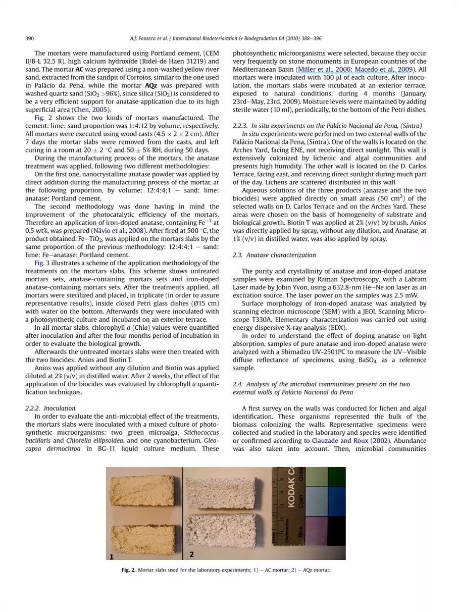
The mortars were manufactured using Portland cement, (CEMII/B-L 32,5 R), high calcium hydroxide (Ridel-de Haen 31219) andsand. Themortar ACwas prepared using a non-washed yellow riversand, extracted from the sandpit of Corroios, similar to the one usedin Palácio da Pena, while the mortar AQz was prepared withwashed quartz sand (SiO2 >96%), since silica (SiO2) is considered tobe a very efficient support for anatase application due to its highsuperficial area (Chen, 2005).
Fig. 2 shows the two kinds of mortars manufactured. Thecement: lime: sand proportion was 1:4:12 by volume, respectively.All mortars were executed using wood casts (4.5 � 2 � 2 cm). After7 days the mortar slabs were removed from the casts, and leftcuring in a room at 20 � 2 �C and 50 � 5% RH, during 50 days.
During the manufacturing process of the mortars, the anatasetreatment was applied, following two different methodologies:
On the first one, nanocrystalline anatase powder was applied bydirect addition during the manufacturing process of the mortar, atthe following proportion, by volume: 12:4:4:1 e sand: lime:anatase: Portland cement.
The second methodology was done having in mind theimprovement of the photocatalytic efficiency of the mortars.Therefore an application of iron-doped anatase, containing Feþ3 at0.5 wt%, was prepared (Návio et al., 2008). After fired at 500 �C, theproduct obtained, FeeTiO2, was applied on the mortars slabs by thesame proportion of the previous methodology: 12:4:4:1 e sand:lime: Feeanatase: Portland cement.
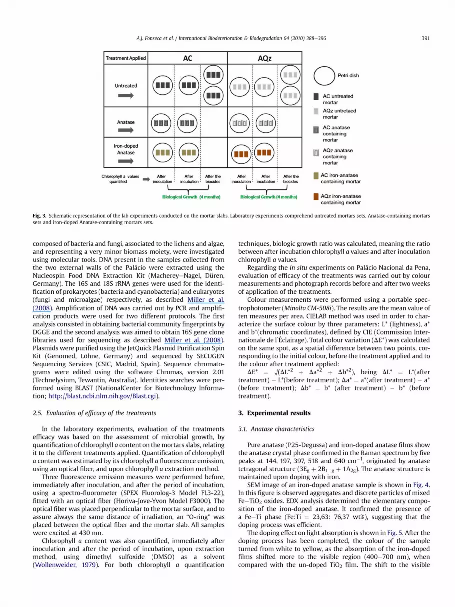
Fig. 3 illustrates a scheme of the application methodology of thetreatments on the mortars slabs. This scheme shows untreatedmortars sets, anatase-containing mortars sets and iron-dopedanatase-containing mortars sets. After the treatments applied, allmortars were sterilized and placed, in triplicate (in order to assurerepresentative results), inside closed Petri glass dishes (Ø15 cm)with water on the bottom. Afterwards they were inoculated witha photosynthetic culture and incubated on an exterior terrace.
In all mortar slabs, chlorophyll a (Chla) values were quantifiedafter inoculation and after the four months period of incubation inorder to evaluate the biological growth.
Afterwards the untreated mortars slabs were then treated withthe two biocides: Anios and Biotin T.
Anios was applied without any dilution and Biotin was applieddiluted at 2% (v/v) in distilled water. After 2 weeks, the effect of theapplication of the biocides was evaluated by chlorophyll a quanti-fication techniques.
2.2.2. InoculationIn order to evaluate the anti-microbial effect of the treatments,
the mortars slabs were inoculated with a mixed culture of photo-synthetic microorganisms: two green microalga, Stichococcusbacillaris and Chlorella ellipsoidea, and one cyanobacterium, Gleo-capsa dermochroa in BG-11 liquid culture medium. These
Fig. 2. Mortar slabs used for the laboratory expe
photosynthetic microorganisms were selected, because they occurvery frequently on stone monuments in European countries of theMediterranean Basin (Miller et al., 2006; Macedo et al., 2009). Allmortars were inoculated with 100 ml of each culture. After inocu-lation, the mortars slabs were incubated at an exterior terrace,exposed to natural conditions, during 4 months (January,23rdeMay, 23rd, 2009). Moisture levels weremaintained by addingsterile water (10 ml), periodically, to the bottom of the Petri dishes.
2.2.3. In situ experiments on the Palácio Nacional da Pena, (Sintra)In situ experiments were performed on two external walls of the
Palácio Nacional da Pena, (Sintra). One of the walls is located on theArches Yard, facing ENE, not receiving direct sunlight. This wall isextensively colonized by lichenic and algal communities andpresents high humidity. The other wall is located on the D. CarlosTerrace, facing east, and receiving direct sunlight during much partof the day. Lichens are scattered distributed in this wall
Aqueous solutions of the three products (anatase and the twobiocides) were applied directly on small areas (50 cm2) of theselected walls on D. Carlos Terrace and on the Arches Yard. Theseareas were chosen on the basis of homogeneity of substrate andbiological growth. Biotin T was applied at 2% (v/v) by brush. Anioswas directly applied by spray, without any dilution, and Anatase, at1% (v/v) in distilled water, was also applied by spray.
2.3. Anatase characterization
The purity and crystallinity of anatase and iron-doped anatasesamples were examined by Raman Spectroscopy, with a LabramLaser made by Jobin Yvon, using a 632.8-nm HeeNe ion laser as anexcitation source. The laser power on the samples was 2.5 mW.
Surface morphology of iron-doped anatase was analyzed byscanning electron microscope (SEM) with a JEOL Scanning Micro-scope T330A. Elementary characterization was carried out usingenergy dispersive X-ray analysis (EDX).
In order to understand the effect of doping anatase on lightabsorption, samples of pure anatase and iron-doped anatase wereanalyzed with a Shimadzu UV-2501PC to measure the UVeVisiblediffuse reflectance of specimens, using BaSO4, as a referencesample.
2.4. Analysis of the microbial communities present on the twoexternal walls of Palácio Nacional da Pena
A first survey on the walls was conducted for lichen and algalidentification. These organisms represented the bulk of thebiomass colonizing the walls. Representative specimens werecollected and studied in the laboratory and species were identifiedor confirmed according to Clauzade and Roux (2002). Abundancewas also taken into account. Then, microbial communities
Fig. 3. Schematic representation of the lab experiments conducted on the mortar slabs. Laboratory experiments comprehend untreated mortars sets, Anatase-containing mortarssets and iron-doped Anatase-containing mortars sets.
A.J. Fonseca et al. / International Biodeterioration & Biodegradation 64 (2010) 388e396 391
composed of bacteria and fungi, associated to the lichens and algae,and representing a very minor biomass moiety, were investigatedusing molecular tools. DNA present in the samples collected fromthe two external walls of the Palácio were extracted using theNucleospin Food DNA Extraction Kit (MachereyeNagel, Düren,Germany). The 16S and 18S rRNA genes were used for the identi-fication of prokaryotes (bacteria and cyanobacteria) and eukaryotes(fungi and microalgae) respectively, as described Miller et al.(2008). Amplification of DNA was carried out by PCR and amplifi-cation products were used for two different protocols. The firstanalysis consisted in obtaining bacterial community fingerprints byDGGE and the second analysis was aimed to obtain 16S gene clonelibraries used for sequencing as described Miller et al. (2008).Plasmids were purified using the JetQuick Plasmid Purification SpinKit (Genomed, Löhne, Germany) and sequenced by SECUGENSequencing Services (CSIC, Madrid, Spain). Sequence chromato-grams were edited using the software Chromas, version 2.01(Technelysium, Tewantin, Australia). Identities searches were per-formed using BLAST (NationalCenter for Biotechnology Informa-tion; http://blast.ncbi.nlm.nih.gov/Blast.cgi).
2.5. Evaluation of efficacy of the treatments
In the laboratory experiments, evaluation of the treatmentsefficacy was based on the assessment of microbial growth, byquantification of chlorophyll a content on themortars slabs, relatingit to the different treatments applied. Quantification of chlorophylla content was estimated by its chlorophyll a fluorescence emission,using an optical fiber, and upon chlorophyll a extraction method.
Three fluorescence emission measures were performed before,immediately after inoculation, and after the period of incubation,using a spectro-fluorometer (SPEX Fluorolog-3 Model FL3-22),fitted with an optical fiber (Horiva-Jove-Yvon Model F3000). Theoptical fiber was placed perpendicular to the mortar surface, and toassure always the same distance of irradiation, an “O-ring” wasplaced between the optical fiber and the mortar slab. All sampleswere excited at 430 nm.
Chlorophyll a content was also quantified, immediately afterinoculation and after the period of incubation, upon extractionmethod, using dimethyl sulfoxide (DMSO) as a solvent(Wollenweider, 1979). For both chlorophyll a quantification
techniques, biologic growth ratio was calculated, meaning the ratiobetween after incubation chlorophyll a values and after inoculationchlorophyll a values.
Regarding the in situ experiments on Palácio Nacional da Pena,evaluation of efficacy of the treatments was carried out by colourmeasurements and photograph records before and after two weeksof application of the treatments.
Colour measurements were performed using a portable spec-trophotometer (Minolta CM-508i). The results are the mean value often measures per area. CIELAB method was used in order to char-acterize the surface colour by three parameters: L* (lightness), a*and b*(chromatic coordinates), defined by CIE (Commission Inter-nationale de l`Éclairage). Total colour variation (DE*) was calculatedon the same spot, as a spatial difference between two points, cor-responding to the initial colour, before the treatment applied and tothe colour after treatment applied:
Pure anatase (P25-Degussa) and iron-doped anatase films showthe anatase crystal phase confirmed in the Raman spectrum by fivepeaks at 144, 197, 397, 518 and 640 cm�1, originated by anatasetetragonal structure (3Eg þ 2B1�g þ 1A2g). The anatase structure ismaintained upon doping with iron.
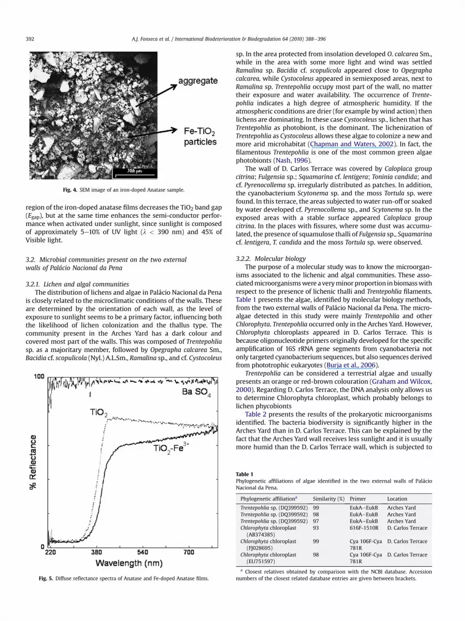
SEM image of an iron-doped anatase sample is shown in Fig. 4.In this figure is observed aggregates and discrete particles of mixedFeeTiO2 oxides. EDX analysis determined the elementary compo-sition of the iron-doped anatase. It confirmed the presence ofa FeeTi phase (Fe:Ti ¼ 23,63: 76,37 wt%), suggesting that thedoping process was efficient.
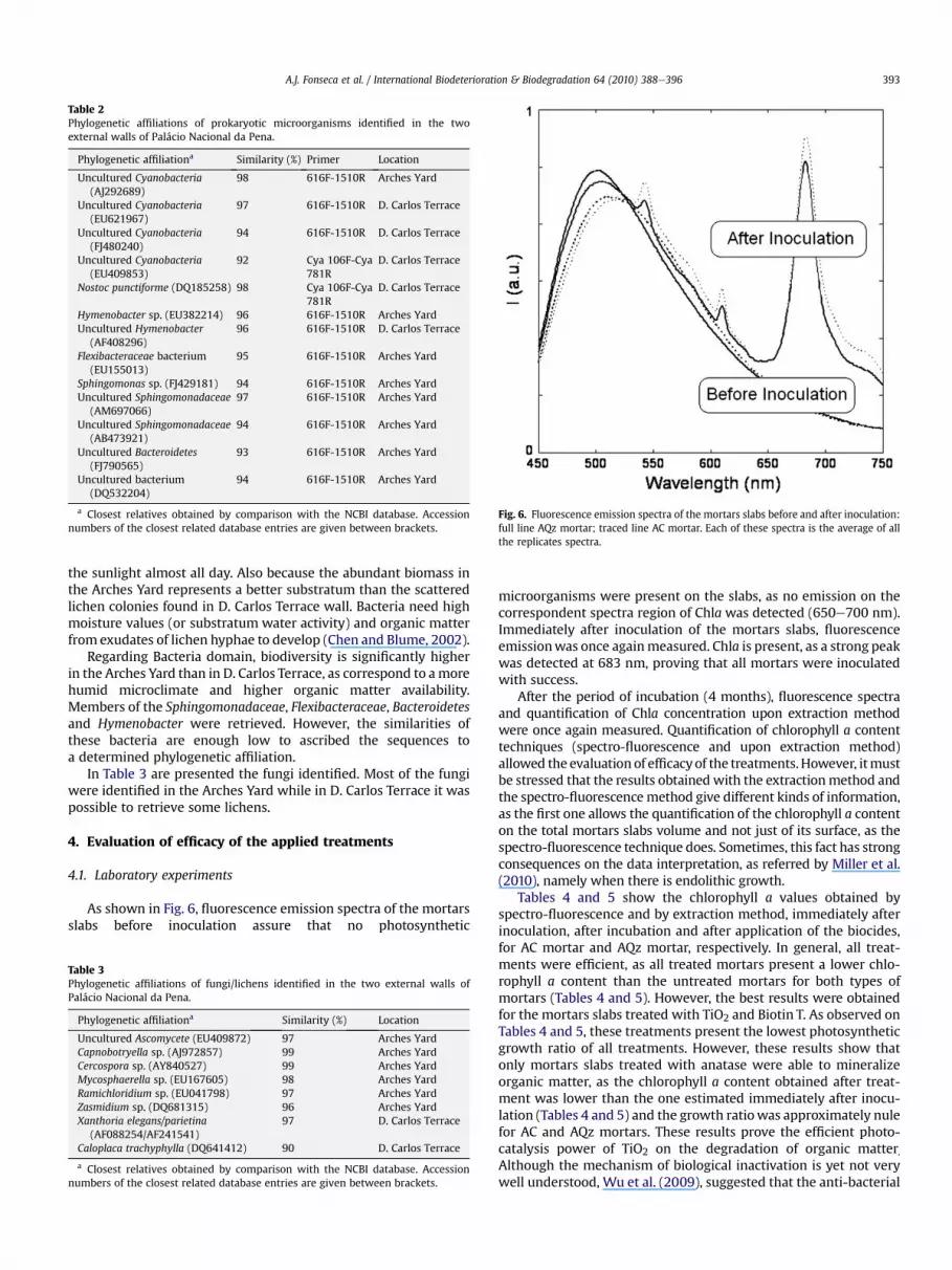
The doping effect on light absorption is shown in Fig. 5. After thedoping process has been completed, the colour of the sampleturned from white to yellow, as the absorption of the iron-dopedfilms shifted more to the visible region (400e700 nm), whencompared with the un-doped TiO2 film. The shift to the visible
Fig. 4. SEM image of an iron-doped Anatase sample.
A.J. Fonseca et al. / International Biodeterioration & Biodegradation 64 (2010) 388e396392
region of the iron-doped anatase films decreases the TiO2 band gap(Egap), but at the same time enhances the semi-conductor perfor-mance when activated under sunlight, since sunlight is composedof approximately 5e10% of UV light (l < 390 nm) and 45% ofVisible light.
3.2. Microbial communities present on the two externalwalls of Palácio Nacional da Pena
3.2.1. Lichen and algal communitiesThe distribution of lichens and algae in Palácio Nacional da Pena
is closely related to the microclimatic conditions of the walls. Theseare determined by the orientation of each wall, as the level ofexposure to sunlight seems to be a primary factor, influencing boththe likelihood of lichen colonization and the thallus type. Thecommunity present in the Arches Yard has a dark colour andcovered most part of the walls. This was composed of Trentepohliasp. as a majoritary member, followed by Opegrapha calcarea Sm.,Bacidia cf. scopulicola (Nyl.) A.L.Sm., Ramalina sp., and cf. Cystocoleus
Fig. 5. Diffuse reflectance spectra of Anatase and Fe-doped Anatase films.
sp. In the area protected from insolation developed O. calcarea Sm.,while in the area with some more light and wind was settledRamalina sp. Bacidia cf. scopulicola appeared close to Opegraphacalcarea, while Cystocoleus appeared in semiexposed areas, next toRamalina sp. Trentepohlia occupy most part of the wall, no mattertheir exposure and water availability. The occurrence of Trente-pohlia indicates a high degree of atmospheric humidity. If theatmospheric conditions are drier (for example bywind action) thenlichens are dominating. In these case Cystocoleus sp., lichen that hasTrentepohlia as photobiont, is the dominant. The lichenization ofTrentepohlia as Cystocoleus allows these algae to colonize a new andmore arid microhabitat (Chapman and Waters, 2002). In fact, thefilamentous Trentepohlia is one of the most common green algaephotobionts (Nash, 1996).
The wall of D. Carlos Terrace was covered by Caloplaca groupcitrina; Fulgensia sp.; Squamarina cf. lentigera; Toninia candida; andcf. Pyrenocollema sp. irregularly distributed as patches. In addition,the cyanobacterium Scytonema sp. and the moss Tortula sp. werefound. In this terrace, the areas subjected towater run-off or soakedby water developed cf. Pyrenocollema sp., and Scytonema sp. In theexposed areas with a stable surface appeared Caloplaca groupcitrina. In the places with fissures, where some dust was accumu-lated, the presence of squamulose thalli of Fulgensia sp., Squamarinacf. lentigera, T. candida and the moss Tortula sp. were observed.
3.2.2. Molecular biologyThe purpose of a molecular study was to know the microorgan-
isms associated to the lichenic and algal communities. These asso-ciatedmicroorganismswere averyminorproportion inbiomasswithrespect to the presence of lichenic thalli and Trentepohlia filaments.Table 1 presents the algae, identified by molecular biology methods,from the two external walls of Palácio Nacional da Pena. The micro-algae detected in this study were mainly Trentepohlia and otherChlorophyta. Trentepohlia occurred only in the Arches Yard. However,Chlorophyta chloroplasts appeared in D. Carlos Terrace. This isbecause oligonucleotide primers originally developed for the specificamplification of 16S rRNA gene segments from cyanobacteria notonly targeted cyanobacterium sequences, but also sequences derivedfrom phototrophic eukaryotes (Burja et al., 2006).
Trentepohlia can be considered a terrestrial algae and usuallypresents an orange or red-brown colouration (Graham and Wilcox,2000). Regarding D. Carlos Terrace, the DNA analysis only allows usto determine Chlorophyta chloroplast, which probably belongs tolichen phycobionts
Table 2 presents the results of the prokaryotic microorganismsidentified. The bacteria biodiversity is significantly higher in theArches Yard than in D. Carlos Terrace. This can be explained by thefact that the Arches Yard wall receives less sunlight and it is usuallymore humid than the D. Carlos Terrace wall, which is subjected to
Table 1Phylogenetic affiliations of algae identified in the two external walls of PalácioNacional da Pena.
Phylogenetic affiliationa Similarity (%) Primer Location
a Closest relatives obtained by comparison with the NCBI database. Accessionnumbers of the closest related database entries are given between brackets.
a Closest relatives obtained by comparison with the NCBI database. Accessionnumbers of the closest related database entries are given between brackets.
Fig. 6. Fluorescence emission spectra of the mortars slabs before and after inoculation:full line AQz mortar; traced line AC mortar. Each of these spectra is the average of allthe replicates spectra.
A.J. Fonseca et al. / International Biodeterioration & Biodegradation 64 (2010) 388e396 393
the sunlight almost all day. Also because the abundant biomass inthe Arches Yard represents a better substratum than the scatteredlichen colonies found in D. Carlos Terrace wall. Bacteria need highmoisture values (or substratum water activity) and organic matterfrom exudates of lichen hyphae to develop (Chen and Blume, 2002).
Regarding Bacteria domain, biodiversity is significantly higherin the Arches Yard than in D. Carlos Terrace, as correspond to amorehumid microclimate and higher organic matter availability.Members of the Sphingomonadaceae, Flexibacteraceae, Bacteroidetesand Hymenobacter were retrieved. However, the similarities ofthese bacteria are enough low to ascribed the sequences toa determined phylogenetic affiliation.
In Table 3 are presented the fungi identified. Most of the fungiwere identified in the Arches Yard while in D. Carlos Terrace it waspossible to retrieve some lichens.
4. Evaluation of efficacy of the applied treatments
4.1. Laboratory experiments
As shown in Fig. 6, fluorescence emission spectra of the mortarsslabs before inoculation assure that no photosynthetic
Table 3Phylogenetic affiliations of fungi/lichens identified in the two external walls ofPalácio Nacional da Pena.
Caloplaca trachyphylla (DQ641412) 90 D. Carlos Terrace
a Closest relatives obtained by comparison with the NCBI database. Accessionnumbers of the closest related database entries are given between brackets.
microorganisms were present on the slabs, as no emission on thecorrespondent spectra region of Chla was detected (650e700 nm).Immediately after inoculation of the mortars slabs, fluorescenceemissionwas once againmeasured. Chla is present, as a strong peakwas detected at 683 nm, proving that all mortars were inoculatedwith success.
After the period of incubation (4 months), fluorescence spectraand quantification of Chla concentration upon extraction methodwere once again measured. Quantification of chlorophyll a contenttechniques (spectro-fluorescence and upon extraction method)allowed the evaluation of efficacyof the treatments.However, itmustbe stressed that the results obtained with the extractionmethod andthe spectro-fluorescencemethod give different kinds of information,as the first one allows the quantification of the chlorophyll a contenton the total mortars slabs volume and not just of its surface, as thespectro-fluorescence technique does. Sometimes, this fact has strongconsequences on the data interpretation, as referred by Miller et al.(2010), namely when there is endolithic growth.
Tables 4 and 5 show the chlorophyll a values obtained byspectro-fluorescence and by extraction method, immediately afterinoculation, after incubation and after application of the biocides,for AC mortar and AQz mortar, respectively. In general, all treat-ments were efficient, as all treated mortars present a lower chlo-rophyll a content than the untreated mortars for both types ofmortars (Tables 4 and 5). However, the best results were obtainedfor the mortars slabs treated with TiO2 and Biotin T. As observed onTables 4 and 5, these treatments present the lowest photosyntheticgrowth ratio of all treatments. However, these results show thatonly mortars slabs treated with anatase were able to mineralizeorganic matter, as the chlorophyll a content obtained after treat-ment was lower than the one estimated immediately after inocu-lation (Tables 4 and 5) and the growth ratiowas approximately nulefor AC and AQz mortars. These results prove the efficient photo-catalysis power of TiO2 on the degradation of organic matter.Although the mechanism of biological inactivation is yet not verywell understood, Wu et al. (2009), suggested that the anti-bacterial
Table 4Chlorophyll a values (mean of triplicates) obtained by spectro-fluorescence and by extractionmethod for ACmortar after inoculation, after incubation and after the applicationof the biocides.
Biotin T After Inoculation 5.6 � 105 0 8.9 � 10�5 0.2After the 2 weeks 7.5 � 104 6.2 � 10�5
% growth ratio values are always referent to the untreated slabs of each mortar. Being the growth ratio of untreated mortars a maximum value ¼ 100%.
A.J. Fonseca et al. / International Biodeterioration & Biodegradation 64 (2010) 388e396394
effect of TiO2 is attributed to the destruction of the bacterial cellwall andmembrane by the photocatalytic oxidation process of TiO2.There is yet a lack of data regarding the specific mechanism of celldeath, however what we know is that hydroxyl radicals generatedby the anatase photocatalyst are very potent oxidants that can infact mineralize organic matter:
HO� þ organic matter / xCO2 þ yH2O
Iron-doped anatase (Feeanatase) treatment showed goodresults but not as good as those estimated for pure TiO2 treatment.This may be due to an excessive addition of iron on the crystallattice, producing large aggregates of hematite that decreased theanatase photocatalytic activity (Asilturk et al., 2009). Concerningthe conventional biocides treatments, results in Tables 4 and 5show that application of Biotin T was much more efficient thanAnios, for both types of mortars.
Therefore the following index of efficacy of treatment (indecreasing order) is presented, on the basis of chlorophyll a contentobtained by extraction method.
Anatase > Biotin T > AnataseeFe > Anios
4.2. In situ experiments in Palácio Nacional da Pena
After application of treatments, chromatic changes weredetected on both selected areas. Tables 6 and 7 show colour vari-ation on D. Carlos Terrace and Arches Yard, respectively.
Total colour variation (DE*) of the different treatments appliedappears to show the same pattern: application of anatase treatmentappears to be more efficient than Biotin T, and much more efficientthat Anios.
Increase of L* parameter after anatase application is moresignificant that the two biocides treatments. The L* parameter is
Table 5Chlorophyll a values (mean of triplicates) obtained by spectro-fluorescence and by exapplication of the biocides.
AQz mortar Spectro-Fluorescence(cps at 683 nm)
Untreated After Inoculation 2.6 � 105
After 4 months 5.8 � 106
Anatase After Inoculation 8.5 � 105
After 4 months 9.0 � 104
FeeAnatase After Inoculation 5.5 � 105
After 4 months 4.7 � 105
Anios D.D.S.H After Inoculation 2.6 � 105
After the biocide 1.1 � 106
Biotin After Inoculation 2.6 � 105
After the biocide 1.1 � 105
% growth ratio values are always referent to the untreated slabs of each mortar. Being t
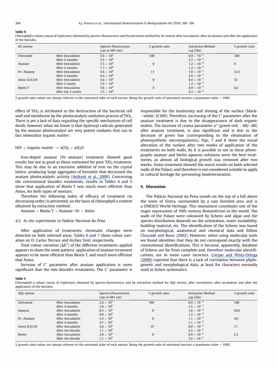
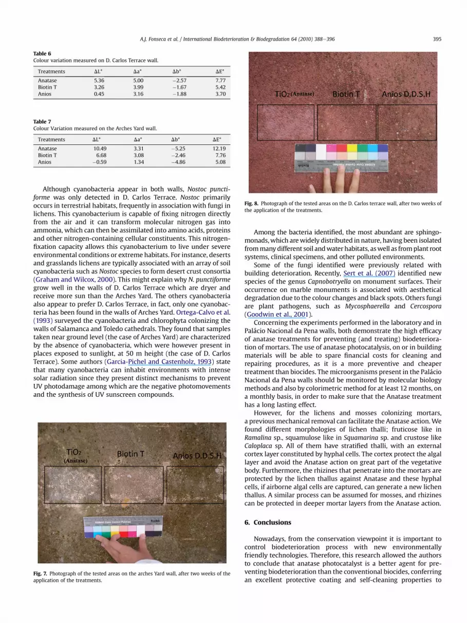
responsible for the luminosity and shining of the surface (black-ewhite; 0/100). Therefore, increasing of the L* parameter after theanatase treatment is due to the disappearance of dark organicmatter. The increase of croma parameter a* (green-red; �60/þ60)after anatase treatment, is also significant and is due to thedecrease of green hue (corresponding to the elimination ofphotosynthetic microorganisms). Figs. 7 and 8 show the visualalteration of the surface after two weeks of application of thetreatments on both walls. As it is possible to see in these photo-graphs anatase and biotin aqueous solutions were the best treat-ments, as almost all biological growth was removed after twoweeks. Anios treatment showed the worst results on both selectedwalls of the Palace, and therefore is not considered suitable to applyin cultural heritage for preventing biodeterioration.
5. Discussion
The Palácio Nacional da Pena stands on the top of a hill abovethe town of Sintra, surrounded by a vast forested area and isa UNESCO World Heritage. This monument constitutes one of themajor expressions of 19th century Romanticism in the world. Thewalls of the Palace were colonized by lichens and algae and thespecies distribution depends on the orientation, water availability,building material, etc. The identification of the lichens was basedon morphological, anatomical and chemical data and followClauzade and Roux (2002). However, when using molecular toolswe found identities that they do not correspond exactly with theconventional identifications. This is because, apparently, databaseof lichens are far from complete and, therefore molecular identifi-cations, are in some cases incorrect. Crespo and Pérez-Ortega(2009) reported that there is a lack of correlation between phylo-genetic and morphological data, at least for characters normallyused in lichen systematics.
traction method for AQz mortar, after inoculation, after incubation and after the
% growth ratio Extraction Method(mg Chla)
% growth ratio
100 6.9 � 10�5 1001.5 � 10�1
0 1.6 � 10�4 0.031.2 � 10�4
0 1.1 � 10�4 4.61.1 � 10�2
19 6.9 � 10�5 172.5 � 10�2
0 6.9 � 10�5 2.33.4 � 10�3
he growth ratio of untreated mortars a maximum value ¼ 100%.
A.J. Fonseca et al. / International Biodeterioration & Biodegradation 64 (2010) 388e396 395
Although cyanobacteria appear in both walls, Nostoc puncti-forme was only detected in D. Carlos Terrace. Nostoc primarilyoccurs in terrestrial habitats, frequently in associationwith fungi inlichens. This cyanobacterium is capable of fixing nitrogen directlyfrom the air and it can transform molecular nitrogen gas intoammonia, which can then be assimilated into amino acids, proteinsand other nitrogen-containing cellular constituents. This nitrogen-fixation capacity allows this cyanobacterium to live under severeenvironmental conditions or extreme habitats. For instance, desertsand grasslands lichens are typically associated with an array of soilcyanobacteria such as Nostoc species to form desert crust consortia(Graham andWilcox, 2000). This might explain why N. punctiformegrow well in the walls of D. Carlos Terrace which are dryer andreceive more sun than the Arches Yard. The others cyanobacteriaalso appear to prefer D. Carlos Terrace, in fact, only one cyanobac-teria has been found in the walls of Arches Yard. Ortega-Calvo et al.(1993) surveyed the cyanobacteria and chlorophyta colonizing thewalls of Salamanca and Toledo cathedrals. They found that samplestaken near ground level (the case of Arches Yard) are characterizedby the absence of cyanobacteria, which were however present inplaces exposed to sunlight, at 50 m height (the case of D. CarlosTerrace). Some authors (Garcia-Pichel and Castenholz, 1993) statethat many cyanobacteria can inhabit environments with intensesolar radiation since they present distinct mechanisms to preventUV photodamage among which are the negative photomovementsand the synthesis of UV sunscreen compounds.
Fig. 7. Photograph of the tested areas on the arches Yard wall, after two weeks of theapplication of the treatments.
Among the bacteria identified, the most abundant are sphingo-monads,which arewidely distributed in nature, having been isolatedfrommanydifferent soil andwaterhabitats, aswell as fromplant rootsystems, clinical specimens, and other polluted environments.
Some of the fungi identified were previously related withbuilding deterioration. Recently, Sert et al. (2007) identified newspecies of the genus Capnobotryella on monument surfaces. Theiroccurrence on marble monuments is associated with aestheticaldegradation due to the colour changes and black spots. Others fungiare plant pathogens, such as Mycosphaerella and Cercospora(Goodwin et al., 2001).
Concerning the experiments performed in the laboratory and inPalácio Nacional da Pena walls, both demonstrate the high efficacyof anatase treatments for preventing (and treating) biodeteriora-tion of mortars. The use of anatase photocatalysis, on or in buildingmaterials will be able to spare financial costs for cleaning andrepairing procedures, as it is a more preventive and cheapertreatment than biocides. Themicroorganisms present in the PalácioNacional da Pena walls should be monitored by molecular biologymethods and also by colorimetric method for at least 12months, ona monthly basis, in order to make sure that the Anatase treatmenthas a long lasting effect.
However, for the lichens and mosses colonizing mortars,a previousmechanical removal can facilitate the Anatase action.Wefound different morphologies of lichen thalli; fruticose like inRamalina sp., squamulose like in Squamarina sp. and crustose likeCaloplaca sp. All of them have stratified thalli, with an externalcortex layer constituted by hyphal cells. The cortex protect the algallayer and avoid the Anatase action on great part of the vegetativebody. Furthermore, the rhizines that penetrate into the mortars areprotected by the lichen thallus against Anatase and these hyphalcells, if airborne algal cells are captured, can generate a new lichenthallus. A similar process can be assumed for mosses, and rhizinescan be protected in deeper mortar layers from the Anatase action.
6. Conclusions
Nowadays, from the conservation viewpoint it is important tocontrol biodeterioration process with new environmentallyfriendly technologies. Therefore, this research allowed the authorsto conclude that anatase photocatalyst is a better agent for pre-venting biodeterioration than the conventional biocides, conferringan excellent protective coating and self-cleaning properties to
A.J. Fonseca et al. / International Biodeterioration & Biodegradation 64 (2010) 388e396396
building materials. Moreover it is non-toxic and by consequencea good alternative to conventional biocides.
Although, it will be necessary to develop a further scientificresearch on anatase photocatalysis self-cleaning applications, thepotential of combined nanotechnology and preventive conserva-tionwill allow a positive compromise for an environmental methodof preventing biodeterioration of building materials.
Acknowledgements
The authorswish to thank Parques de SintraeMonte da Lua for thepossibility to study this beautiful monument that is Palácio Nacionalda Pena. We also wish to thank the Departamento de Quimica -REQUIMTE for technical and experimental support. Acknowledge-ments are also due to the CICEGe-FCT/UNL, for SEM-EDX facilities,and to the DCT-FCT/UNL, for the use of the terrace for externalconditions experiments. This is a TCP CSD2007-00058 paper.
References
Asilturk, M., Sayilkan, F., Arpaç, E., 2009. Effect of Feþ3 dopping to TiO2 on thephotocatalytic degradation of Malachite Green dye under UV and vis-irradia-tion. Journal of Photochemistry and Photobiology A: Chemistry 203, 64e71.
Burja, A.M., Tamagnini, P., Bustard, M.T., Wright, P.C., 2006. Identification of thegreen alga, Chlorella vulgaris (SDC1) using cyanobacteria derived 16S rDNAprimers: targeting the chloroplast. FEMS. Microbiology Letters 202, 195e203.
Caneva, G., Nugari, M.P., Pinna, D., Salvadori, O., 1996. Il Controllo del degradobiológico e i biocidi nel restauro dei materiali lapidei. Nardini Editore, Fiesole.
Chapman, R.L., Waters, D.A., 2002. Lichenization of the Trentepohliales. In:Seckbach, J. (Ed.), Symbiosis: Mechanisms and Model Systems. Kluwer, Dor-drecht, pp. 359e371.
Chen, J., Blume, H.-P., 2002. Rock-weathering by lichens in Antarctic: patterns andmechanisms. Journal of Geographical Science 12, 387e396.
Chen, J., Poon, C., 2009. Photocatalytic construction and building materials: fromfundamentals to applications. Building and Environment 44, 1899e1906.
Chen, X., 2005. Synthesis and investigation of novel materials for improved pho-tocatalysis, PhD thesis, Case Western Reserve University, USA.
Clauzade, G., Roux, C., 2002. Likenoj de Okcidenta E�uropo, Traduction des clés dedétermination par P. Ravel. Association Française de Lichénologie, Paris.
Crespo, A., Pérez-Ortega, S., 2009. Cryptic species and species pairs in lichens:a discussion on the relationship between molecular phylogenies andmorphological characters. Anales del Jardin Botanico de Madrid 66S1, 71e81.
Diamanti, M.V., Ormellese, M., Pedeferri, M., 2008. Characterization of photo-catalytic and superhydrophilic properties of mortars containing titaniumdioxide. Cement and Concrete Research 38, 1349e1353.
Fu, G., Vary, P.S., Lin, C., 2005. Anatase TiO2 Nanocomposites for AntimicrobialCoatings. Journal of Physical Chemistry B 109, 8889e8898.
Garcia-Pichel, F., Castenholz, R.W., 1993. Occurrence of UV-absorbing, mycosporine-like compounds among cyanobacterial isolates and an estimate of theirscreening capacity. Applied and Environmental Microbiology 59, 163e169.
Goodwin, S.B., Dunkle, L.D., Zismann, V.L., 2001. Phylogenetic analysis of Cercosporaand Mycosphaerella based on the internal transcribed spacer region of ribo-somal DNA. Phytopathology 91, 648e658.
Graham, L.E., Wilcox, L.W., 2000. Algae. Prentice Hall, Upper Saddle River, NJ.640 pp.
Kelerher, J., Bashant, J., Heldt, N., Johnson, L., Li, Y., 2002. Photo-catalytic preparationof silver-coated TiO2 particles for antibacterial applications. Journal of Micro-biology and Biotechnology 18, 133e139.
Kurth, J.C., 2008. Mitigating biofilm growth through the modification of concretedesign and practice. Master thesis, Georgia Institute of Technology, USA.
Macedo, M.F., Miller, A.Z., Dionisio, A., Saiz-Jimenez, C., 2009. Biodiversity of cya-nobacteria and green algae on monuments in the Mediterranean Basin: anoverview. Microbiology 155, 3476e3490.
Maury Ramirez, A., Demeestere, K., Belie, N., Mantyla, T., Levanen, E., 2010. Titaniumdioxide coated cementitious materials for air purifying purposes: preparation,characterization and toluene removal potential. Building and Environment 45,832e838.
Miller, A.Z., Dionísio, A., Macedo, M.F., 2006. Primary bioreceptivity: a comparativestudy of different Portuguese lithotypes. International Biodeterioration andBiodegradation 57, 136e142.
Miller, A.Z., Laiz, L., Gonzalez, J.M., Dionísio, A., Macedo, M.F., Saiz-Jimenez, C., 2008.Reproducing stone monument photosynthetic-based colonization under labo-ratory conditions. Science of the Total Environment 405, 278e285.
Miller, A.Z., Leal, N., Laiz, L., Rogerio-Candelera, M.A., Silva, R.J.C., Dionisio, A.,Macedo, M.F., Saiz-Jimenez, C., 2010. Primary bioreceptivity of limestones usedin Southern Europe monuments. In: Smith, B.J., Gomez-Heras, M., Viles, H.A.,Cassar, J. (Eds.), Limestone in the Built Environment: Present Day Challenges forthe Preservation of the Past. Geological Society of London, Special Publications,vol. 331, pp. 79e92.
Nash III, T.H., 1996. Lichen Biology. CambridgeUniversity Press.Návio, J.A., Macias, M., Gárcia-Gómez, M., Pradera, M.A., 2008. Functionalisation
versus mineralisation of some N-heterocyclic compounds upon UV-illumina-tion in the presence of un-doped and iron-doped TiO2 photocatalysts. AppliedCatalysis B: Environmental 82, 225e232.
Nugari, M.P., SaIvadori, O., 2003. Biodeterioration control in cultural heritage:methods and products. In: Saiz-Jimenez, C. (Ed.), Molecular Biology and CulturalHeritage. Balkema, Lisse, pp. 233e242.
Ortega-Calvo, J.J., Sanchez-Castillo, P.M., Hernandez-Marine, M., Saiz-Jimenez, C.,1993. Isolation and characterization of epilithic chrorophytes and cyanobac-teria from two cathedrals (Salamanca and Toledo). Nova Hedwigia 57,239e253.
Pereira, T.A.R., 2008. Optimização das características de humedecimento e secagemde argamassas. Master thesis, Faculdade de Ciências e Tecnologia, UniversidadeNova de Lisboa, Lisboa, Portugal.
Russel, A.D., Chopra, I., 1990. Understanding Antibacterial Action and Resistance.Horwood Ltd, Chichester.
Saiz-Jimenez, C., 1999. Biogeochemistry of weathering processes in monuments.Geomicrobiology Journal 16, 27e37.
Sert, H., Sümbül, H., Sterflinge, r K., 2007. A new specis of Capnobotryella frommonument surfaces. Mycological Research 111, 1235e1241.
Silva, S., 2002. Caracterização de argamassas antigas - casos paradigmáticos. In:Cadernos edifícios 02- Revestimentos de paredes em edifícios antigos. LNEC,Lisboa.
Tiano, P., 1998. Biodeterioration of monumental rocks: decay mechanisms andcontrol methods. Science and Technology for Cultural Heritage 7, 19e38.
Tomaselli, L., Lamenti, G., Tiano, P., Bosco, M., 2000. Biodiverity of photosyntheticmicro-organisms dwelling on stone monuments. International Biodeteriorationand Biodegradation 46, 251e258.
Wollenweider, R.A., 1979. A Manual on Methods for Measuring Primary Productionin Aquatic Environments. Blackwell Scientific Publications, Oxford, UK.
Wu, D., Long, M., Zhou, J., Cai, W., Zhu, X., Chen, C., Wu, Y., 2009. Synthesis andcharacterization of self-cleaning cotton fabrics modified by TiO2 through a facileapproach. Surface & Coatings Technology 203, 3728e3733.