ORIGINAL RESEARCH published: 13 June 2017 doi: 10.3389/fmicb.2017.01088 Edited by: Axel Cloeckaert, Institut National de la Recherche Agronomique (INRA), France Reviewed by: Alessandra Occhialini, Université de Montpellier, France Roy Martin Roop II, East Carolina University, United States Renee M. Tsolis, University of California, Davis, United States *Correspondence: Jean-Jacques Letesson [email protected]Specialty section: This article was submitted to Infectious Diseases, a section of the journal Frontiers in Microbiology Received: 20 April 2017 Accepted: 30 May 2017 Published: 13 June 2017 Citation: Barbier T, Machelart A, Zúñiga-Ripa A, Plovier H, Hougardy C, Lobet E, Willemart K, Muraille E, De Bolle X, Van Schaftingen E, Moriyón I and Letesson J-J (2017) Erythritol Availability in Bovine, Murine and Human Models Highlights a Potential Role for the Host Aldose Reductase during Brucella Infection. Front. Microbiol. 8:1088. doi: 10.3389/fmicb.2017.01088 Erythritol Availability in Bovine, Murine and Human Models Highlights a Potential Role for the Host Aldose Reductase during Brucella Infection Thibault Barbier 1 , Arnaud Machelart 1 , Amaia Zúñiga-Ripa 2 , Hubert Plovier 1 , Charlotte Hougardy 1 , Elodie Lobet 1 , Kevin Willemart 1 , Eric Muraille 3 , Xavier De Bolle 1 , Emile Van Schaftingen 4 , Ignacio Moriyón 2 and Jean-Jacques Letesson 1 * 1 Research Unit in Biology of Microorganisms, Department of Veterinary Medicine, University of Namur, Namur, Belgium, 2 Departamento de Microbiología y Parasitología, Instituto de Salud Tropical, Instituto de Investigación Sanitaria de Navarra, Universidad de Navarra, Pamplona, Spain, 3 Laboratoire de Parasitologie, Faculté de Médecine, Université Libre de Bruxelles, Brussels, Belgium, 4 WELBIO and de Duve Institute, Université Catholique de Louvain, Brussels, Belgium Erythritol is the preferential carbon source for most brucellae, a group of facultative intracellular bacteria that cause a worldwide zoonosis. Since this polyol is abundant in genital organs of ruminants and swine, it is widely accepted that erythritol accounts at least in part for the characteristic genital tropism of brucellae. Nevertheless, proof of erythritol availability and essentiality during Brucella intracellular multiplication has remained elusive. To investigate this relationship, we compared 1eryH (erythritol- sensitive and thus predicted to be attenuated if erythritol is present), 1eryA (erythritol- tolerant but showing reduced growth if erythritol is a crucial nutrient) and wild type B. abortus in various infection models. This reporting system indicated that erythritol was available but not required for B. abortus multiplication in bovine trophoblasts. However, mice and humans have been considered to lack erythritol, and we found that it was available but not required for B. abortus multiplication in human and murine trophoblastic and macrophage-like cells, and in mouse spleen and conceptus (fetus, placenta and envelopes). Using this animal model, we found that B. abortus infected cells and tissues contained aldose reductase, an enzyme that can account for the production of erythritol from pentose cycle precursors. Keywords: Brucella, erythritol, aldose reductase, murine model, bovine trophoblast, human trophoblast, pentose phosphate cycle, polyol pathway INTRODUCTION To survive and multiply any pathogen must harvest nutrients and consequently adapt to the carbon, energy and nitrogen sources that are available in the host. Pathogenesis is therefore not only a matter of virulence determinants, metabolism also enables virulence (de Lorenzo, 2014; Nataro, 2015). In this context, a paradigm of the correlation between metabolism and pathogenicity has been the preferential use of erythritol by the brucellae (McCullough and Beal, 1951; Smith et al., 1962), a group of facultative intracellular bacteria that cause brucellosis, a worldwide extended Frontiers in Microbiology | www.frontiersin.org 1 June 2017 | Volume 8 | Article 1088
Transcript
fmicb-08-01088 June 10, 2017 Time: 15:44 # 1
ORIGINAL RESEARCHpublished: 13 June 2017
doi: 10.3389/fmicb.2017.01088
Edited by:Axel Cloeckaert,
Institut National de la RechercheAgronomique (INRA), France
Reviewed by:Alessandra Occhialini,
Université de Montpellier, FranceRoy Martin Roop II,
East Carolina University, United StatesRenee M. Tsolis,
Erythritol Availability in Bovine,Murine and Human ModelsHighlights a Potential Role for theHost Aldose Reductase duringBrucella InfectionThibault Barbier1, Arnaud Machelart1, Amaia Zúñiga-Ripa2, Hubert Plovier1,Charlotte Hougardy1, Elodie Lobet1, Kevin Willemart1, Eric Muraille3, Xavier De Bolle1,Emile Van Schaftingen4, Ignacio Moriyón2 and Jean-Jacques Letesson1*
1 Research Unit in Biology of Microorganisms, Department of Veterinary Medicine, University of Namur, Namur, Belgium,2 Departamento de Microbiología y Parasitología, Instituto de Salud Tropical, Instituto de Investigación Sanitaria de Navarra,Universidad de Navarra, Pamplona, Spain, 3 Laboratoire de Parasitologie, Faculté de Médecine, Université Libre deBruxelles, Brussels, Belgium, 4 WELBIO and de Duve Institute, Université Catholique de Louvain, Brussels, Belgium
Erythritol is the preferential carbon source for most brucellae, a group of facultativeintracellular bacteria that cause a worldwide zoonosis. Since this polyol is abundant ingenital organs of ruminants and swine, it is widely accepted that erythritol accountsat least in part for the characteristic genital tropism of brucellae. Nevertheless, proofof erythritol availability and essentiality during Brucella intracellular multiplication hasremained elusive. To investigate this relationship, we compared 1eryH (erythritol-sensitive and thus predicted to be attenuated if erythritol is present), 1eryA (erythritol-tolerant but showing reduced growth if erythritol is a crucial nutrient) and wild typeB. abortus in various infection models. This reporting system indicated that erythritol wasavailable but not required for B. abortus multiplication in bovine trophoblasts. However,mice and humans have been considered to lack erythritol, and we found that it wasavailable but not required for B. abortus multiplication in human and murine trophoblasticand macrophage-like cells, and in mouse spleen and conceptus (fetus, placenta andenvelopes). Using this animal model, we found that B. abortus infected cells and tissuescontained aldose reductase, an enzyme that can account for the production of erythritolfrom pentose cycle precursors.
To survive and multiply any pathogen must harvest nutrients and consequently adapt to thecarbon, energy and nitrogen sources that are available in the host. Pathogenesis is therefore not onlya matter of virulence determinants, metabolism also enables virulence (de Lorenzo, 2014; Nataro,2015). In this context, a paradigm of the correlation between metabolism and pathogenicity hasbeen the preferential use of erythritol by the brucellae (McCullough and Beal, 1951; Smith et al.,1962), a group of facultative intracellular bacteria that cause brucellosis, a worldwide extended
Frontiers in Microbiology | www.frontiersin.org 1 June 2017 | Volume 8 | Article 1088
zoonosis (Zinsstag et al., 2011). A relevant part of thesymptomatology of this disease is related to the particular tropismof the pathogen for the reproductive tract of ungulates and swine,which results in orchitis, epididymitis, abortion and infertility(Moreno and Moriyón, 2006). Massive intratrophoblasticcolonization occurs in brucellosis by B. abortus in cows,B. melitensis in goats and sheep, B. ovis in sheep, B. suis in sowsand B. canis in bitches (reviewed in Anderson et al., 1986b), andthe infection of trophoblasts is a key step in the loss of integrityof the placenta that leads to abortion (Samartino and Enright,1993) and subsequent dissemination. These events are criticalin the biology of Brucella as these bacteria do not survive longin the environment and are transmitted mostly by contact withaborted tissues and fluids as well as venereally and congenitally(Moreno and Moriyón, 2006). Although not detected in earlystudies, more recent literature from endemic areas report acorrelation between adverse pregnancy outcomes and Brucellainfection (Khan et al., 2001; Karcaaltincaba et al., 2010; Al-Tawfiqand Memish, 2013; Vilchez et al., 2015), and epididymo-orchitisoccurs in up to 20% of infected males (Navarro-Martinez et al.,2001).
The reasons for the preferential colonization of reproductiveorgans by the brucellae are not fully understood, and they mayinvolve nutritional, immune, and hormonal factors (Samartinoand Enright, 1993; Letesson et al., 2017). One of the molecularbases that is proposed to account, at least partially, for thistropism is the existence of erythritol in the target organs ofungulates (Smith et al., 1962). Present in substantial amountsin fetal fluids, placenta, seminal vesicles and semen of severalungulate species (Smith et al., 1962; Keppie et al., 1965; Clarket al., 1967), this four carbon polyol promotes Brucella growthat low concentrations and is also a preferred carbon source(McCullough and Beal, 1951; Smith et al., 1962). Bovine fetaltissues that were obtained from 6 to 7 months pregnant cattle(the time after which Brucella abortion often occurs) (Williamset al., 1962) and chorioallantoic membrane explants (Enright andSamartino, 1994) have been described to produce high amountsof erythritol. Nevertheless, other observations are not consistentwith erythritol being the only factor in Brucella localizationin vivo. First, although B. ovis and B. canis are unable to catabolizeerythritol, they cause genital infections and abortion in sheepand dogs, respectively (Blasco, 1990; Carmichael, 1990). Second,vaccine B. abortus S19 is inhibited by erythritol (Jones et al.,1965) (see also below) and can cause genital infections andabortion in cattle. Third, high erythritol concentrations are notfound in human or rodents (Keppie et al., 1965), hosts in whosereproductive tracts Brucella can localize and even multiply incognate trophoblastic cell lines (Bosseray, 1980, 1983; Tobiaset al., 1993; Kim et al., 2005; Salcedo et al., 2013). This lastevidence, however, may need to be reinterpreted because B. suismutants eryB and eryC (see below), which cannot catabolizeerythritol, are attenuated in human macrophage-like THP-1cells, murine J774 cells and BALB/C mice (Köhler et al., 2002;Burkhardt et al., 2005). As discussed below, the phenotypeof these mutants suggests the presence of erythritol in thesecells in amounts that are high enough to effect on Brucellamultiplication.
The attenuation observed for some ery mutants has beenattributed to the strong growth inhibition caused by erythritolthat can be observed in vitro (Burkhardt et al., 2005). Thisbacteriostatic effect was identified very early as one of themarkers of vaccine B. abortus S19 (Jones et al., 1965) and itwas traced to an interrupted assimilation pathway (see below)(Sperry and Robertson, 1975). The mechanism of erythritolcatabolism has been recently revised (Barbier et al., 2014)and proceeds through a five step pathway (Figure 1A). Thefirst step, catalyzed by the EryA kinase, is an ATP-dependentphosphorylation that, while highly efficient, becomes futilewhen the downstream pathway is interrupted (i.e., when eitherEryB, EryC, EryH or EryI are not functional), as occurs inB. abortus S19. The consequence is that the ATP that wasinvested cannot be recovered and becomes depleted, hence theobserved growth inhibition (Sperry and Robertson, 1975). This“toxicity” phenotype has prevented the carrying out of mutant-based analyses to investigate whether erythritol is actually usedduring infection and to what extent it contributes to Brucellamultiplication during infection.
To clarify these issues, we exploited the phenotypes causedby mutations in different steps of the erythritol pathway as areporting system. This system was based on a comparison ofB. abortus 2308 wild type (WT) and mutants eryA (erythritolkinase) and eryH (isomerase; blocked in D-3-tetrulose-4-P/D-erythrulose-4-P conversion) (Figure 1A). As discussed above,the erythritol kinase mutant is erythritol-tolerant but, since itcannot metabolize erythritol, it should be attenuated if erythritolis a crucially needed nutrient (i.e., a major or even the only Csource) in the replicative niche. However, the eryH mutant ispredicted to be erythritol-sensitive, and therefore, it should beattenuated if erythritol is present. Using this reporting system, wefound that erythritol was available but not required for B. abortusmultiplication in bovine trophoblastic cells and, notably, also inhuman trophoblastic cells, in murine and human macrophage-like cells and in the spleen and conceptus of mice. These resultsled us to hypothesize that there should be a source of erythritolin tissues of mammals other than ungulates, and we presentevidence for the involvement of the host aldose reductase (AR),an enzyme in the polyol pathway that can catalyze the synthesisof a plethora of polyols including erythritol.
RESULTS
B. abortus 2308 1eryA and 1eryHMutants Are Erythritol-Tolerant andSensitive, RespectivelyWe first constructed the non-polar mutants 1eryA and 1eryHof B. abortus 2308. Using a chemically defined medium, weconfirmed for 1eryH (Barbier et al., 2014) and demonstrated for1eryA their inability to grow with erythritol as the sole carbonsource (Supplementary Figure S1). We found no growth defectin a rich medium that lacked erythritol (2YT [10% yeast extract,1% tryptone, 5% NaCl]) (Dozot et al., 2010), a result that makesbroader metabolic defects in these mutants unlikely. We made
Frontiers in Microbiology | www.frontiersin.org 2 June 2017 | Volume 8 | Article 1088
FIGURE 1 | Brucella abortus 1eryA and 1eryH are erythritol-tolerant and erythritol-sensitive. (A) Revised erythritol catabolic pathway (the mutants used in thisinvestigation are circled); (B) Growth of B. abortus 2308 WT, 1eryA, 1eryH and complemented 1eryH mutants in 2YT medium supplemented with increasingconcentrations of erythritol. Values are the average of biological and technical triplicates plus the standard deviation (∗p < 0.05; ∗∗∗p < 0.001 [Student’s t-test]).
the same observation using a 1eryI mutant (not shown), whichwas as expected.
A critical requirement for using these mutants as reportersof erythritol availability was that 1eryA and 1eryH shouldbe erythritol-tolerant and erythritol-sensitive, respectively. Toprove these correlation, we grew 1eryA and 1eryH in 2YTmedium that was supplemented with increasing concentrationsof erythritol (Figure 1B). Whereas erythritol did not affect thegrowth of the mutant 1eryA at the highest concentration tested,it markedly inhibited the growth of the mutant 1eryH at aconcentration as low as 0.05 g/L, which is in the inhibitory rangefor vaccine S19 (Keppie et al., 1967) and for an eryC mutantof B. suis 1330 (Burkhardt et al., 2005). Since we could restorethe WT phenotype of the 1eryH mutant by complementation(Figure 1B), we concluded that deletion of eryH caused both theinability to grow on erythritol and its toxicity. As expected fromthe activity of EryI (downstream of EryA, Figure 1A), the 1eryImutant was also inhibited by erythritol (not shown) and, as itphenocopied 1eryH, below we present only the data obtainedwith the latter mutant.
Another critical requirement of any reporter system isspecificity. To test the specificity, we studied the growth of WTand 1eryH in 2YT medium supplemented with polyols (0.1 g/L)with structures close to erythritol (Supplementary Figure S2)that have been reported in male or pregnant female genital organs(Clark et al., 1967; Brusati et al., 2005; Jauniaux, 2005; Regnaultet al., 2010; Larose et al., 2012). Since we did not detect anyinhibitory effects for glycerol, ribitol, arabitol, xylitol, sorbitol,mannitol or dulcitol, the toxicity was specific for erythritol.
Erythritol Is Available But Not Essentialfor B. abortus Multiplication in Bovineand Human Trophoblastic CellsSince brucellae multiply intensively in bovine trophoblasts(Anderson and Cheville, 1986; Anderson et al., 1986a), we tested
our reporting system in a suitable bovine trophoblastic cell line(Samartino et al., 1994). As shown in Figure 2A, no differencescould be evidenced between the WT and the mutants at 2 hpost infection (p.i.), a time when bacteria have not yet reachedtheir replicative niche (Samartino et al., 1994). At later times,1eryH (erythritol-sensitive) but not 1eryA (erythritol-tolerant)failed to multiply at the level of the WT strain. These resultsare consistent with the availability of erythritol in the replicativeniche of B. abortus in bovine trophoblasts.
These observations and the recent description that Brucellacan colonize human trophoblastic cell lines (Salcedo et al., 2013;Fernandez et al., 2016) prompted us to test our reporting systemin trophoblastic cells other than those of bovine origin. Whenwe infected human BeWo trophoblastic cells with B. abortus2308 WT, 1eryA or 1eryH, we observed that the mutantswere indistinguishable from the WT 2 h after infection andthat only the multiplication of the erythritol-sensitive mutant1eryH was significantly reduced at later times (Figure 2B).These observations are in apparent conflict with previous studiesthat reported only very low erythritol concentrations in humanfetal tissues (Keppie et al., 1965; Clark et al., 1967; Amin andWilsmore, 1997). However, results obtained with cell lines do notnecessarily reflect the in vivo situation and the parallelism withthe results in bovine trophoblastic cells indicated that, while notbeing required for optimal bacterial replication, erythritol shouldbe available in human BeWo cells at a concentration above thatwhich is toxic for the sensitive mutant.
Erythritol Is Available But Not Essentialfor B. abortus Multiplication in RAW264.7 and THP-1 Macrophage-Like CellsMacrophages are another cell type in which Brucella multipliesextensively. For this reason, RAW 264.7 murine macrophagesand THP-1 human macrophage-like cells were infected withthe erythritol catabolic mutants (Figure 3). Although both were
Frontiers in Microbiology | www.frontiersin.org 3 June 2017 | Volume 8 | Article 1088
FIGURE 2 | Brucella abortus 1eryH but not B. abortus 2308 1eryA or the complemented B. abortus 1eryH (eryH comp.) are attenuated in bovine (A) and humantrophoblastic (B) cells. Values are the average of biological and technical triplicates plus the standard deviation (∗p < 0.05; ∗∗p < 0.01; ∗∗∗p < 0.001 [Student’st-test]).
able to invade these cells to the same extent as the WT (i.e.,resulted in the same CFU/ml at 2 h p.i.), the erythritol-sensitivemutant was found in lower numbers 24 and 48 h after infection.These results are in agreement with those of Burkhardt et al.(2005) who showed that a B. suis 1330 1eryC mutant waserythritol-sensitive and attenuated in macrophages. Indeed, theseauthors also reported 1eryC attenuation in mice and suggestedan availability of erythritol in vivo.
Erythritol Availability and Essentiality AreTime-Dependent in C57BL/6 MouseSpleensIn C57BL/6 mice that were infected intraperitoneally with eitherB. abortus 2308 WT or the erythritol catabolic mutants, the CFUnumbers/spleen of both 1eryA and 1eryH were significantlylower than those of the WT 3 days after infection (Figure 4). After9 days, only the erythritol-sensitive mutant showed significantlyreduced CFU numbers, and consistent with this, splenomegalywas lower in the corresponding group of mice. Althoughsplenomegaly was reduced in the 1eryH-infected group, nodifferences in the CFU numbers of the mutants were observed30 days after infection. These results are also in line with those
of Burkhardt et al. (2005) who also found reduced CFU/spleenfor the B. suis 1eryC at early times (7 days or less) but not28 days after infection. On the other hand, they are only inpartial agreement with those of Sangari et al. (1998). Theseauthors found that the CFU/spleen of a B. abortus 2308 erythritolsensitive Tn5 mutant and the parental strain were similar 7, 14,and 28 days after infection. The discrepancy is thus limited to theresults obtained 7 days after infection but as earlier times were notstudied and the CFU/spleen at longer times coincide with the ourobservations, it seems possible that the differences could relateto the protocols (breed of mice [BalB/c versus C57BL/6], Tn5polarity effects on regulators downstream and possibly others).
Bearing in mind the properties of the mutants in our reportingsystem, these results suggest that the availability and importanceof erythritol as a carbon source in the spleen of mice changesduring the course of infection. During a splenic infectioneverything including the bacterial load, the type of infected cells,the type of recruited cells, the splenic microarchitecture and theimmune environment is evolving in a dynamic way. Therefore,it may be not so surprising that 3 days after infection (that is tosay before the peak of splenic infection and before any detectablesplenomegaly) Brucella is in a compartment where erythritol isavailable (attenuation of 1eryH) but also required (attenuation
Frontiers in Microbiology | www.frontiersin.org 4 June 2017 | Volume 8 | Article 1088
FIGURE 3 | Brucella abortus 1eryH but not B. abortus 2308 1eryA or the complemented B. abortus 1eryH (eryH comp.) are attenuated in human (A) and murine(B) macrophage-like cells. Values are the average plus the standard deviation of three experiments for THP-1 and of one representative replicate for RAW 264.7(∗p < 0.05; ∗∗p < 0.01; ∗∗∗p < 0.001 [Student’s t-test]).
of 1eryA); later on, changes in one or several parameters (i.e.,infected cells or immune environment) might lead to a changein the nutrients that are available which makes erythritol, whileavailable (attenuation of 1eryH) less needed (no attenuationof 1eryA). Afterward, erythritol availability would progressivelydwindle because both mutants reached a WT-like bacterial loadat 30 days p.i.
Erythritol Is also Available in theB. abortus-Infected Murine ConceptusThe well-known genital tropism of Brucella prompted us toinvestigate erythritol availability in the mouse conceptus. Weinfected C57BL/6 mice in either early (6 days) or late (14 days)pregnancy with B. abortus WT, 1eryA and 1eryH (Bosseray,1980). Then, we determined the CFU in fetuses, placenta and fetalenvelopes 1 and 9 days p.i., which in both groups correspondsto post-conception day 15. The results (Figure 5) showed asystemic distribution of the three strains in the conceptus withthe placenta being be the most heavily infected organ, as expectedfrom its barrier function (Bosseray, 1980). For the mice that wereinfected at day 6 post-conception, only the erythritol-sensitivestrain 1eryH was present in significantly lower numbers in the
fetus and placenta. When mice were infected late in pregnancy,only 1eryH was attenuated, and attenuation was observed in alltissues. These results strongly suggest that erythritol is availablebut not crucially required for B. abortus to multiply in the murineconceptus.
Aldose Reductase Is Expressed in RAW264.7 Macrophages and Tissues Infectedby B. abortusThe polyol pathway has been suggested to be involved in thesynthesis of erythritol in mammalian tissues (Pearce et al., 1962)and the key enzyme, AR, can catalyze the conversion of erythroseinto erythritol (Hayman and Kinoshita, 1965; Kardon et al.,2008). Thus, we examined whether there was a connectionbetween AR and the ability of Brucella to multiply in cells andtissues.
First, we studied whether AR is expressed in some of thecells in which our reporting system indicated the presenceof erythritol. We found that the enzyme was detectable byimmunofluorescence and that AR gene expression was observedin uninfected RAW 264.7 macrophages that were culturedin standard media; no noticeable changes were noted when
Frontiers in Microbiology | www.frontiersin.org 5 June 2017 | Volume 8 | Article 1088
FIGURE 4 | The B. abortus erythritol catabolic mutants 1eryA and 1eryH are attenuated in C57BL/6 mice. Mice were infected with 5 × 10⁴ CFU, and CFU in thespleen (A) and spleen weights (B) determined at the indicated intervals. Each dot represents the CFU counts of one individual mouse, and bars themean ± standard deviation (∗p < 0.01; ∗∗p < 0.001; Mann–Whitney test).
these cells were infected by Brucella (data not shown). SinceAR is induced by hyperglycemia (≥20 mM glucose; 5 mMbeing normoglycemic) (González et al., 1984; Tawata et al.,1992) and the DMEM-HG culture medium contains 25 mMglucose, we also measured the dependence of the expressionof gene Akr1b3 (which codes for mouse AR) on glucoseas an indirect and complementary test for AR activity. InRAW 264.7 macrophages that were grown in a range (2.8[0.5 g/L], 5.6 [1.0 g/L] and 25 mM [4.5 g/L]) of glucoseconcentrations, we found that expression of Akr1b3 wasmodulated by glucose (Figure 6A), a result that strongly suggeststhat AR is active in these cells in culture. Although the glucoseconcentration did not affect the multiplication of the 1eryHmutant during the first 2 h after infection, its inhibitionwas significant at 24 and 48 h (Figure 6B) suggesting thaterythritol is available in cells that are grown in the rangeof physiological glucose concentrations that induce AR geneexpression.
Second, we examined the AR and bacterial distributions inthe conceptuses of mice that were infected intraperitoneally with
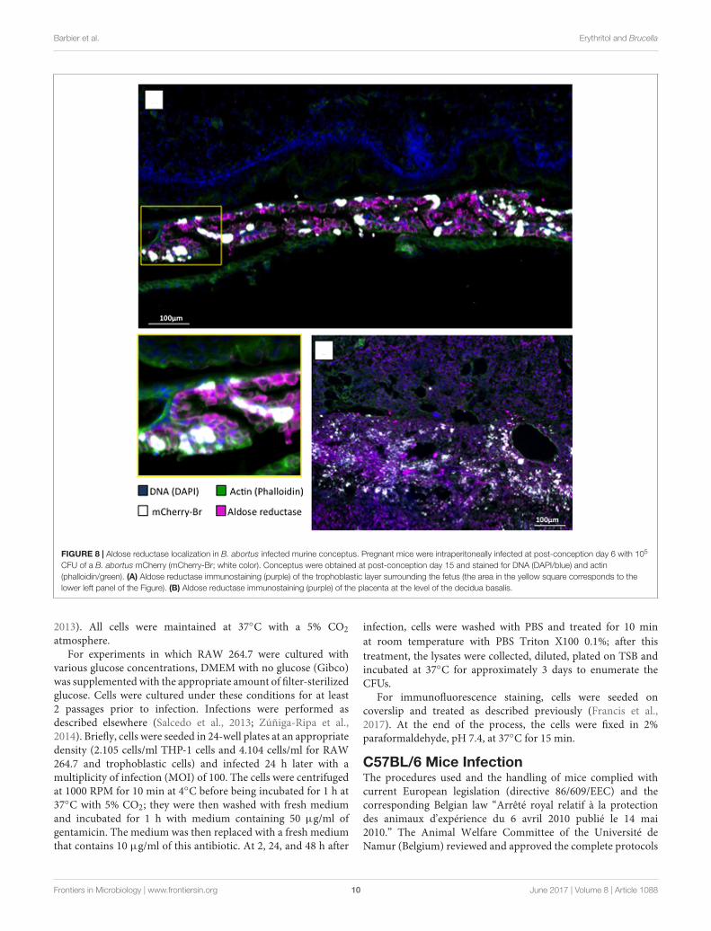
B. abortus 2308-mCherry at 6 days post conception. Bacteriaand AR-positive cells were located preferentially in the junctionalzone of the placenta just underneath the decidua basalis and ina cellular sheet surrounding the fetus apposed on the internalface of the distended decidua parietalis (Figure 7A). Infectedcells were positive for cytokeratin 7 (Figures 7B,C) and thustrophoblastic in nature (Croy et al., 2013), probably beingtrophoblastic giant cells (Kim et al., 2005). As expected, someinfected trophoblasts were also scattered in the decidua aftermid-gestation (Hu and Cross, 2010). Bacteria were found almostexclusively in AR-positive cells (Figure 8).
Finally, we investigated mouse spleens 9 days after infection,a time at which the erythritol-sensitive strain 1eryH showedattenuation. We found intense AR-staining in the red pulp(Supplementary Figure S3), with small clusters of CD11b-positive cells [often iNOS positive and corresponding togranulomas (Copin et al., 2012)], which are frequently associatedwith AR. In contrast, we hardly detected AR in spleens ofnon-infected mice; when we did, AR was mostly restricted toscattered CD11b-negative cells in the red pulp.
Frontiers in Microbiology | www.frontiersin.org 6 June 2017 | Volume 8 | Article 1088
FIGURE 5 | Brucella abortus1eryH but not 1eryA show reduced ability tocolonize the mouse conceptus. Pregnant C56BL/6 mice were inoculatedintraperitoneally with 105 B. abortus 2308 WT, 1eryH or 1eryA mutant at 6 or14 days post conception (P-C). At day 15 P-C, individual conceptuses weredissected to separate the placenta (P), the fetus (F) and the fetal envelopes (E)and CFU determined. Each dot represents the CFU counts obtained from oneconceptus and bars represent the mean ± standard deviation (∗∗p < 0.01;∗∗∗p < 0.001; Mann–Whitney test).
DISCUSSION
In this work, we set up and validated a reporting systemto detect the presence and catabolism of erythritol in theBrucella replicative niche, and this system demonstratedthe availability of this polyol in infection models of bovine,human and murine origin, extending previous researchin macrophages and mice (Burkhardt et al., 2005) totrophoblastic cell lines. Indeed, because erythritol is presentin comparatively large amounts in the placenta and genitaltissues of ruminants and swine and because Brucella is
found inside trophoblastic cells of ruminants and useserythritol very efficiently, it has been widely assumed thattrophoblasts produce erythritol. However, most evidenceis limited to extracts of fetal allantoic and amniotic fluids,cotyledons, whole placenta, seminal vesicles and testis(Smith et al., 1962; Williams et al., 1962; Clark et al.,1967), and to the best of our knowledge, only one workhas reported the presence of erythritol in trophoblasts ofbovine origin (Enright and Samartino, 1994). Our workconfirms this pattern and provides the first experimentaldata that support the presence of erythritol in human andmurine trophoblastic cells. In addition, we demonstratefor the first time that the catabolism of erythritol is notessential to the infectious processes in these infectionmodels.
The fact that bovine and human trophoblastic cells andmurine models give similar results contrasts with the lowerythritol concentrations that were reported in fetal fluidsof humans and mice (approximately 60 µg/ml in cows andless than 2 µg/ml in humans or mice) (Keppie et al.,1965; Amin and Wilsmore, 1997). Based on our in vitrotoxicity assays in 2YT, it can be speculated that erythritolconcentration should reach 50–100 µg/ml to result in 1eryHattenuation during infection. If correct, these differences inerythritol measurements in fetal fluids could reflect the particularcomposition of the Brucella replicative niche during infection.It is apparently puzzling that, although our reporting systemshows that erythritol is catabolized “in vivo,” it also showsthat erythritol is not an essential carbon source, indicating acomplex nutritional situation in the replicative niche whereerythritol, but also alternative C sources, should be availablein various and evolving proportions based on the time ofinfection, the type of cell infected and other variables. A possibleexplanation for this situation could be the presence of anactive polyol pathway because this pathway (which depends onAR; see below) can supply not only erythritol but also otherpolyols such as glycerol, arabitol, mannitol and inositol thatare in fact found in fetal tissues and reproductive systems ofseveral Brucella hosts (Jauniaux, 2005). These polyols, erythritolincluded, may not be critical individually but could be alternativecarbon sources. Of course, a definite answer needs specificinvestigations carried out in the natural host species, notonly with B. abortus, B. melitensis and B. suis but also withB. ovis and B. canis, the two classical species not stimulated byerythritol.
Since our reporting system showed erythritol in cells inwhich its presence has not been described, we looked forpossible biosynthetic mechanisms in mammal tissues. Over50 years ago, Pearce et al. (1962) proposed that erythritol “mayarise from D-erythrose [ . . .] as an intermediate between D-erythrose and D-erythrulose as sorbitol acts as an intermediatebetween glucose and fructose.” The enzyme responsible for theconversion of glucose to sorbitol is the aldose reductase (AR,AKR1B1 in human and bovines, AKR1B3 in mice) of thepolyol pathway, which oxidizes sorbitol to fructose. Thus, weinvestigated the presence of AR in cells and tissues whereB. abortus multiplied and where the presence of erythritol
Frontiers in Microbiology | www.frontiersin.org 7 June 2017 | Volume 8 | Article 1088
FIGURE 6 | The expression of Aldose reductase gene Akr1b3 in RAW 264.7 macrophages depends on glucose concentration. (A) Expression of gene Akr1b3measured by qRTPCR in macrophages cultured with 0.5, 1, and 4.5 g/L of glucose. (B) Multiplication of B. abortus 2308 WT and 1eryH in macrophages culturedwith 0.5, 1, and 4.5 g/L of glucose. All experiments were performed in biological and technical duplicates (∗p < 0.05; ∗∗p < 0.01; ∗∗∗p < 0.001 [Student’s t-test]).
was detected. We found that AR was present in RAW 264.7macrophages independently of an infection, that 9 days afterinfection there was a sharp increase in AR in the splenicred pulp where clusters of CD11b+ (indicative of Brucella-induced granulomas) cells co-localized with AR and that ARand B. abortus co-localized in the infected murine conceptus.There is abundant indirect evidence that this coexistenceof AR and brucellae in the laboratory models parallels thesituation in the natural hosts. Actually, tissues characteristicallytargeted by brucellae such as the placenta of cows, sheepand pigs, and the epididymis, seminal fluids and oviduct ofpigs, cattle and some rodents, which are among those tissueswhose fructose concentrations are high or are predominantover glucose, contain abundant amount of AR (Clark et al.,1967; Frenette, 2006; Pruneda et al., 2006). Moreover, AR(AKR1B1) has also been recently identified by proteomics asdifferentially produced in bovine chorioallantoic membranesthat were infected by Brucella (Mol et al., 2016). Indeed, ARcan reduce a broad range of aldehydes to their correspondingalcohols (Håstein and Velle, 1968) and significantly its affinityis far higher for erythrose than for glucose (Hayman andKinoshita, 1965; Kardon et al., 2008). Furthermore, a role ofAR in erythritol generation in these tissues is consistent withthe fact that the pentose phosphate pathway, which can supplyD-erythrose, is active in testes, ovaries and placenta (Ferrier,2013). Although further research is necessary, all these indirectevidences together with the data presented here, lend supportto the hypothesis that AR accounts for erythritol productionin cells that have been invaded by brucellae, as well as for theapparently puzzling observation that erythritol is not essentialfor Brucella multiplication. In preliminary experiments, we havefound that treatment of murine macrophages with the potentAR inhibitor Sulindac (Ratliff et al., 1999) impairs B. abortus2308 intracellular multiplication, an observation that is alsoconsistent with our hypothesis. It is also worth commentingthat AR is a moonlighting protein that in addition to itsfunction in the polyol pathway, has been linked to inflammationregulation (Ramana and Srivastava, 2010) and is involved in thehormonal regulation of pregnancy and parturition. Some ARare, as a matter of fact, involved in progesterone degradationand are also the main human, murine and bovine prostaglandin
F2α (PGF2α) synthase (Madore et al., 2003; Kabututu et al.,2008; Bresson et al., 2011, 2012). It is thus tempting tohypothesize that AR could represent an actor in the contextof Brucella–host interaction at the crossroad of metabolism,inflammation and abortion, a possibility that deserves furtherinvestigation.
MATERIALS AND METHODS
Bacterial Strains and Culture ConditionsEscherichia coli DH10B were grown in LB medium. B. abortus2308 NalR and derived strains were grown at 37◦C in richmedium 2YT (16 g/L bacto tryptone 10 g/L yeast extractand 5 g/L NaCl; BD Difco) or in a chemically definedmedium (Barbier et al., 2014) composed of 2.3 g/L K2HPO4;3 g/L KH2PO4; 0.1 g/L Na2S2O3; 5 g/L NaCl; 0.2 g/Lnicotinic acid; 0.2 g/L thiamine; 0.07 g/L pantothenic acid;0.5 g/L (NH4)2SO4; 0.01 g/L MgSO4; 0.1 mg/L MnSO4;0.1 mg/L FeSO4; 0.1 mg/L biotin and 2 g/L of erythritol.Growth was monitored using an automated plate reader(Bioscreen C, Lab Systems) following the OD (600 nm)with continuous shaking at 37◦C. The growth rate wascalculated as follows:
(ln (ODt2)− ln (ODt1)
) /(t2− t1).
The 1t was set for 7 h, i.e., approximately two divisiontimes, and incremented over the log phase every 0.5 h(0–7 h; 0.5–7.5 h,. . .) resulting in a set of values whose meanis the average growth rate, µ. When required, the mediumwas supplemented with chloramphenicol (20 mg/ml), nalidixicacid (25 mg/ml), sucrose (5%), agar (15 g/L, BD Difco)and polyols (concentrations annotated in the manuscript).Unless otherwise stated, reagents were purchased fromSigma–Aldrich.
Construction of an mCherry-producing B. abortus2308 strain was performed following the same procedurethat was validated for B. melitensis (Copin et al., 2012).Construction of the in-frame deletion in eryA was donefollowing a previously described strategy (Barbier et al.,2014). Briefly, approximately 750 bp upstream anddownstream of BAB2_0372 were amplified by PCR fromgenomic DNA of B. abortus 2308. The obtained PCR
Frontiers in Microbiology | www.frontiersin.org 8 June 2017 | Volume 8 | Article 1088
FIGURE 7 | Localization of B. abortus 2308 in murine conceptus. Pregnant mice were infected intraperitoneally at 6 days post-conception with 105 CFU ofB. abortus mCherry (white), and the conceptuses were obtained at post-conception day 15. (A) Mosaic reconstitution (210 individual images taken at 10×magnification) of a sagittal section of an infected murine conceptus stained for DNA (DAPI/blue) and actin (phalloidin/green); dashed squares B and C circumscribethe prototypal zone corresponding to (B,C). (B) Cytokeratine-7 immunostaining (light blue) of trophoblastic cells of the dorsal part of the conceptus with a close upof the infected trophoblastic cells lining the decidua parietalis (yellow square). (C) Cytokeratine-7 immunostaining (light blue) of the placenta at the level of thedecidua basalis. TGC, trophoblast giant cell; ST, spongiotrophoblast; L, labyrinth.
products were, respectively, flanked by SpeI/BamHI (SpeI_F:5′-ACTAGTCTTGGCGGAAACTTGACTGG-3′; BamHI_R: 5′-ATACGCGGATCCGCGATAACGCATGGCTGACACAGG-3′)and BamHI/SphI restriction sites (BamHI_F: 5′-TATCGCGGATCCGCGTATGGCAAATAAGGAAACATTGAATG-3′; SphI_R: 5′-GCATGCGCGCTTGTCGTGGTTCTG-3′).A third PCR joined the two fragments together using primersSpeI_F and SphI_R, which was followed by ligating thisproduct into an EcoRV-digested pGEM plasmid (Promega).After sequence verification (Beckman Coulter Genomics),the ±1500 bp insert was excised as a SpeI – SphI fragmentand cloned into a pNPTS138 suicide vector (KanR, SucS).The acquisition of this vector by Brucella after mating withconjugative S17 E. coli was selected by kanamycin and nalidixicacid resistance. The loss of the plasmid concomitant with eithera deletion or a return to WT phenotype was then selectedon sucrose. Mutants were identified using PCR with primersthat were located external to the deletion. The 1eryH and1eryI strains were previously characterized (Barbier et al.,2014). For complementation, eryH was amplified by PCRfrom genomic DNA of B. abortus 2308 as a BamHI/XhoIfragment (BamHI_F: 5′-gcgggatccatgaccaaattctggattgg-3′;XhoI_R: 5′-ttaattcgcttgaaccttggctcgagccg-3′). Fragments werecloned into an EcoRV-digested pGEM, sequenced and thentransferred into a pBBR1MCS1 (CmR). The 1eryH strain with
the construct was then transformed by conjugation with theconstruction and selected for with the newly acquired resistanceto chloramphenicol.
All Brucellawere handled under BSL-3 containment accordingto the Directive 98/81/CE du Conseil du 26 octobre 1998and to a law of the Gouvernement wallon du 4 juillet2002.
Cell Culture and InfectionRAW 264.7 murine macrophages were routinely culturedin Dulbecco’s modified Eagle’s medium with high glucose(DMEM, Gibco) supplemented with 10% heat-inactivated fetalcalf serum (FCS, Gibco). THP-1 human macrophage-likecells were cultured in RPMI 1640 medium (Gibco) that wassupplemented with 10% FCS and 2 mM L-glutamine. Cellswere differentiated into adherent monocytes by overnighttreatment with 5 nM phorbol myristate (PMA). Bovinetrophoblastic CL2 cells were kindly provided by Pr. CynthiaBaldwin (University of Massachusetts, Amherst, MA, UnitedStates) and cultured in RPMI 1640 supplemented with 10%FCS and 0.05 mM 2-mercaptoethanol (Gibco) as previouslydescribed (Parent et al., 2012). BeWo human trophoblasticcells (ATCC clone CCL-98) were cultured in DMEM-F12Ham medium (Gibco) that was supplemented with 10% FCSand 2 mM L-glutamine as already described (Salcedo et al.,
Frontiers in Microbiology | www.frontiersin.org 9 June 2017 | Volume 8 | Article 1088
FIGURE 8 | Aldose reductase localization in B. abortus infected murine conceptus. Pregnant mice were intraperitoneally infected at post-conception day 6 with 105
CFU of a B. abortus mCherry (mCherry-Br; white color). Conceptus were obtained at post-conception day 15 and stained for DNA (DAPI/blue) and actin(phalloidin/green). (A) Aldose reductase immunostaining (purple) of the trophoblastic layer surrounding the fetus (the area in the yellow square corresponds to thelower left panel of the Figure). (B) Aldose reductase immunostaining (purple) of the placenta at the level of the decidua basalis.
2013). All cells were maintained at 37◦C with a 5% CO2atmosphere.
For experiments in which RAW 264.7 were cultured withvarious glucose concentrations, DMEM with no glucose (Gibco)was supplemented with the appropriate amount of filter-sterilizedglucose. Cells were cultured under these conditions for at least2 passages prior to infection. Infections were performed asdescribed elsewhere (Salcedo et al., 2013; Zúñiga-Ripa et al.,2014). Briefly, cells were seeded in 24-well plates at an appropriatedensity (2.105 cells/ml THP-1 cells and 4.104 cells/ml for RAW264.7 and trophoblastic cells) and infected 24 h later with amultiplicity of infection (MOI) of 100. The cells were centrifugedat 1000 RPM for 10 min at 4◦C before being incubated for 1 h at37◦C with 5% CO2; they were then washed with fresh mediumand incubated for 1 h with medium containing 50 µg/ml ofgentamicin. The medium was then replaced with a fresh mediumthat contains 10 µg/ml of this antibiotic. At 2, 24, and 48 h after
infection, cells were washed with PBS and treated for 10 minat room temperature with PBS Triton X100 0.1%; after thistreatment, the lysates were collected, diluted, plated on TSB andincubated at 37◦C for approximately 3 days to enumerate theCFUs.
For immunofluorescence staining, cells were seeded oncoverslip and treated as described previously (Francis et al.,2017). At the end of the process, the cells were fixed in 2%paraformaldehyde, pH 7.4, at 37◦C for 15 min.
C57BL/6 Mice InfectionThe procedures used and the handling of mice complied withcurrent European legislation (directive 86/609/EEC) and thecorresponding Belgian law “Arrêté royal relatif à la protectiondes animaux d’expérience du 6 avril 2010 publié le 14 mai2010.” The Animal Welfare Committee of the Université deNamur (Belgium) reviewed and approved the complete protocols
Frontiers in Microbiology | www.frontiersin.org 10 June 2017 | Volume 8 | Article 1088
(Permit Number 16/277). All infections were performed at anAnimal Biosafety Level 3 facility.
To obtain the inoculum, bacteria from an overnight cultureof Brucella in rich medium were pelleted, washed with RPMI1640 and diluted in this medium. Intraperitoneal infection wascarried out as previously described (Copin et al., 2012). Briefly,500 µl of suspension (105 CFU) was injected into groups of 8 to12 C57BL/6 mice for each tested strain. Mice were euthanized 3,9, and 30 days post-infection by cervical dislocation, the spleenswere isolated, weighted and homogenized in 1 ml of PBS TritonX100 0.1%, and the CFU were counted on tryptic soy agarplates.
The procedure that was used to infect pregnant mice wasadapted from previous reports (Bosseray, 1982; Kim et al., 2005;Pennington et al., 2012). Estruses of 6–14 weeks old C57BL/6females were synchronized 3 days before mating pairs were setup with males that were 3–4 months old. Then, the presenceof a vaginal plug was checked daily, and potentially fertilizedfemales were isolated. That day corresponds to day 0 post-fecundation (PF). Four to five pregnant females were infectedintraperitoneally with 500 µl of bacterial suspension that wasprepared as previously described (105 bacteria) at day 6 or14 PF. At day 15 PF, mice were anesthetized with isoflurane(Zoetis) and euthanized by cervical dislocation. Conceptuseswere removed from maternal uterine horns and transferredto sterile Petri dishes on ice, where they stayed for 15 min.Placenta, fetuses and surrounding fetal membranes were thenfurther isolated and weighed. Tissues were homogenized in 1 mlPBS Triton X100 0.1% with an Ultra-Turrax homogenizer, thehomogenates were serially diluted in PBS, and their CFU werecounted.
Immunofluorescence MicroscopyFetuses and spleens were fixed for 2 h at room temperature in2% paraformaldehyde (pH 7.4), washed in PBS, and incubatedovernight at 4◦C in a 20% PBS-sucrose solution. The tissueswere then embedded in Tissue-Tek OCT compound (Sakura)and frozen in liquid nitrogen, and cryostat sections (thickness,5 µm for spleens and 10 µm for fetus) were prepared. For thestaining, tissue sections were rehydrated in PBS and incubatedin a PBS solution that contained 1% blocking reagent (PBS-BR1%, Boehringer) for 20 min before they were incubated overnightin PBS-BR 1% containing mAbs or the following reagents:DAPI nucleic acid stain Alexa Fluor 350 or 488 phalloidin(Molecular Probes) to visualize the structure of the organ, and ratbiotin-coupled anti-mouse CD11b (BD Pharmingen), rabbit anti-mouse iNOS (Calbiochem), rabbit anti-mouse Cytokeratin 7 (ab181598, Abcam), and rabbit anti-mouse AR (CPA3124, CohesionBiosciences) to stain the cells of interest. The samples wereincubated with the appropriate secondary reagents [Alexa Fluor568 streptavidin (Molecular Probes) or Alexa Fluor 647-coupleddonkey anti-rabbit IgG (Molecular Probes)] for 2 h. Slideswere mounted in Fluoro-Gel medium (Electron MicroscopySciences, Hatfield, PA, United States). Labeled tissue sectionswere visualized with an Axiovert M200 inverted microscope(Zeiss, Iena, Germany) that was equipped with a high-resolutionmonochrome camera (AxioCam HR, Zeiss).
Images (1384 pixels × 1036 pixels, 0.16 µm/pixel) wereacquired sequentially for each fluorochrome with A-Plan10×/0.25 N.A. and LD-Plan-NeoFluar 63×/0.75 N.A. dryobjectives and recorded as eight-bit gray-level ∗.zvi files. Atleast three slides per organ were analyzed from three differentanimals, and the results are representative of two independentexperiments.
For immunostaining of AR in RAW 264.7 murinemacrophages, the primary antibody that was used was thesame that was used for staining in mice with a goat anti-rabbitIgG Alexa 488 (Life Technologies) as the secondary antibody.
Measurement of the Murine AldoseReductase AKR1B3 Expression in RAW264.7 by qRT-PCRRNA from cells cultivated in a T75 flask was extracted withTriPure isolation reagent (Roche) according to the instructionsof the manufacturer and DNA contamination was eliminatedby incubation with DNase I (Fermentas). Then, RNA was firstreverse transcribed (two steps) by SuperScript II (Invitrogen)into cDNA, which was then amplified in a LightCycler 96Instrument (Roche) with FastStart Universal SYBR GreenMaster (Roche) as the fluorescent dye. The specificity of theSYBR Green assays was assessed by melting-point analysisand gel electrophoresis. The results were normalized using thehousekeeping b-actin gene. Primer sequences are described inSupplementary Table S1.
AUTHOR CONTRIBUTIONS
TB, AZ-R, IM and J-JL conceived the study. AM and EM wereresponsible for the immunofluorescence microscopy analysis.XDB supervised all the molecular approaches. HP, CH, and ELcontributed in the mutant construction, growth curves and testedthem in cells and mice. TB and AZ-R were the main researchersinvolved in mutant and metabolic tests. EVS brought a lot ofinput in the aldose reductase and polyol pathway. J-JL, TB, andIM wrote the paper. All the authors read and commented on thepaper.
FUNDING
J-JL’s team is supported by an FNRS grant (Fonds de laRecherche Fondamentale Collective Grant N◦ 2452110) andby the Interuniversity Attraction Poles Programme initiated bythe Belgian Science Policy Office. TB has a Ph.D. grant as“Aspirant FNRS.” AM and EL have a Ph.D. grant from the FRIA.Research at the University of Navarra is supported by grantsfrom the Ministerio de Economía y Competitividad of Spain(AGL2011-30453-C04-00) and the Institute for Tropical Healt.The E.V.S. laboratory is supported by a Welbio grant of theWalloon Region and by a grant from the Fonds de la RechercheScientifique Médicale. EM is a Research Associate at the Fonds dela Recherche Scientifique (FRS)–FNRS (Belgium).
Frontiers in Microbiology | www.frontiersin.org 11 June 2017 | Volume 8 | Article 1088
The Supplementary Material for this article can be foundonline at: http://journal.frontiersin.org/article/10.3389/fmicb.2017.01088/full#supplementary-material
FIGURE S1 | Brucella abortus 2308 1eryA and 1eryH are not able to growwith erythritol as the only carbon source. The growth of the 1eryA mutantwas monitored in rich medium 2YT or in chemically defined medium with erythritolas the only carbon source and compared to 1eryH {Barbier:2014 cm}. Asexpected, the growth of the deletion strains is abolished when only erythritol isavailable.
FIGURE S2 | The growth of the erythritol-sensitive B. abortus 2308 1eryH strainis not affected by other polyols. Growth was monitored in 2YT supplemented withpolyols structurally close to erythritol that are also found in fetal fluids and tissues.The growth of the 1eryH mutant was not affected by any of the polyols tested
strongly suggesting that the toxicity is specific for erythritol. The experiment wasperformed in biological triplicates and technical duplicates. Values represent theaverage of one representative experiment ± standard deviation (shaded gray anddashed lines).
FIGURE S3 | Aldose reductase localization in the spleen of mice infected withB. abortus 2308. Mice were infected intraperitoneally with 105 CFU of B. abortus2308, spleens obtained at day 9 post infection and stained for actin(phalloidin/gray) and CD11b (green). (A) iNOS immunostaining (blue) of spleensections of non-infected (control, Left), and infected (Br.9d, Right), mice. The red
pulp shows small clusters of CD11b-positive cells (often iNOS positive) thatcorrespond to Brucella granulomas {Copin:2012ee}. (B) Aldose reductase
immunostaining (purple) of spleen sections of non-infected and infected mice. Inthe non-infected mice (control, Left), aldose reductase was scarcely detected andwas restricted to a few and CD11b-negative cells of the red pulp. In infected mice9 days post-infection (Br.9d, Right), aldose reductase was abundant in the redpulp in the same areas where the Brucella granulomas developed.
REFERENCESAl-Tawfiq, J. A., and Memish, M. A. (2013). Pregnancy associated
brucellosis. Recent Pat. Antiinfect. Drug Discov. 8, 47–50. doi: 10.2174/1574891X11308010009
Amin, J. D., and Wilsmore, A. J. (1997). The effects of crude placental extractand erythritol on growth of Chlamydia psittaci (ovis) in McCoy cells. Vet. Res.Commun. 21, 431–435. doi: 10.1023/A:1005807402736
Anderson, T. D., and Cheville, N. F. (1986). Ultrastructural morphometric analysisof Brucella abortus-infected trophoblasts in experimental placentitis. Bacterialreplication occurs in rough endoplasmic reticulum. Am. J. Pathol. 124,226–237.
Anderson, T. D., Cheville, N. F., and Meador, V. P. (1986a). Pathogenesis ofplacentitis in the goat inoculated with Brucella abortus. II. Ultrastructuralstudies. Vet. Pathol. 23, 227–239.
Anderson, T. D., Meador, V. P., and Cheville, N. F. (1986b). Pathogenesis ofplacentitis in the goat inoculated with Brucella abortus. I. Gross and histologiclesions. Vet. Pathol. 23, 219–226.
Barbier, T., Collard, F., Zúñiga-Ripa, A., Moriyón, I., Godard, T., Becker, J.,et al. (2014). Erythritol feeds the pentose phosphate pathway via threenew isomerases leading to D-erythrose-4-phosphate in Brucella. Proc.Natl. Acad. Sci. U.S.A. 111, 17815–17820. doi: 10.1073/pnas.1414622111
Blasco, J. M. (1990). “Brucella ovis,” in Animal Brucellosis, eds K. H. Nielsen andJ. R. Ducan (Boca Raton, FL: CRC Press), 352–378.
Bosseray, N. (1980). Colonization of mouse placentas by Brucella abortusinoculated during pregnancy. Br. J. Exp. Pathol. 61, 361–368.
Bosseray, N. (1982). Mother to young transmission of Brucella abortus infection inmouse model. Ann. Rech. Vét. 13, 341–349.
Bosseray, N. (1983). Kinetics of placental colonization of mice inoculatedintravenously with Brucella abortus at day 15 of pregnancy. Br. J. Exp. Pathol.64, 612–616.
Bresson, E., Boucher-Kovalik, S., Chapdelaine, P., Madore, E., Harvey, N., Laberge,P. Y., et al. (2011). The human aldose reductase AKR1B1 qualifies as the primaryprostaglandin F synthase in the endometrium. J. Clin. Endocrinol. Metab. 96,210–219. doi: 10.1210/jc.2010-1589
Bresson, E., Lacroix-Pépin, N., Boucher-Kovalik, S., Chapdelaine, P., and Fortier,M. A. (2012). The prostaglandin F synthase activity of the human aldosereductase AKR1B1 brings new lenses to look at pathologic conditions. Front.Pharmacol. 3:98. doi: 10.3389/fphar.2012.00098/abstract
Brusati, V., Józwik, M., Józwik, M., Teng, C., Paolini, C., Marconi, A. M.,et al. (2005). Fetal and maternal Non-glucose carbohydrates and polyolsconcentrations in normal human pregnancies at term. Pediatr. Res. 58, 700–704.doi: 10.1203/01.PDR.0000180549.86614.73
Burkhardt, S., Jiménez de Bagüés, M. P., Liautard, J.-P., and Köhler, S. (2005).Analysis of the behavior of eryC mutants of Brucella suis attenuated inmacrophages. Infect. Immun. 73, 6782–6790. doi: 10.1128/IAI.73.10.6782-6790.2005
Carmichael, L. E. (1990). “Brucella canis,” in Animal Brucellosis, eds K. H. Nielsenand J. R. Ducan (Boca Raton, FL: CRC Press), 336–350.
Clark, J. B., Graham, E. F., Lewis, B. A., and Smith, F. (1967). D-mannitol, erythritoland glycerol in bovine semen. J. Reprod. Fertil. 13, 189–197. doi: 10.1530/jrf.0.0130189
Copin, R., Vitry, M.-A., Hanot Mambres, D., Machelart, A., De Trez, C.,Vanderwinden, J.-M., et al. (2012). In situ microscopy analysis reveals localinnate immune response developed around Brucella infected cells in resistantand susceptible mice. PLoS Pathog. 8:e1002575. doi: 10.1371/journal.ppat.1002575.g009
Croy, B. A., Yamada, A. T., DeMayo, F. J., and Adamson, S. L. (2013). The Guide toInvestigation of Mouse Pregnancy. Amsterdam: Academic Press.
de Lorenzo, V. (2014). From the selfish gene to selfish metabolism: revisiting thecentral dogma. Bioessays 36, 226–235. doi: 10.1002/bies.201300153
Dozot, M., Poncet, S., Nicolas, C., Copin, R., Bouraoui, H., Mazé, A., et al. (2010).Functional characterization of the incomplete phosphotransferase system(PTS) of the intracellular pathogen Brucella melitensis. PLoS ONE 5:e12679.doi: 10.1371/journal.pone.0012679
Enright, F. M., and Samartino, L. E. (1994). Mechanisms of abortion in Brucellaabortus infected cattle. Proc. Annu. Meet U.S. Anim. Health Assoc. 98, 55–63.doi: 10.1186/1471-2164-14-426
Fernandez, A. G., Ferrero, M. C., Hielpos, M. S., Fossati, C. A., and Baldi, P. C.(2016). Proinflammatory response of human trophoblastic cells to Brucellaabortus infection and upon interactions with infected phagocytes. Biol. Reprod.94, 48. doi: 10.1095/biolreprod.115.131706
Ferrier, D. R. (ed.) (2013). Biochemistry, 6 Edn. Hagerstwon: Lippincott Williams &Wilkins.
Francis, N., Poncin, K., Fioravanti, A., Vassen, V., Willemart, K., Ong, T. A. P.,et al. (2017). CtrA controls cell division and outer membrane composition ofthe pathogen Brucella abortus. Mol. Microbiol. 103, 780–797. doi: 10.1111/mmi.13589
Frenette, G. (2006). Polyol pathway in human epididymis and semen. J. Androl. 27,233–239. doi: 10.2164/jandrol.05108
González, R. G., Barnett, P., Aguayo, J., Cheng, H. M., and Chylack, L. T. (1984).Direct measurement of polyol pathway activity in the ocular lens. DiabetesMetab. Res. Rev. 33, 196–199. doi: 10.2337/diab.33.2.196
Håstein, T., and Velle, W. (1968). Placental aldose reductase activity andfoetal blood fructose during bovine pregnancy. J. Reprod. Fertil. 15, 47–52.doi: 10.1530/jrf.0.0150047
Hayman, S., and Kinoshita, J. H. (1965). Isolation and properties of lens aldosereductase. J. Biol. Chem. 240, 877–882.
Hu, D., and Cross, J. C. (2010). Development and function of trophoblast giantcells in the rodent placenta. Int. J. Dev. Biol. 54, 341–354. doi: 10.1387/ijdb.082768dh
Jauniaux, E. (2005). Polyol concentrations in the fluid compartments of thehuman conceptus during the first trimester of pregnancy: maintenance ofredox potential in a low oxygen environment. J. Clin. Endocrinol. Metab. 90,1171–1175. doi: 10.1210/jc.2004-1513
Frontiers in Microbiology | www.frontiersin.org 12 June 2017 | Volume 8 | Article 1088
Jones, L. M., Montgomery, V., and And Wilson, J. B. (1965). Characteristics ofcarbon dioxide-independent cultures of Brucella abortus isolated from cattlevaccinated with strain 19. J. Inf. Dis. 115, 312–320. doi: 10.1093/infdis/115.3.312
Kabututu, Z., Manin, M., Pointud, J. C., Maruyama, T., Nagata, N., Lambert, S.,et al. (2008). Prostaglandin F2 synthase activities of aldo-keto reductase 1B1,1B3 and 1B7. J. Biochem. 145, 161–168. doi: 10.1093/jb/mvn152
Karcaaltincaba, D., Sencan, I., Kandemir, O., Guvendag Guven, E. S., andYalvac, S. (2010). Does brucellosis in human pregnancy increase abortion risk?Presentation of two cases and review of literature. J. Obstet. Gynaecol. Res. 36,418–423. doi: 10.1111/j.1447-0756.2009.01156.x
Kardon, T., Stroobant, V., Veiga-da-Cunha, M., and Van Schaftingen, E.(2008). Characterization of mammalian sedoheptulokinase and mechanismof formation of erythritol in sedoheptulokinase deficiency. FEBS Lett. 582,3330–3334. doi: 10.1016/j.febslet.2008.08.024
Keppie, J., Williams, A., Witt, K., and Smith, H. (1965). The role oferythritol in the tissue localization of the brucellae. Br. J. Exp. Pathol. 46,104–108.
Keppie, J., Witt, K., and Smith, H. (1967). The effect of erythritol on the growth ofS19 and other attenuated strains of Brucella abortus. Res. Vet. Sci. 8, 294–296.
Khan, M. Y., Mah, M. W., and Memish, Z. A. (2001). Brucellosis in pregnantwomen. Clin. Infect. Dis. 32, 1172–1177. doi: 10.1086/319758
Kim, S., Lee, D., Watanabe, K., Furuoka, H., Suzuki, H., and Watarai, M. (2005).Interferon-γ promotes abortion due to Brucella infection in pregnant mice.BMCMicrobiol. 5:22. doi: 10.1186/1471-2180-5-22
Köhler, S., Foulongne, V., Ouahrani-Bettache, S., Bourg, G., Teyssier, J., Ramuz, M.,et al. (2002). The analysis of the intramacrophagic virulome of Brucella suisdeciphers the environment encountered by the pathogen inside the macrophagehost cell. Proc. Natl. Acad. Sci. U.S.A. 99, 15711–15716. doi: 10.1073/pnas.232454299
Larose, J., Laflamme, J., Côté, I., Lapointe, J., Frenette, G., Sullivan, R., et al. (2012).The polyol pathway in the bovine oviduct. Mol. Reprod. Dev. 79, 603–612.doi: 10.1002/mrd.22067
Letesson, J.-J., Barbier, T., Zúñiga-Ripa, A., Godfroid, J., De Bolle, X., andMoriyón, I. (2017). Brucella genital tropism: what’s on the menu. Front.Microbiol. 8:506. doi: 10.3389/fmicb.2017.00506
Madore, E., Harvey, N., Parent, J., Chapdelaine, P., Arosh, J. A., and Fortier, M. A.(2003). An aldose reductase with 20 -hydroxysteroid dehydrogenase activity ismost likely the enzyme responsible for the production of prostaglandin F2 inthe bovine endometrium. J. Biol. Chem. 278, 11205–11212. doi: 10.1074/jbc.M208318200
McCullough, W. G., and Beal, G. A. (1951). Growth and manometric studies oncarbohydrate utilization of Brucella. J. Infect. Dis. 89, 266–271. doi: 10.1093/infdis/89.3.266
Mol, J. P. S., Pires, S. F., Chapeaurouge, A. D., Perales, J., Santos, R. L., Andrade,H. M., et al. (2016). Proteomic profile of Brucella abortus-infected bovinechorioallantoic membrane explants. PLoS ONE 11:e0154209. doi: 10.1371/journal.pone.0154209
Moreno, E., and Moriyón, I. (2006). “The genus Brucella,” in The Prokaryotes,eds M. Dworkin, S. Falkow, E. Rosenberg, K.-H. Schleifer, and E. Stackebrandt(New York, NY: Springer), doi: 10.1007/0-387-30745-1_17
Nataro, J. P. (2015). “Pathogenesis — Thoughts from the front line,” in Metabolismand Bacterial Pathogenesis, eds T. Conway and P. S. Cohen (Washington, DC:American Society of Microbiology), 17–26. doi: 10.1128/microbiolspec.MBP-0012-2014
Navarro-Martinez, A., Solera, J., Corredoira, J., Beato, J. L., Martinez, A. E.,Atienzar, M., et al. (2001). Epididymoorchitis due to Brucella melitensis: aretrospective study of 59 patients. Clin. Infect. Dis. 33, 2017–2022. doi: 10.1086/324489
Parent, M. A., Bellaire, B. H., Murphy, E. A., Roop, R. M., Elzer, P. H., and Baldwin,C. L. (2012). Brucella abortus siderophore 2,3-dihydroxybenzoic acid (DHBA)facilitates intracellular survival of the bacteria. Microb. Pathog. 32, 239–248.doi: 10.1006/mpat.2002.0500
Pearce, J., Williams, A., Harris-Smith, P. W., Fitzgeorge, R., and Smith, H. (1962).The chemical basis of the virulence of Brucella abortus: II. Erythritol, aconstituent of bovine foetal fluids which stimulates the growth of Br. abortusin bovine phagocytes. Br. J. Exp. Pathol. 43, 31–37.
Pennington, K. A., Schlitt, J. M., and Schulz, L. C. (2012). Isolation of primarymouse trophoblast cells and trophoblast invasion assay. J. Vis. Exp. 59:e3202.doi: 10.3791/3202
Pruneda, A., Pinart, E., Bonet, S., Yeung, C.-H., and Cooper, T. G. (2006). Study ofthe polyol pathway in the porcine epididymis. Mol. Reprod. Dev. 73, 859–865.doi: 10.1002/mrd.20481
Ramana, K. V., and Srivastava, S. K. (2010). Aldose reductase: a novel therapeutictarget for inflammatory pathologies. Int. J. Biochem. Cell Biol. 42, 17–20.doi: 10.1016/j.biocel.2009.09.009
Ratliff, D. M., Martinez, F. J., Vander Jagt, T. J., Schimandle, C. M., Robinson, B.,Hunsaker, L. A., et al. (1999). Inhibition of human aldose and aldehydereductases by non-steroidal anti-inflammatory drugs. Adv. Exp. Med. Biol. 463,493–499. doi: 10.1007/978-1-4615-4735-8_62
Regnault, T. R. H., Teng, C., de Vrijer, B., Galan, H. L., Wilkening, R. B., andBattaglia, F. C. (2010). The tissue and plasma concentration of polyols andsugars in sheep intrauterine growth retardation. Exp. Biol. Med. 235, 999–1006.doi: 10.1258/ebm.2010.009360
Salcedo, S. P., Chevrier, N., Lacerda, T. L. S., Ben Amara, A., Gerart, S., Gorvel,V. A., et al. (2013). Pathogenic brucellae replicate in human trophoblasts.J. Infect. Dis. 207, 1075–1083. doi: 10.1093/infdis/jit007
Samartino, L. E., and Enright, F. M. (1993). Pathogenesis of abortion of bovinebrucellosis. Comp. Immunol. Microbiol. Infect. Dis. 16, 95–101. doi: 10.1016/0147-9571(93)90001-L
Samartino, L. E., Traux, R. E., and Enright, F. M. (1994). Invasion andreplication of Brucella abortus in three different trophoblastic cell lines.Zentralbl. Veterinarmed. B 41, 229–236. doi: 10.1111/j.1439-0450.1994.tb00223.x
Sangari, F. J., Grilló, M. J., Jimenez de Bagues, M. P., González-Carreró, M. I.,García-Lobo, J. M., Blasco, J. M., et al. (1998). The defect in the metabolismof erythritol of the Brucella abortus B19 vaccine strain is unrelated withits attenuated virulence in mice. Vaccine 16, 1640–1645. doi: 10.1016/S0264-410X(98)00063-2
Smith, H., Williams, A., Pearce, E. J., Keppie, J., Harris-Smith, P. W., Fitz-George, R. B., et al. (1962). Foetal erythritol: a cause of the localization ofBrucella abortus in bovine contagious abortion.Nature 193, 47–49. doi: 10.1038/193047a0
Sperry, J. F., and Robertson, D. C. (1975). Inhibition of growth by erythritolcatabolism in Brucella abortus. J. Bacteriol. 124, 391–397.
Tawata, M., Ohtaka, M., Hosaka, Y., and Onaya, T. (1992). Aldose reductasemRNA expression and its activity are induced by glucose in fetal rat aorticsmooth muscle (A10) cells. Life Sci. 51, 719–726. doi: 10.1016/0024-3205(92)90480-D
Tobias, L., Cordes, D. O., and Schurig, G. G. (1993). Placental pathology of thepregnant mouse inoculated with Brucella abortus strain 2308. Vet. Pathol. 30,119–129. doi: 10.1177/030098589303000204
Vilchez, G., Espinoza, M., D’Onadio, G., Saona, P., and Gotuzzo, E. (2015).Brucellosis in pregnancy: clinical aspects and obstetric outcomes. Int. J. Infect.Dis. 38, 95–100. doi: 10.1016/j.ijid.2015.06.027
Williams, A. E., Keppie, J., and Smith, H. (1962). The chemical basis of the virulenceof Brucella abortus. III. Foetal erythritol a cause of the localisation of Brucellaabortus in pregnant cows. Br. J. Exp. Pathol. 43, 530–537.
Zinsstag, J., Schelling, E., Solera, J., Blasco, M. J., and Moriyón, I. (2011).“Brucellosis,” in Handbook of Zoonoses, eds R. S. Palmer, L. Soulsby, R. P.Torgeson, and G. D. Brown (Oxford: Oxford University Press), 54–62.
Zúñiga-Ripa, A., Barbier, T., Conde-Alvarez, R., Martínez-Gómez, E., Palacios-Chaves, L., Gil-Ramírez, Y., et al. (2014). Brucella abortus depends on pyruvatephosphate dikinase and malic enzyme but not on Fbp and GlpX fructose-1,6-bisphosphatases for full virulence in laboratory models. J. Bacteriol. 196,3045–3057. doi: 10.1128/JB.01663-14
Conflict of Interest Statement: The authors declare that the research wasconducted in the absence of any commercial or financial relationships that couldbe construed as a potential conflict of interest.